Abstract
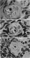
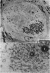
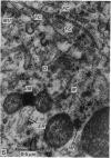
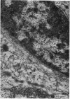
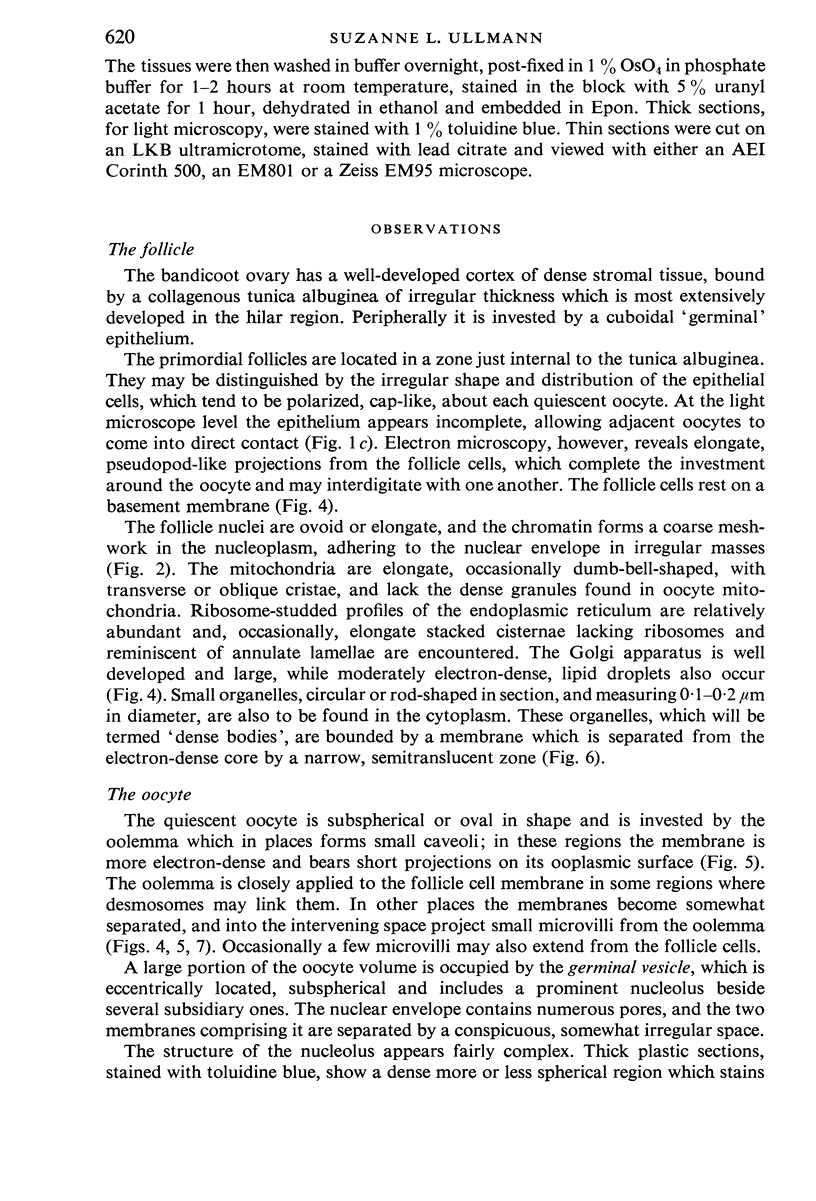
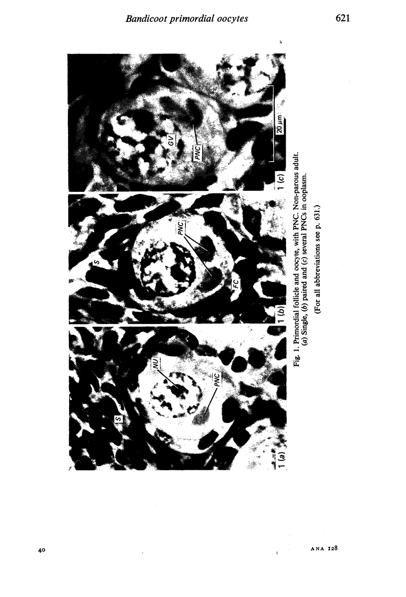
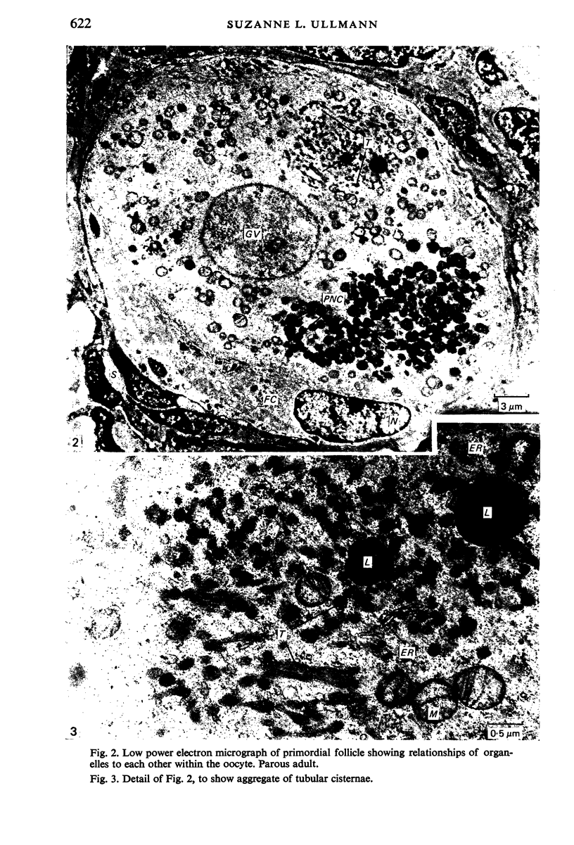
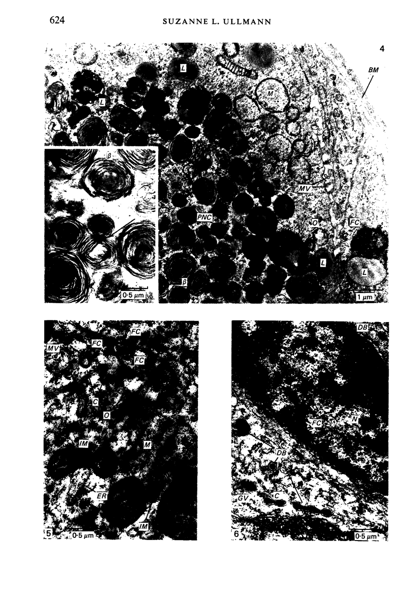
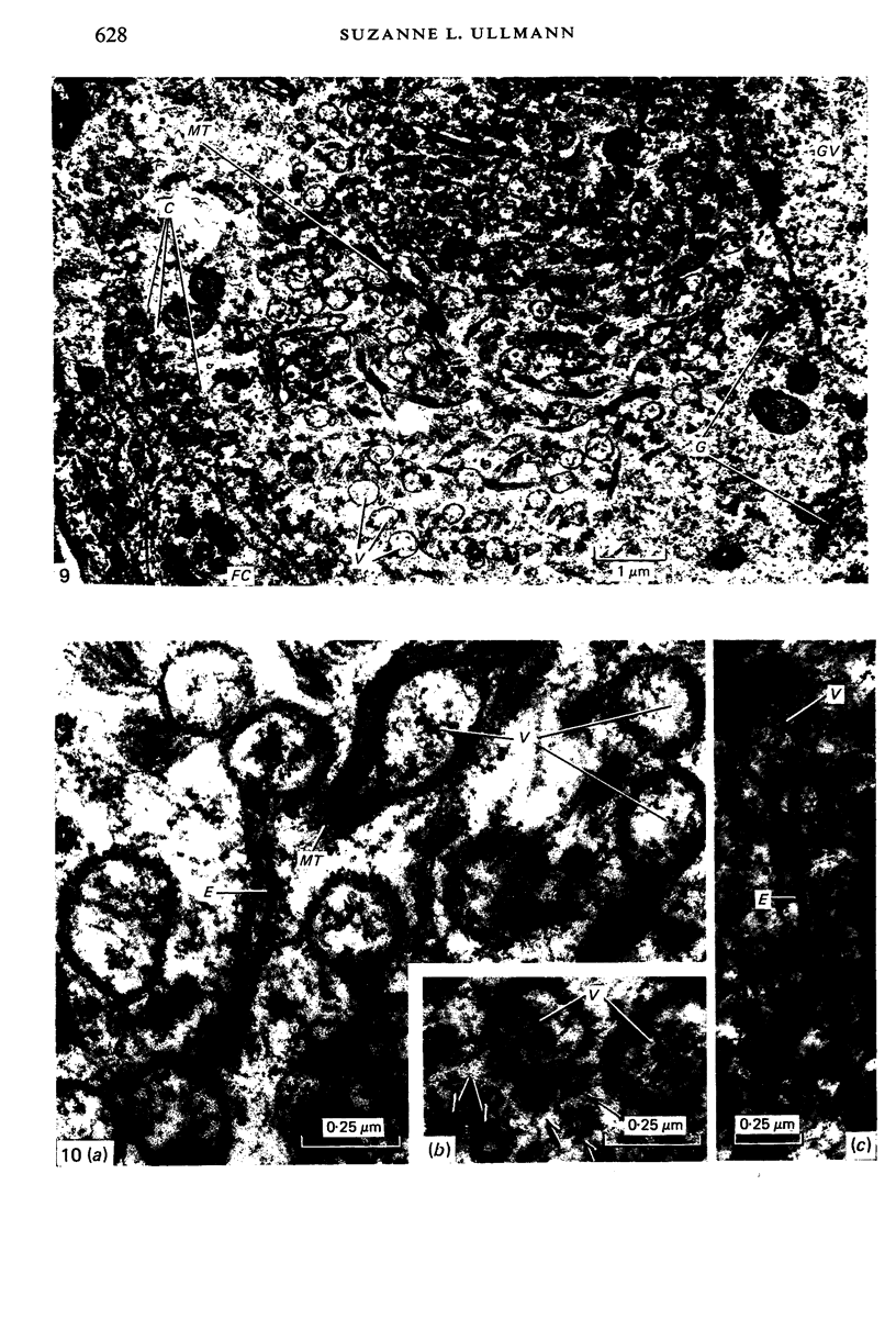
An ultrastructural study of bandicoot primordial follicles and oocytes was undertaken, as information on this subject is lacking in marsupials. Conspiculous features of the ooplasm are a paranuclear complex (PNC), a vesicle-microtubule complex (VMC) and an aggregate of tubular cisternae (ATC). The PNCappears as one or, more rarely, several homogeneous eosinophil bodies at the light microscope level. Ultrastructurally it is particulate, consisting of five distinct types of bodies, most of which are composed of concentric fibrillar whorls, but others appear homogeneous, granular or crystalline. Embedded among the particles is a group of Golgi-like vesicles. The bandicoot PNC-unlike similar structures found in the ooplasm of a variety of vertebrates, and known variously as "Balbiani body", "yolk nucleus", etc.-totally lacks nitochondria. The VMC consists of vesicle-like organelles which may be drawn out into tubular extensions, while the bounding membrane may be decorated with granules. Bundles of microtubules ramify between the vesicles, from which they appear to originate. The vesicles contain a matrix similar to the ooplasm. The ATC contains a homogeneous substance more electron-dense than the surrounding ooplasm. 'Dense bodies' occur in the cytoplasm of both the follicle cells and the oocytes. These are elongate membrane-bound organelles, circular in cross section. An electron-dense core is separated from the membrane by a narrow, less dense zone. The genesis and morphogenetic significance of these various organelles is unknown.
Full text
PDF
Images in this article
Selected References
These references are in PubMed. This may not be the complete list of references from this article.
- ADAMS E. C., HERTIG A. T. STUDIES ON GUINEA PIG OOCYTES. I. ELECTRON MICROSCOPIC OBSERVATIONS ON THE DEVELOPMENT OF CYTOPLASMIC ORGANELLES IN OOCYTES OF PRIMORDIAL AND PRIMARY FOLLICLES. J Cell Biol. 1964 Jun;21:397–427. doi: 10.1083/jcb.21.3.397. [DOI] [PMC free article] [PubMed] [Google Scholar]
- ANDERSON E., BEAMS H. W. Cytological observations on the fine structure of the guinea pig ovary with special reference to the oogonium, primary oocyte and associated follicle cells. J Ultrastruct Res. 1960 Jun;3:432–446. doi: 10.1016/s0022-5320(60)90021-6. [DOI] [PubMed] [Google Scholar]
- ANDRE J. [Contribution to the knowledge of the chondriome. Study of its ultrastructural changes during spermatogenesis]. J Ultrastruct Res. 1962 May;Suppl 3:1–185. [PubMed] [Google Scholar]
- Anderson E. Comparative aspects of the ultrastructure of the female gamete. Int Rev Cytol. 1974;Suppl 4:1–70. [PubMed] [Google Scholar]
- GURAYA S. S. HISTOCHEMICAL STUDIES ON THE YOLK NUCLEUS IN THE OOGENESIS OF MAMMALS. Am J Anat. 1964 Mar;114:283–289. doi: 10.1002/aja.1001140208. [DOI] [PubMed] [Google Scholar]
- Guraya S. S. Further morphological and histochemical studies on the yolk nucleus and associated cell components in the developing oocyte of the Indian wall lizard. J Morphol. 1968 Mar;124(3):283–294. doi: 10.1002/jmor.1051240303. [DOI] [PubMed] [Google Scholar]
- Guraya S. S. Histochemical study of developing ovarian oocyte of the American opossum. Acta Embryol Morphol Exp. 1968 Jul;10(2):181–191. [PubMed] [Google Scholar]
- Guraya S. S. Morphology, histochemistry, and biochemistry of human oogenesis and ovulation. Int Rev Cytol. 1974;37(0):121–151. doi: 10.1016/s0074-7696(08)61358-3. [DOI] [PubMed] [Google Scholar]
- Hadek R. The structure of the mammalian egg. Int Rev Cytol. 1965;18:29–71. doi: 10.1016/s0074-7696(08)60551-3. [DOI] [PubMed] [Google Scholar]
- Hertig A. T., Adams E. C. Studies on the human oocyte and its follicle. I. Ultrastructural and histochemical observations on the primordial follicle stage. J Cell Biol. 1967 Aug;34(2):647–675. doi: 10.1083/jcb.34.2.647. [DOI] [PMC free article] [PubMed] [Google Scholar]
- Hertig A. T. The primary human oocyte: some observations on the fine structure of Balbiani's vitelline body and the origin of the annulate lamellae. Am J Anat. 1968 Jan;122(1):107–137. doi: 10.1002/aja.1001220107. [DOI] [PubMed] [Google Scholar]
- KARASAKI S. Studies on amphibian yolk 1. The ultrastructure of the yolk platelet. J Cell Biol. 1963 Jul;18:135–151. doi: 10.1083/jcb.18.1.135. [DOI] [PMC free article] [PubMed] [Google Scholar]
- Lintern-Moore S., Moore G. P., Tyndale-Biscoe C. H., Poole W. E. The growth of the oocyte and follicle in the ovaries of monotremes and marsupials. Anat Rec. 1976 Jul;185(3):325–332. doi: 10.1002/ar.1091850306. [DOI] [PubMed] [Google Scholar]
- Norrevang A. Electron microscopic morphology of oogenesis. Int Rev Cytol. 1968;23:113–186. [PubMed] [Google Scholar]
- ODOR D. L. THE ULTRASTRUCTURE OF UNILAMINAR FOLLICLES OF THE HAMSTER OVARY. Am J Anat. 1965 May;116:493–521. doi: 10.1002/aja.1001160304. [DOI] [PubMed] [Google Scholar]
- Paulson J. L., Rosenberg M. D. Formation of lining bodies and oocyte bodies during avian oogenesis. Dev Biol. 1974 Oct;40(2):366–371. doi: 10.1016/0012-1606(74)90137-7. [DOI] [PubMed] [Google Scholar]
- SOTELO J. R. An electron microscope study on the cytoplasmic and nuclear components of rat primary oocytes. Z Zellforsch Mikrosk Anat. 1959;50:749–765. doi: 10.1007/BF00342364. [DOI] [PubMed] [Google Scholar]
- Srivastava M. D. Cytoplasmic inclusions in oogenesis. Int Rev Cytol. 1965;18:73–98. doi: 10.1016/s0074-7696(08)60552-5. [DOI] [PubMed] [Google Scholar]
- Weakley B. S. Electron microscopy of the oocyte and granulosa cells in the developing ovarian follicles of the golden hamster (Mesocricetus auratus). J Anat. 1966 Jul;100(Pt 3):503–534. [PMC free article] [PubMed] [Google Scholar]
- al-Mukhtar K. A., Webb A. C. An ultrastructural study of primordial germ cells, oogonia and early oocytes in Xenopus laevis. J Embryol Exp Morphol. 1971 Oct;26(2):195–217. [PubMed] [Google Scholar]


