Abstract
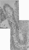
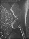
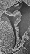
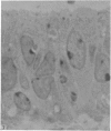
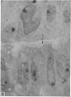
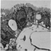
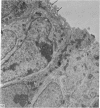
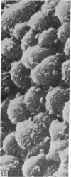
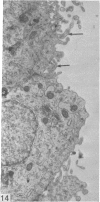
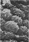
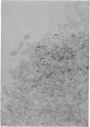
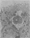
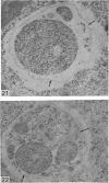
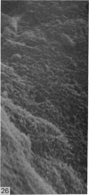
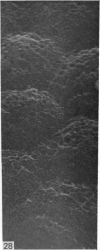
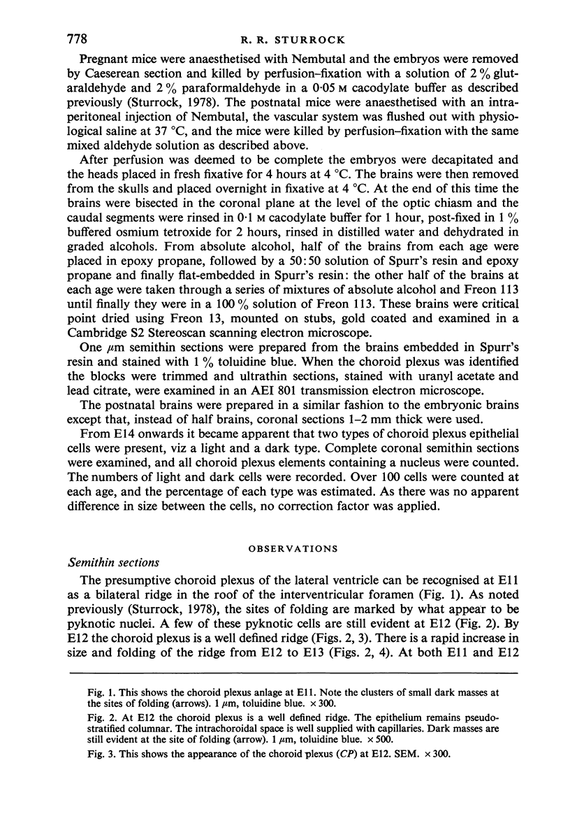
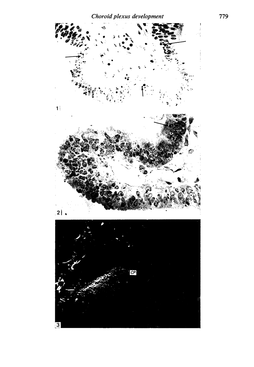
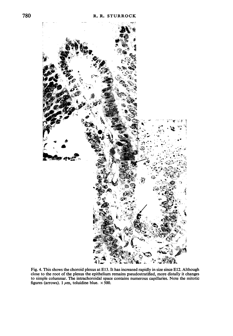
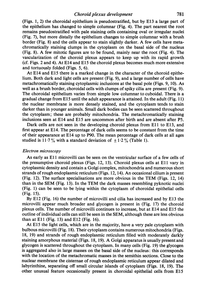
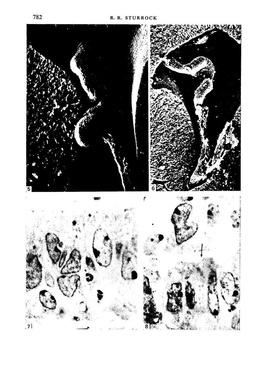
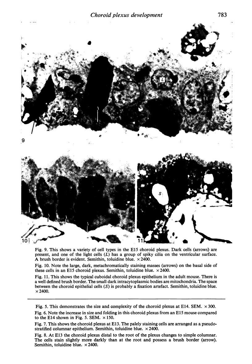
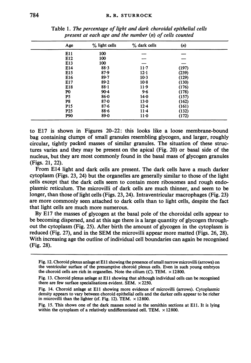
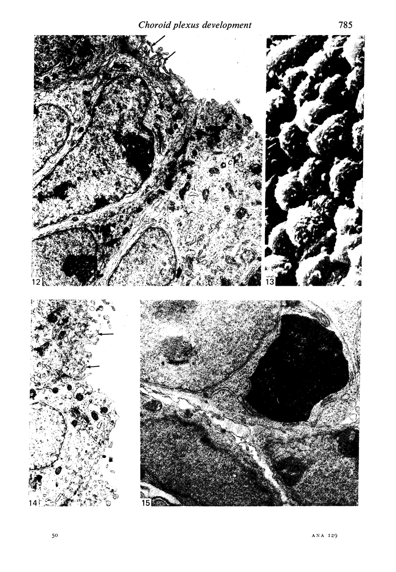
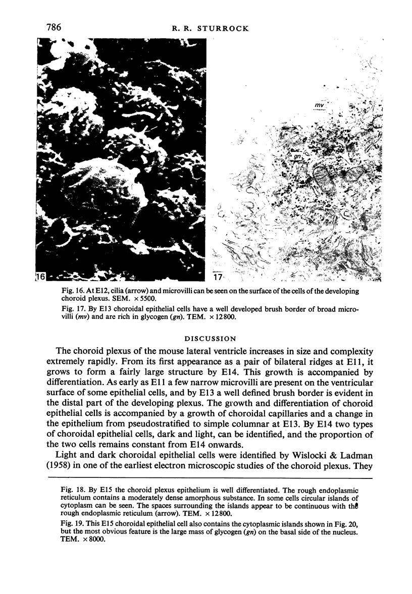
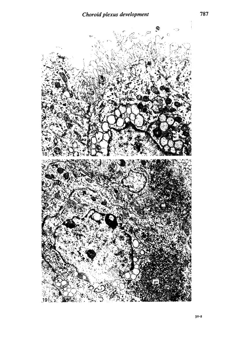
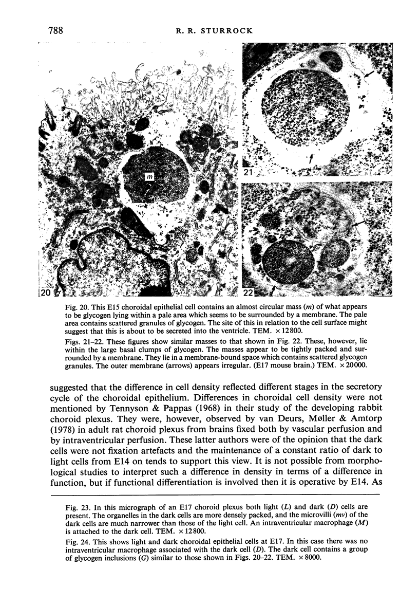
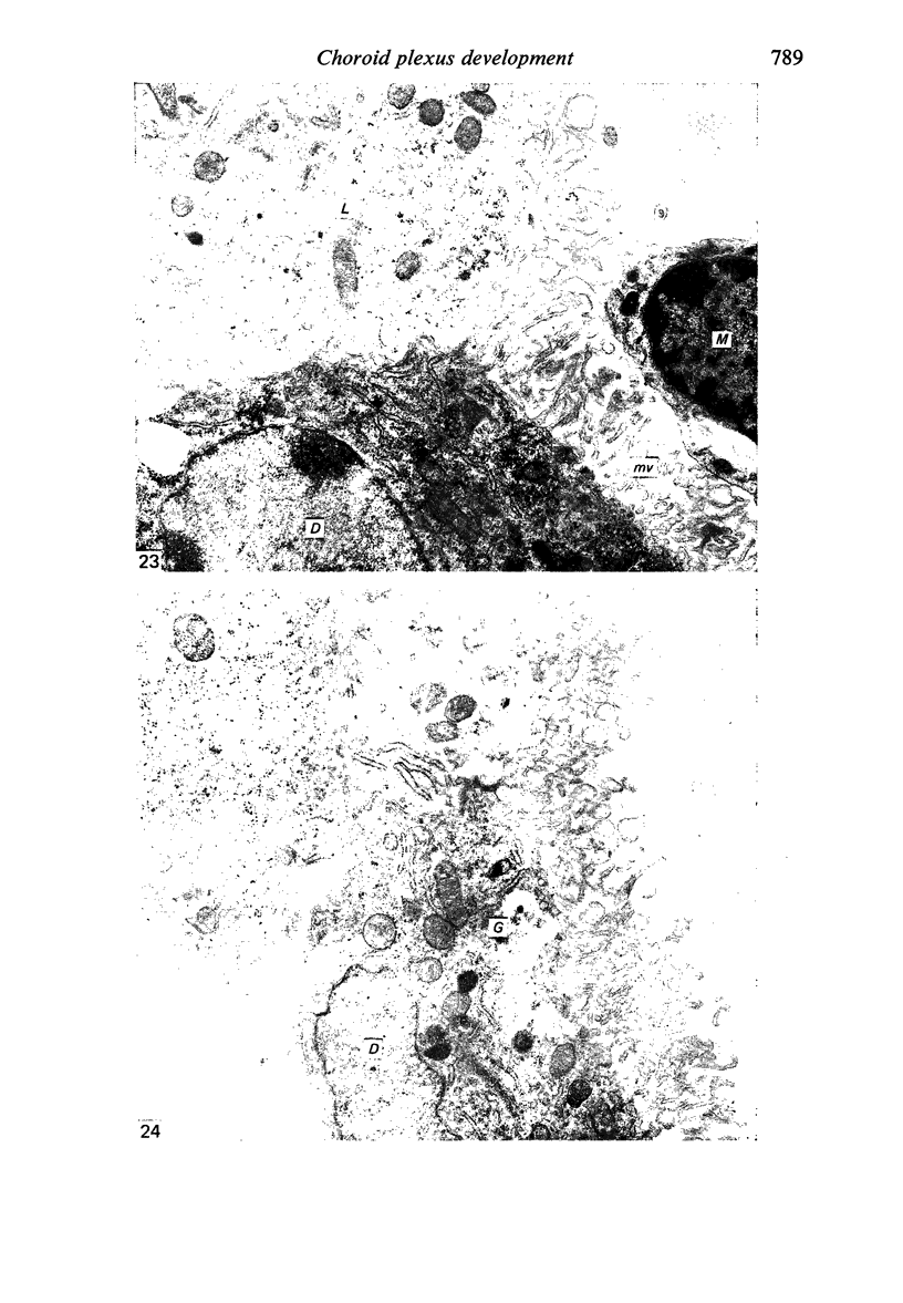
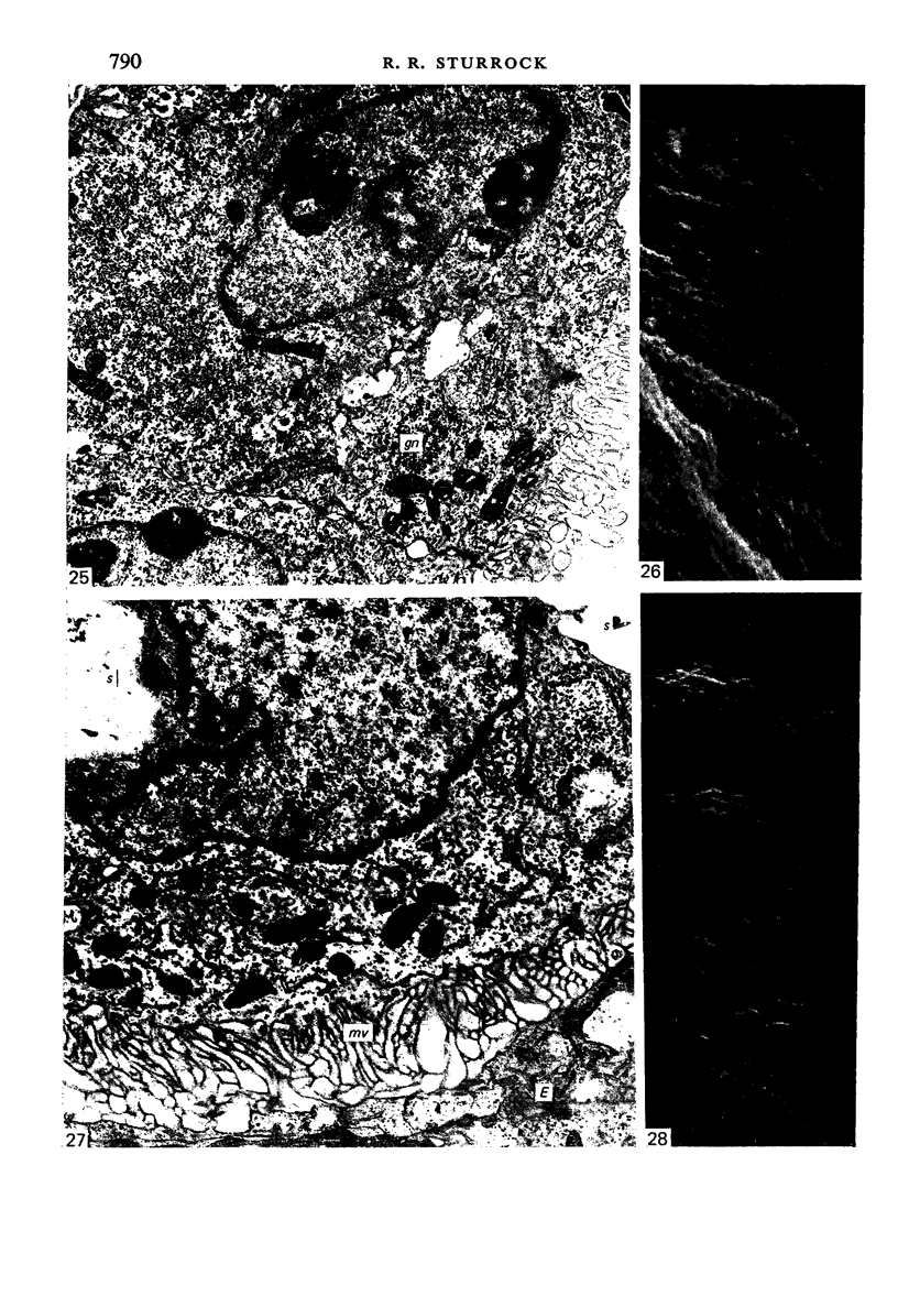
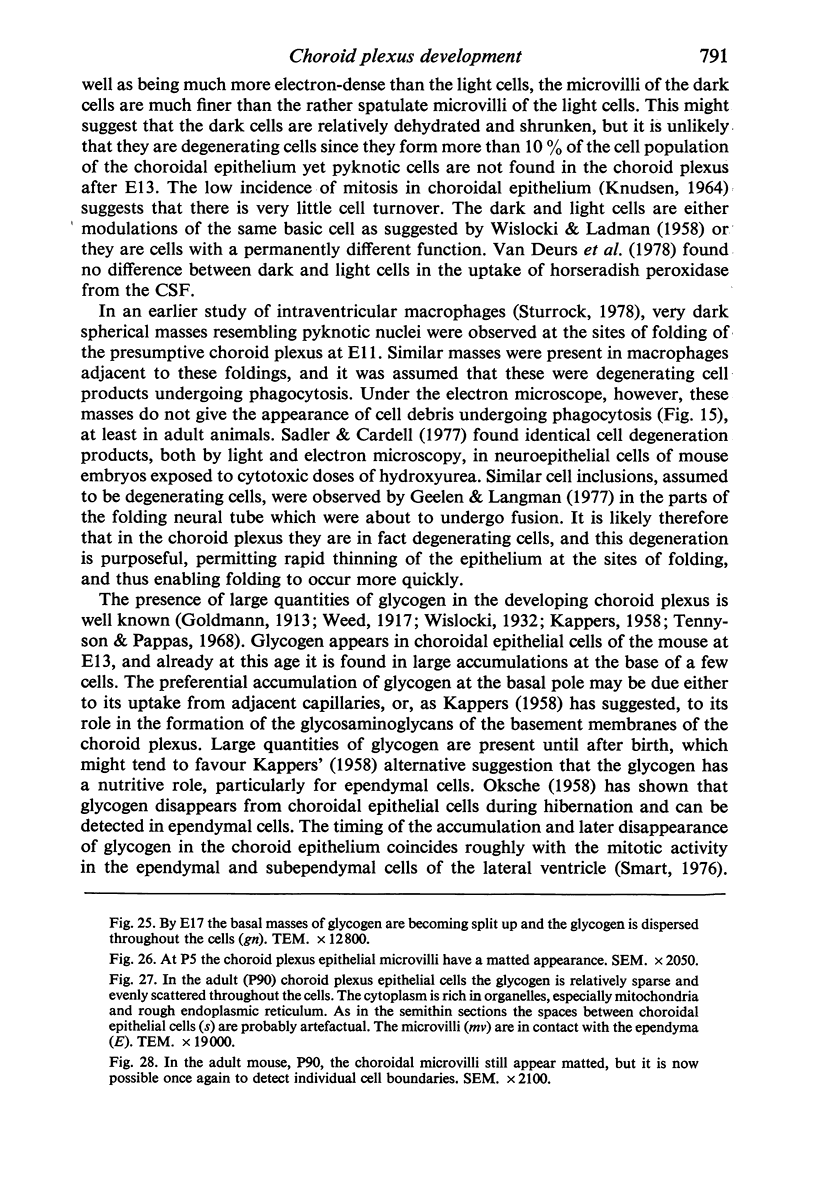
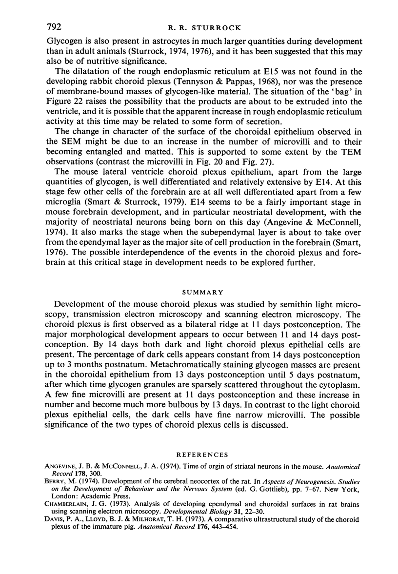
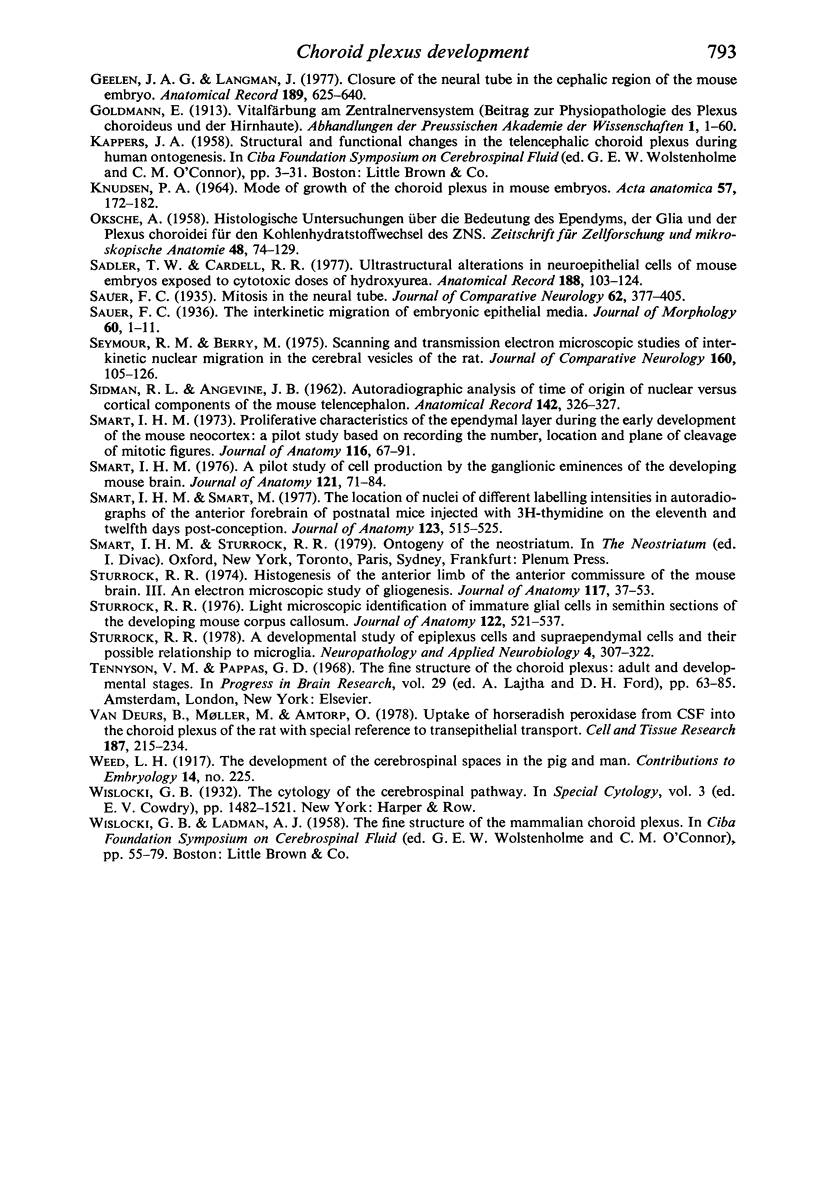
Development of the mouse choroid plexus was studied by semithin light microscopy, transmission electron microscopy and scanning electron microscopy. The choroid plexus is first observed as a bilateral ridge at 11 days postconception. The major morphological development appears to occur between 11 and 14 days postconception. By 14 days both dark and light choroid plexus epithelial cells are present. The percentage of dark cells appears constant from 14 days postconception up to 3 months postnatum. Metachromatically staining glycogen masses are present in the choroidal epithelium from 13 days postconception until 5 days postnatum, after which time glcogen granules are sparsely scattered throughout the cytoplasm. A few fine microvilli are present at 11 days postconception and these increase in number and become much more bulbous by 13 days. In contrast to the light choroid plexus epithelial cells, the dark cells have fine narrow microvilli. The possible significance of the two types of choroid plexus cells is discussed.
Full text
PDF
Images in this article
Selected References
These references are in PubMed. This may not be the complete list of references from this article.
- Chamberlain J. G. Analysis of developing ependymal and choroidal surfaces in rat brains using scanning electron microscopy. Dev Biol. 1973 Mar;31(1):22–30. doi: 10.1016/0012-1606(73)90317-5. [DOI] [PubMed] [Google Scholar]
- Davis D. A., Lloyd B. J., Jr, Milhorat T. H. A comparative ultrastructural study of the choroid plexuses of the immature pig. Anat Rec. 1973 Aug;176(4):443–454. doi: 10.1002/ar.1091760407. [DOI] [PubMed] [Google Scholar]
- Geelen J. A., Langman J. Closure of the neural tube in the cephalic region of the mouse embryo. Anat Rec. 1977 Dec;189(4):625–640. doi: 10.1002/ar.1091890407. [DOI] [PubMed] [Google Scholar]
- KNUDSEN P. A. MODE OF GROWTH OF THE CHOROID PLEXUS IN MOUSE EMBRYOS. Acta Anat (Basel) 1964;57:172–182. doi: 10.1159/000142545. [DOI] [PubMed] [Google Scholar]
- OKSCHE A. Histologische Untersuchungen über die Bedeutung des Ependyms, der Glia und der Plexus chorioidei für den Kohlenhydratstoffwechsel des ZNS. Z Zellforsch Mikrosk Anat. 1958;48(1):74–129. [PubMed] [Google Scholar]
- Sadler T. W., Cardell R. R. Ultrastructural alterations in neuroepithelial cells of mouse embryos exposed to cytotoxic doses of hydroxyurea. Anat Rec. 1977 May;188(1):103–123. doi: 10.1002/ar.1091880110. [DOI] [PubMed] [Google Scholar]
- Seymour R. M., Berry M. Scanning and transmission electron microscope studies of interkinetic nuclear migration in the cerebral vesicles of the rat. J Comp Neurol. 1975 Mar 1;160(1):105–125. doi: 10.1002/cne.901600107. [DOI] [PubMed] [Google Scholar]
- Smart I. H. A pilot study of cell production by the ganglionic eminences of the developing mouse brain. J Anat. 1976 Feb;121(Pt 1):71–84. [PMC free article] [PubMed] [Google Scholar]
- Smart I. H. Proliferative characteristics of the ependymal layer during the early development of the mouse neocortex: a pilot study based on recording the number, location and plane of cleavage of mitotic figures. J Anat. 1973 Oct;116(Pt 1):67–91. [PMC free article] [PubMed] [Google Scholar]
- Smart I. H., Smart M. The location of nuclei of different labelling intensities in autoradiographs of the anterior forebrain of postnatial mice injected with [3H]thymidine on the eleventh and twelfth days post-conception. J Anat. 1977 Apr;123(Pt 2):515–525. [PMC free article] [PubMed] [Google Scholar]
- Sturrock R. R. A developmental study of epiplexus cells and supraependymal cells and their possible relationship to microglia. Neuropathol Appl Neurobiol. 1978 Sep-Oct;4(5):307–322. doi: 10.1111/j.1365-2990.1978.tb01345.x. [DOI] [PubMed] [Google Scholar]
- Sturrock R. R. Histogenesis of the anterior limb of the anterior commissure of the mouse brain. 3. An electron microscopic study of gliogenesis. J Anat. 1974 Feb;117(Pt 1):37–53. [PMC free article] [PubMed] [Google Scholar]
- Sturrock R. R. Light microscopic identification of immature glial cells in semithin sections of the developing mouse corpus callosum. J Anat. 1976 Dec;122(Pt 3):521–537. [PMC free article] [PubMed] [Google Scholar]
- Tennyson V. M., Appas G. D. The fine structure of the choroid plexus adult and developmental stages. Prog Brain Res. 1968;29:63–85. doi: 10.1016/s0079-6123(08)64149-7. [DOI] [PubMed] [Google Scholar]
- van Deurs B., Møller M., Amtorp O. Uptake of horseradish peroxidase from CSF into the choroid plexus of the rat, with special reference to transepithelial transport. Cell Tissue Res. 1978 Feb 24;187(2):215–234. doi: 10.1007/BF00224366. [DOI] [PubMed] [Google Scholar]


