Abstract
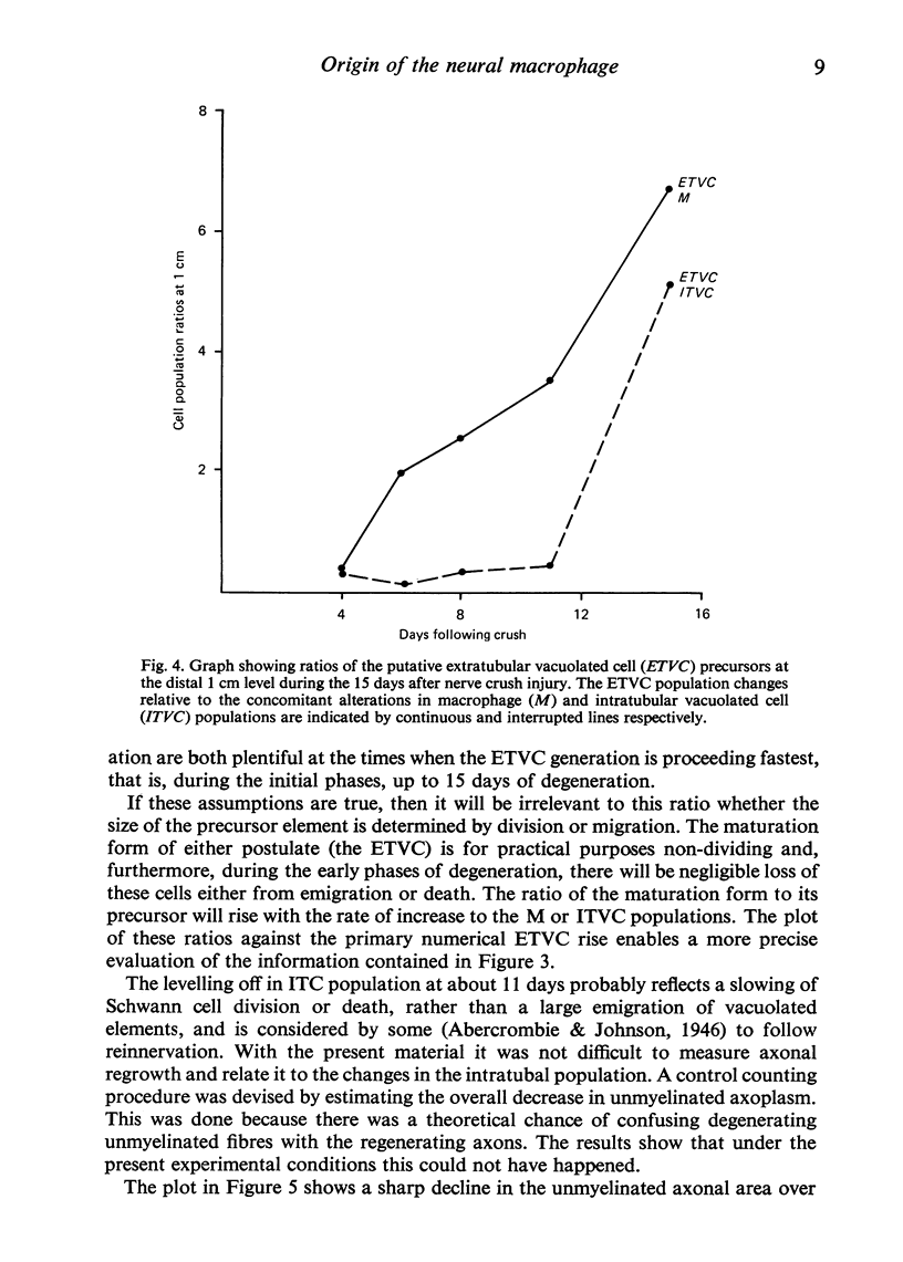
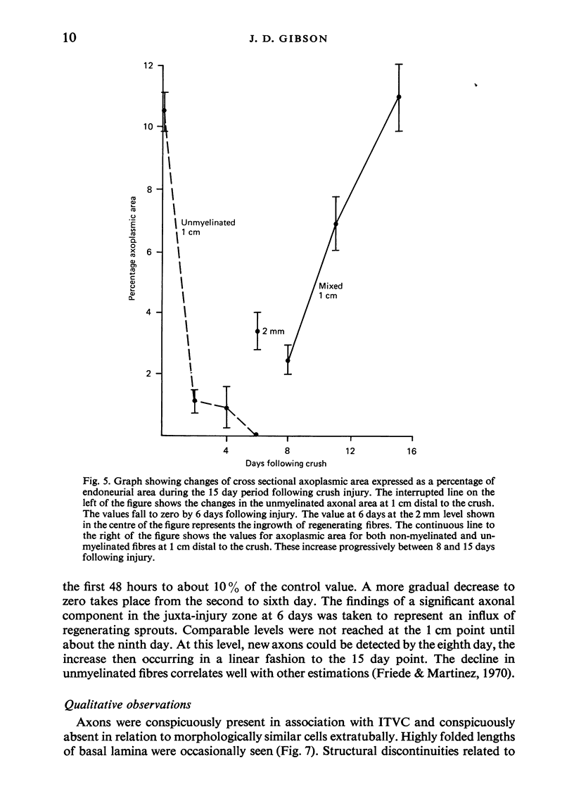
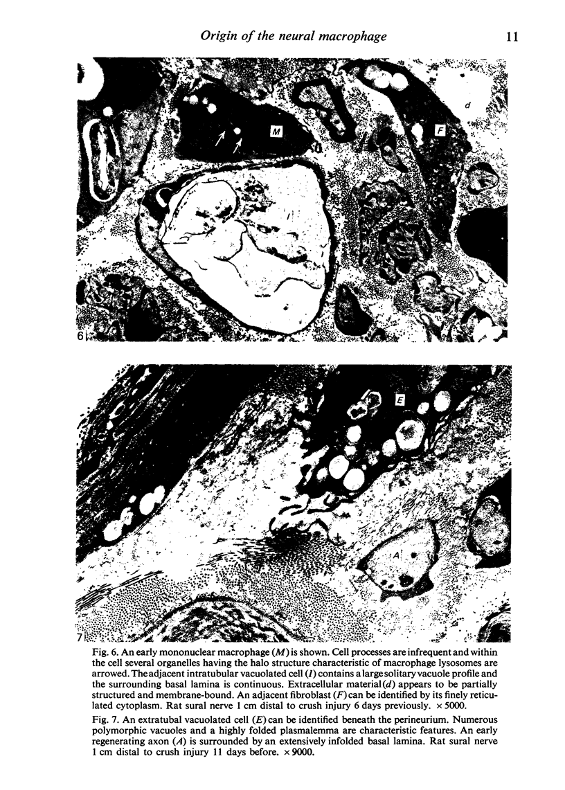
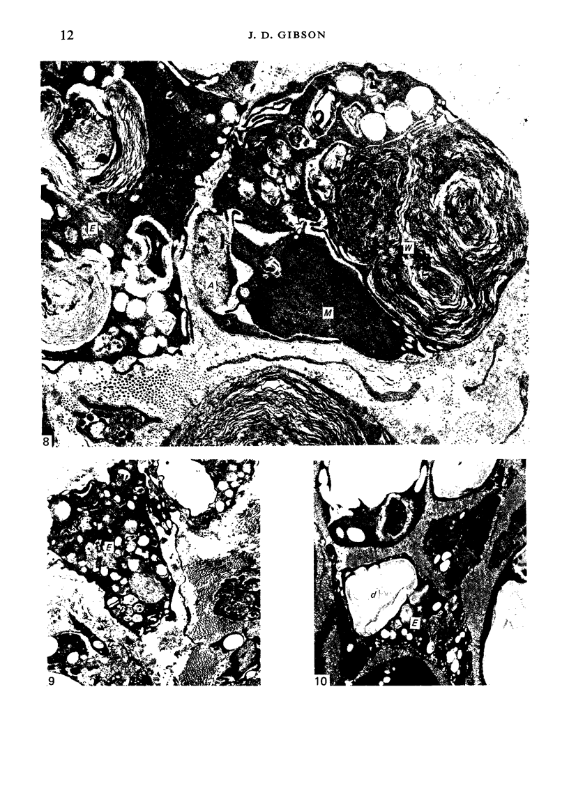
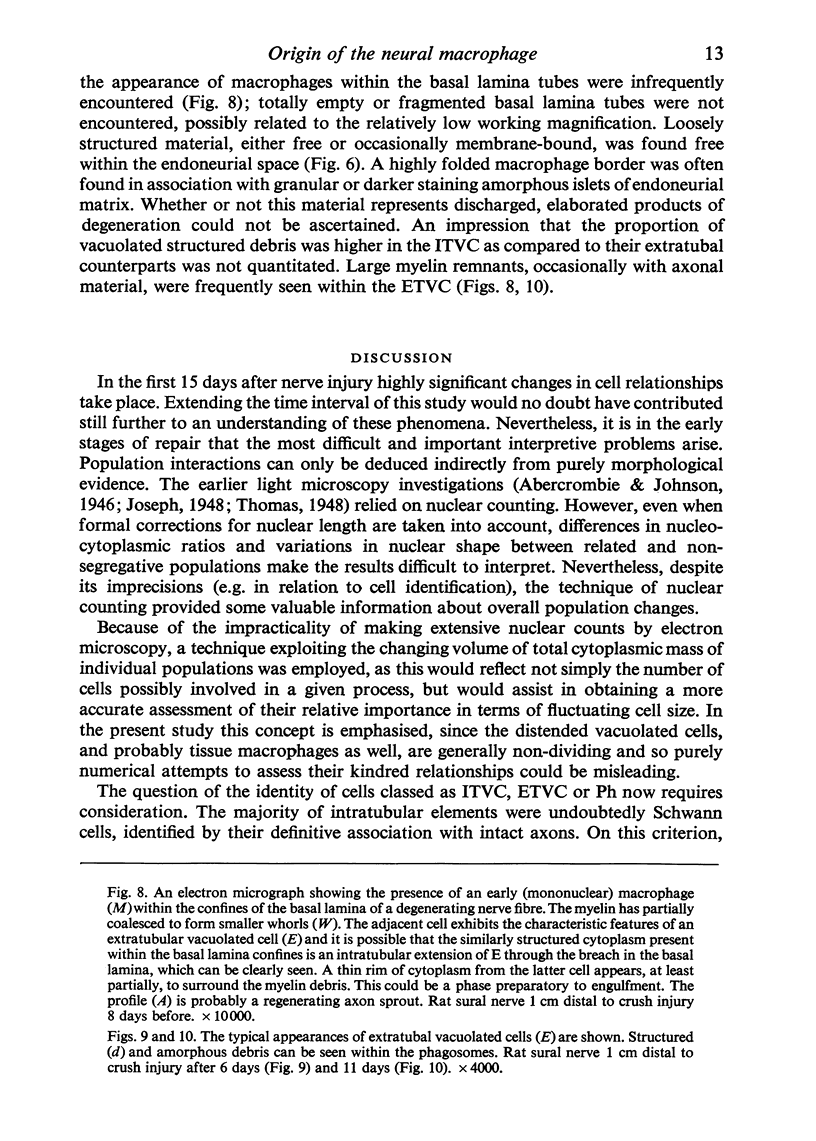
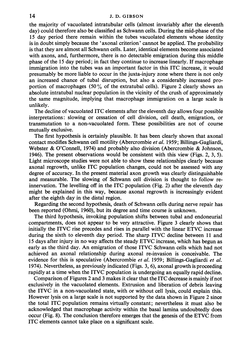
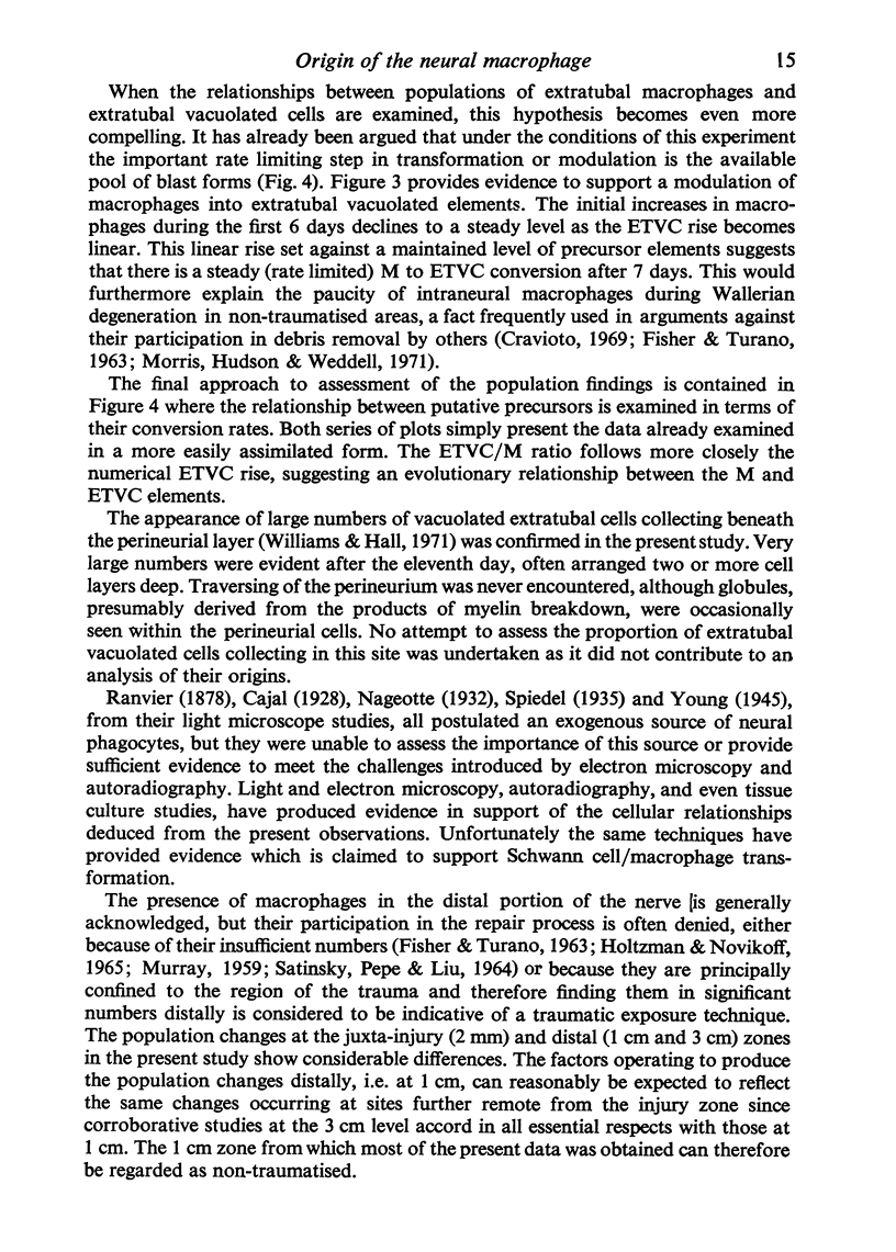
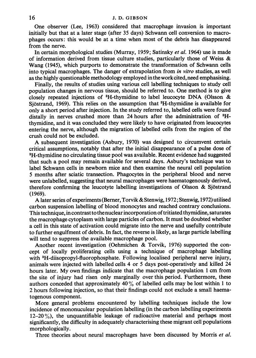
The changes that follow a localised crush injury to the rat sural nerve have been used to study endoneurial populations during Wallerian degeneration. The removal of products of degeneration, and in particular myelin debris, is accomplished by globule-laden cells which appear in the endoneurium during the first few days of repair. The origin of these cells has been investigated using a quantitative ultrastructural technique. Serial planimetric measurements of all populations, identifiable in terms of criteria that did not pre-judge their true nature, were made at intervals over a period of 15 days. Cell counts obtained immediately below the site of injury and 1 and 3 cm distally were compared, and graphs of endoneurial population changes constructed from these measurements. Additional descriptive evidence was invoked to assist in establishing the actual identity of the extratubal vacuolated cells which had been classified and measured empirically. Comparing the changes in the number of these cells with that of the intratubal vascuolated cell population, and taking account of the presence of immature macrophages in both proximal and distal situations, lead to the conclusion that the extratubal vacuolated cells are mostly derived from the bloodstream, the rest being of local intratubal origin. There was no evidence to support the notion that Schwann cells transform into macrophages.
Full text
PDF
Images in this article
Selected References
These references are in PubMed. This may not be the complete list of references from this article.
- ABERCROMBIE M., EVANS D. H., MURRAY J. G. Nuclear multiplication and cell migration in degenerating unmyelinated nerves. J Anat. 1959 Jan;93(1):9–14. [PMC free article] [PubMed] [Google Scholar]
- Abercrombie M., Johnson M. L. Quantitative histology of Wallerian degeneration: I. Nuclear population in rabbit sciatic nerve. J Anat. 1946 Jan;80(Pt 1):37–50. [PMC free article] [PubMed] [Google Scholar]
- BARTON A. A. An electron microscope study of degeneration and regeneration of nerve. Brain. 1962 Dec;85:799–808. doi: 10.1093/brain/85.4.799. [DOI] [PubMed] [Google Scholar]
- Berner A., Torvik A., Stenwig A. E. Origin of macrophages in traumatic lesions and Wallerian degeneration in peripheral nerves. Acta Neuropathol. 1973;25(3):228–236. doi: 10.1007/BF00685202. [DOI] [PubMed] [Google Scholar]
- Billings-Gagliardi S., Webster H. F., O'Connell M. F. In vivo and electron microscopic observations on Schwann cells in developing tadpole nerve fibers. Am J Anat. 1974 Nov;141(3):375–391. doi: 10.1002/aja.1001410308. [DOI] [PubMed] [Google Scholar]
- Bradley W. G., Asbury A. K. Duration of synthesis phase in neuilemma cells in mouse sciatic nerve during degeneration. Exp Neurol. 1970 Feb;26(2):275–282. doi: 10.1016/0014-4886(70)90125-1. [DOI] [PubMed] [Google Scholar]
- Bray G. M., Aguayo A. J., Martin J. B. Immunosympathectomy: late effects on the composition of rat cervical sympathetic trunks and influence on axonal regeneration after crush. Acta Neuropathol. 1973 Dec 3;26(4):345–352. doi: 10.1007/BF00688081. [DOI] [PubMed] [Google Scholar]
- Cotran R. S., Karnovsky M. J. Ultrastructural studies on the permeability of the mesothelium to horseradish peroxidase. J Cell Biol. 1968 Apr;37(1):123–137. doi: 10.1083/jcb.37.1.123. [DOI] [PMC free article] [PubMed] [Google Scholar]
- Cravioto H. Wallerian degeneration: ultrastructural and histochemical studies. Bull Los Angeles Neurol Soc. 1969 Oct;34(4):233–253. [PubMed] [Google Scholar]
- ESTABLE C., ACOSTA-FERREIRA W., SOTELO J. R. An electron microscope study of the regenerating nerve fibers. Z Zellforsch Mikrosk Anat. 1957;46(4):387–399. doi: 10.1007/BF00345052. [DOI] [PubMed] [Google Scholar]
- FISHER E. R., TURANO A. Schwann cells in wallerian degeneration. Arch Pathol. 1963 May;75:517–527. [PubMed] [Google Scholar]
- Friede R. L., Martinez A. J. Analysis of axon-sheath relations during early Wallerian degeneration. Brain Res. 1970 Apr 14;19(2):199–212. doi: 10.1016/0006-8993(70)90434-8. [DOI] [PubMed] [Google Scholar]
- GUTH L. Regeneration in the mammalian peripheral nervous system. Physiol Rev. 1956 Oct;36(4):441–478. doi: 10.1152/physrev.1956.36.4.441. [DOI] [PubMed] [Google Scholar]
- Holtzman E., Novikoff A. B. Lysomes in the rat sciatic nerve following crush. J Cell Biol. 1965 Dec;27(3):651–669. doi: 10.1083/jcb.27.3.651. [DOI] [PMC free article] [PubMed] [Google Scholar]
- Joseph J. Changes in nuclear population following twenty-one days' degeneration in a nerve consisting of small myelinated fibres. J Anat. 1948 Jul;82(Pt 3):146–152.1. [PMC free article] [PubMed] [Google Scholar]
- LEE J. C. Electron microscopy of Wallerian degeneration. J Comp Neurol. 1963 Feb;120:65–79. doi: 10.1002/cne.901200107. [DOI] [PubMed] [Google Scholar]
- Morris J. H., Hudson A. R., Weddell G. A study of degeneration and regeneration in the divided rat sciatic nerve based on electron microscopy. I. The traumatic degeneration of myelin in the proximal stump of the divided nerve. Z Zellforsch Mikrosk Anat. 1972;124(1):76–102. [PubMed] [Google Scholar]
- NATHANIEL E. J. PEASE DC: DEGENERATIVE CHANGES IN RAT DORSAL ROOTS DURING WALLERIAN DEGENERATION. J Ultrastruct Res. 1963 Dec;52:511–532. doi: 10.1016/s0022-5320(63)80082-9. [DOI] [PubMed] [Google Scholar]
- NATHANIEL E. J., PEASE D. C. REGENERATIVE CHANGES IN RAT DORSAL ROOTS FOLLOWING WALERIAN DEGENERATION. J Ultrastruct Res. 1963 Dec;52:533–549. doi: 10.1016/s0022-5320(63)80083-0. [DOI] [PubMed] [Google Scholar]
- OHMI S. Electron microscopic study of wallerian degeneration of the peripheral nerve. Z Zellforsch Mikrosk Anat. 1961;54:39–67. doi: 10.1007/BF00384198. [DOI] [PubMed] [Google Scholar]
- Oehmichen M., Torvik A. The origin of reactive cells in retrograde and Wallerian degeneration. Experiments with intravenous injection of 3H-DFP-labeled macrophages. Cell Tissue Res. 1976 Oct 13;173(3):343–348. doi: 10.1007/BF00220322. [DOI] [PubMed] [Google Scholar]
- Olsson Y., Sjöstrand J. Origin of macrophages in Wallerian degereration of peripheral nerves demonstrated autoradiographically. Exp Neurol. 1969 Jan;23(1):102–112. doi: 10.1016/0014-4886(69)90037-5. [DOI] [PubMed] [Google Scholar]
- Romine J. S., Bray G. M., Aguayo A. J. Schwann cell multiplication after crush injury of unmyelinated fibers. Arch Neurol. 1976 Jan;33(1):49–54. doi: 10.1001/archneur.1976.00500010051008. [DOI] [PubMed] [Google Scholar]
- SATINSKY D., PEPE F. A., LIU C. N. THE NEURILEMMA CELL IN PERIPHERAL NERVE DEGENERATION AND REGENERATION. Exp Neurol. 1964 Jun;9:441–451. doi: 10.1016/0014-4886(64)90052-4. [DOI] [PubMed] [Google Scholar]
- Stenwig A. E. The origin of brain macrophages in traumatic lesions, Wallerian degeneration, and retrograde degeneration. J Neuropathol Exp Neurol. 1972 Oct;31(4):696–704. doi: 10.1097/00005072-197210000-00011. [DOI] [PubMed] [Google Scholar]
- THOMAS P. K. CHANGES IN THE ENDONEURIAL SHEATHS OF PERIPHERAL MYELINATED NERVE FIBRES DURING WALLERIAN DEGENERATION. J Anat. 1964 Apr;98:175–182. [PMC free article] [PubMed] [Google Scholar]
- Thomas G. A. Quantitative histology of Wallerian degeneration: II. Nuclear population in two nerves of different fibre spectrum. J Anat. 1948 Jul;82(Pt 3):135–145. [PMC free article] [PubMed] [Google Scholar]
- WETTSTEIN R., SOTELO J. R. Electron microscope study on the regenerative process of peripheral nerves of mice. Z Zellforsch Mikrosk Anat. 1963;59:708–730. doi: 10.1007/BF00319067. [DOI] [PubMed] [Google Scholar]
- Weibel E. R. Stereological principles for morphometry in electron microscopic cytology. Int Rev Cytol. 1969;26:235–302. doi: 10.1016/s0074-7696(08)61637-x. [DOI] [PubMed] [Google Scholar]
- Williams P. L., Hall S. M. Chronic Wallerian degeneration--an in vivo and ultrastructural study. J Anat. 1971 Sep;109(Pt 3):487–503. [PMC free article] [PubMed] [Google Scholar]


