

Abstract
The natural history of the subependymal layer around the lateral ventricle of the mouse brain was studied from its appearance at E11 up to 22 months postnatum. In the young embryo four regions of the ventricle can be recognized by their histological characteristics: (1) the ventricular roof, (2) the medial roof, (3) the ventricular elevations and (4) the medial wall. The characteristics of the ventricular roof and ventricular elevations were examined in detail. The ventricular roof appears to be the main site of production of cortical neurons while the subependymal layer of the ventricular elevations seems to be the main site of origin of forebrain glia. The age of differentiation of the ependyma differs for each region, with the medial roof differentiating first, followed by the ventricular roof and medial wall, and ventricular elevations or lateral wall last. Differentiation begins with a change from pseudostratified columnar epithelium to simple columnar epithelium and the appearance of cilia in large numbers.
Full text
PDF


Images in this article


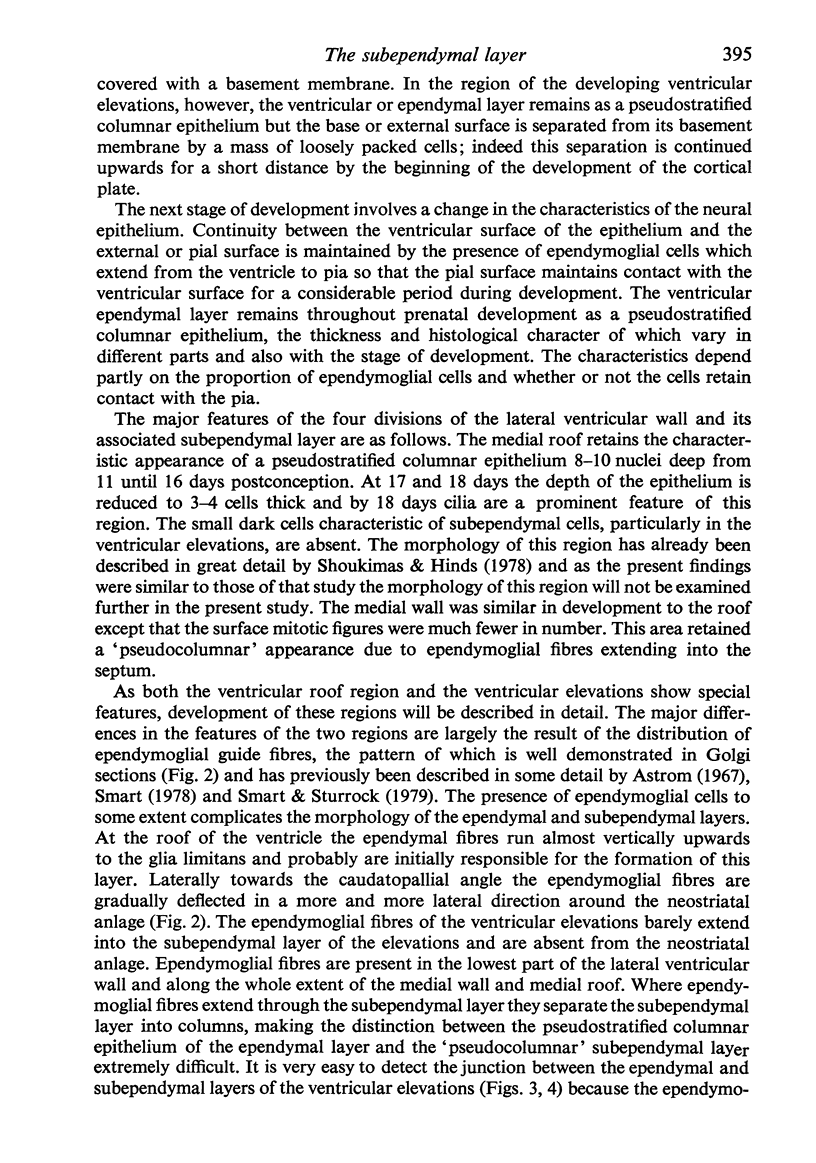
Selected References
These references are in PubMed. This may not be the complete list of references from this article.
- Aström K. E. On the early development of the isocortex in fetal sheep. Prog Brain Res. 1967;26:1–59. [PubMed] [Google Scholar]
- Blakemore W. F. The ultrastructure of the subependymal plate in the rat. J Anat. 1969 May;104(Pt 3):423–433. [PMC free article] [PubMed] [Google Scholar]
- Cammermeyer J. The hypependymal microglia cell. Z Anat Entwicklungsgesch. 1965;124(6):543–561. doi: 10.1007/BF00520846. [DOI] [PubMed] [Google Scholar]
- Glees P., Hasan M. Lipofuscin in neuronal aging and diseases. Norm Pathol Anat (Stuttg) 1976;32:1–68. [PubMed] [Google Scholar]
- Henrikson C. K., Vaughn J. E. Fine structural relationships between neurites and radial glial processes in developing mouse spinal cord. J Neurocytol. 1974 Dec;3(6):659–675. doi: 10.1007/BF01097190. [DOI] [PubMed] [Google Scholar]
- Hinds J. W., Ruffett T. L. Cell proliferation in the neural tube: an electron microscopic and golgi analysis in the mouse cerebral vesicle. Z Zellforsch Mikrosk Anat. 1971;115(2):226–264. doi: 10.1007/BF00391127. [DOI] [PubMed] [Google Scholar]
- Imamoto K., Leblond C. P. Radioautographic investigation of gliogenesis in the corpus callosum of young rats. II. Origin of microglial cells. J Comp Neurol. 1978 Jul 1;180(1):139–163. doi: 10.1002/cne.901800109. [DOI] [PubMed] [Google Scholar]
- Imamoto K., Paterson J. A., Leblond C. P. Radioautographic investigation of gliogenesis in the corpus callosum of young rats. I. Sequential changes in oligodendrocytes. J Comp Neurol. 1978 Jul 1;180(1):115-28, 132-7. doi: 10.1002/cne.901800108. [DOI] [PubMed] [Google Scholar]
- Lewis P. D. Glial reactions to cerebral injury. Proc R Soc Med. 1974 Feb;67(2):130–131. [PMC free article] [PubMed] [Google Scholar]
- Lewis P. D. The fate of the subependymal cell in the adult rat brain, with a note on the origin of microglia. Brain. 1968;91(4):721–736. doi: 10.1093/brain/91.4.721. [DOI] [PubMed] [Google Scholar]
- Paterson J. A., Privat A., Ling E. A., Leblond C. P. Investigation of glial cells in semithin sections. 3. Transformation of subependymal cells into glial cells, as shown by radioautography after 3 H-thymidine injection into the lateral ventricle of the brain of young rats. J Comp Neurol. 1973 May 1;149(1):83–102. doi: 10.1002/cne.901490106. [DOI] [PubMed] [Google Scholar]
- Privat A., Leblond C. P. The subependymal layer and neighboring region in the brain of the young rat. J Comp Neurol. 1972 Nov;146(3):277–302. doi: 10.1002/cne.901460302. [DOI] [PubMed] [Google Scholar]
- Proceedings of the Anatomical Society of Great Britain and Ireland. November 1978. Abstracts. J Anat. 1979 Mar;128(Pt 2):411–443. [PMC free article] [PubMed] [Google Scholar]
- Seymour R. M., Berry M. Scanning and transmission electron microscope studies of interkinetic nuclear migration in the cerebral vesicles of the rat. J Comp Neurol. 1975 Mar 1;160(1):105–125. doi: 10.1002/cne.901600107. [DOI] [PubMed] [Google Scholar]
- Shoukimas G. M., Hinds J. W. The development of the cerebral cortex in the embryonic mouse: an electron microscopic serial section analysis. J Comp Neurol. 1978 Jun 15;179(4):795–830. doi: 10.1002/cne.901790407. [DOI] [PubMed] [Google Scholar]
- Smart I. H. A pilot study of cell production by the ganglionic eminences of the developing mouse brain. J Anat. 1976 Feb;121(Pt 1):71–84. [PMC free article] [PubMed] [Google Scholar]
- Smart I. H., Smart M. The location of nuclei of different labelling intensities in autoradiographs of the anterior forebrain of postnatial mice injected with [3H]thymidine on the eleventh and twelfth days post-conception. J Anat. 1977 Apr;123(Pt 2):515–525. [PMC free article] [PubMed] [Google Scholar]
- Stensaas L. J., Gilson B. C. Ependymal and subependymal cells of the caudato-pallial junction in the lateral ventricle of the neonatal rabbit. Z Zellforsch Mikrosk Anat. 1972;132(3):297–322. doi: 10.1007/BF02450711. [DOI] [PubMed] [Google Scholar]
- Sturrock R. R. A developmental study of epiplexus cells and supraependymal cells and their possible relationship to microglia. Neuropathol Appl Neurobiol. 1978 Sep-Oct;4(5):307–322. doi: 10.1111/j.1365-2990.1978.tb01345.x. [DOI] [PubMed] [Google Scholar]
- Sturrock R. R. Development of the indusium griseum. II. A semithin light microscopic and electron microscopic study. J Anat. 1978 Mar;125(Pt 3):433–445. [PMC free article] [PubMed] [Google Scholar]
- Sturrock R. R. Histogenesis of the anterior limb of the anterior commissure of the mouse brain. 3. An electron microscopic study of gliogenesis. J Anat. 1974 Feb;117(Pt 1):37–53. [PMC free article] [PubMed] [Google Scholar]
- Sturrock R. R. Light microscopic identification of immature glial cells in semithin sections of the developing mouse corpus callosum. J Anat. 1976 Dec;122(Pt 3):521–537. [PMC free article] [PubMed] [Google Scholar]