Abstract
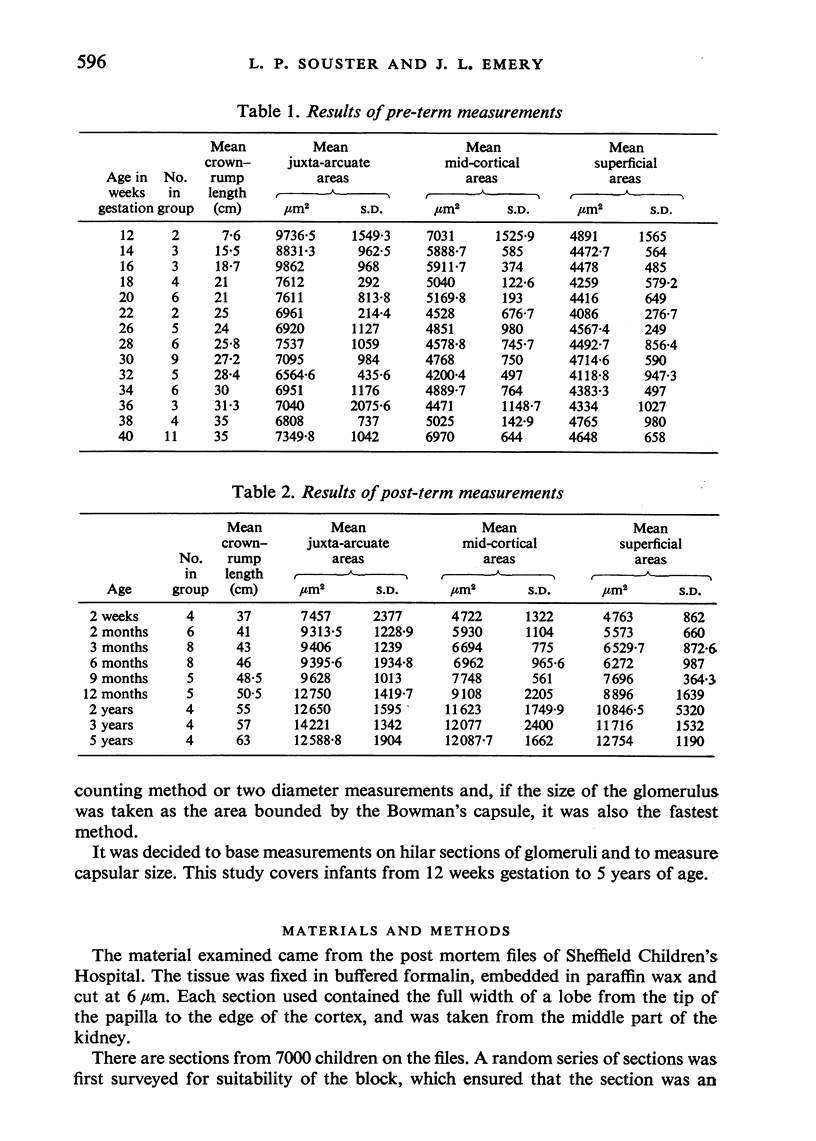
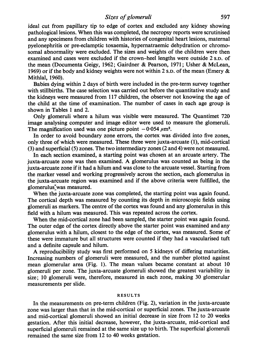
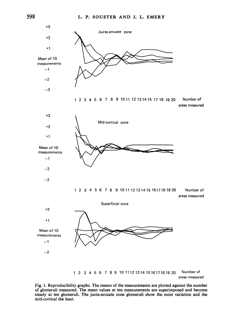
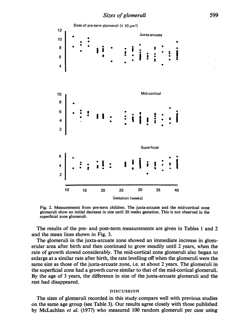
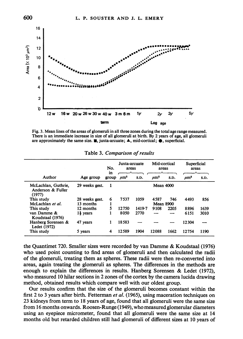
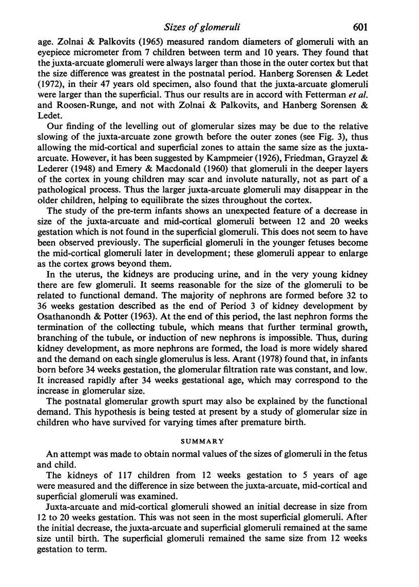
An attempt was made to obtain normal values of the sizes of glomeruli in the fetus and child. The kidneys of 117 children from 12 weeks gestation to 5 years of age were measured and the difference in size between the juxta-arcuate, mid-cortical and superficial glomeruli was examined. Juxta-arcuate and mid-cortical glomeruli showed an initial decrease in size from 12 to 20 weeks gestation. This was not seen in the most superficial glomeruli. After the initial decrease, the juxta-arcuate and superficial glomeruli remained at the same size until birth. The superficial glomeruli remained the same size from 12 weeks gestation to term. There was an immediate increase in size after birth in all three groups which slowed down after 2 years, when all three groups became the same size. The changes in size of the juxta-arcuate and mid-cortical glomeruli may be explained by functional demand.
Full text
PDF
Selected References
These references are in PubMed. This may not be the complete list of references from this article.
- ABRAMS R. L., LIPKIN L. E., HENNIGAR G. R. A quantitative estimation of variation among human renal glomeruli. Lab Invest. 1963 Jan;12:69–76. [PubMed] [Google Scholar]
- Arant B. S., Jr Developmental patterns of renal functional maturation compared in the human neonate. J Pediatr. 1978 May;92(5):705–712. doi: 10.1016/s0022-3476(78)80133-4. [DOI] [PubMed] [Google Scholar]
- Darmady E. M., Offer J., Woodhouse M. A. The parameters of the ageing kidney. J Pathol. 1973 Mar;109(3):195–207. doi: 10.1002/path.1711090304. [DOI] [PubMed] [Google Scholar]
- Dunnill M. S., Halley W. Some observations on the quantitative anatomy of the kidney. J Pathol. 1973 Jun;110(2):113–121. doi: 10.1002/path.1711100202. [DOI] [PubMed] [Google Scholar]
- EMERY J. L., MACDONALD M. S. Involuting and scarred glomeruli in the kidneys of infants. Am J Pathol. 1960 Jun;36:713–723. [PMC free article] [PubMed] [Google Scholar]
- EMERY J. L., MITHAL A. The weights of kidneys in late intrautrine life and childhood. J Clin Pathol. 1960 Nov;13:490–493. doi: 10.1136/jcp.13.6.490. [DOI] [PMC free article] [PubMed] [Google Scholar]
- FETTERMAN G. H., SHUPLOCK N. A., PHILIPP F. J., GREGG H. S. THE GROWTH AND MATURATION OF HUMAN GLOMERULI AND PROXIMAL CONVOLUTIONS FROM TERM TO ADULTHOOD: STUDIES BY MICRODISSECTION. Pediatrics. 1965 Apr;35:601–619. [PubMed] [Google Scholar]
- Friedman H. H., Grayzel D. M., Lederer M. Kidney Lesions in Stillborn and Newborn Infants: "Congenital Glomerulosclerosis. Am J Pathol. 1942 Jul;18(4):699–713. [PMC free article] [PubMed] [Google Scholar]
- Gairdner D., Pearson J. A growth chart for premature and other infants. Arch Dis Child. 1971 Dec;46(250):783–787. doi: 10.1136/adc.46.250.783. [DOI] [PMC free article] [PubMed] [Google Scholar]
- Kawano K., McCoy J., Wenzi J., Porch J., Howard C., Goddard M., Kimmelstiel P. Quantitation of glomerular structure. A study of methodology. Lab Invest. 1971 Oct;25(4):343–348. [PubMed] [Google Scholar]
- McLachlan M. S., Guthrie J. C., Anderson C. K., Fulker M. J. Vascular and glomerular changes in the ageing kidney. J Pathol. 1977 Feb;121(2):65–78. doi: 10.1002/path.1711210202. [DOI] [PubMed] [Google Scholar]
- OSATHANONDH V., POTTER E. L. DEVELOPMENT OF HUMAN KIDNEY AS SHOWN BY MICRODISSECTION. III. FORMATION AND INTERRELATIONSHIP OF COLLECTING TUBULES AND NEPHRONS. Arch Pathol. 1963 Sep;76:290–302. [PubMed] [Google Scholar]
- Sorensen F. H., Ledet T. Quantitative studies of the renal corpuscles. II. A methodological study. Acta Pathol Microbiol Scand A. 1972;80(6):721–728. doi: 10.1111/j.1699-0463.1972.tb00342.x. [DOI] [PubMed] [Google Scholar]
- Sorensen F. H. Quantitative studies of the renal corpuscles. I. Intraglomerular, interglomerular and interfocal variation in the normal kidney. Acta Pathol Microbiol Scand A. 1972;80(1):115–124. [PubMed] [Google Scholar]
- Usher R., McLean F. Intrauterine growth of live-born Caucasian infants at sea level: standards obtained from measurements in 7 dimensions of infants born between 25 and 44 weeks of gestation. J Pediatr. 1969 Jun;74(6):901–910. doi: 10.1016/s0022-3476(69)80224-6. [DOI] [PubMed] [Google Scholar]
- Zolnai B., Palkovits M. Glomerulometrics. 3. Data referring to the growth of the glomeruli in man. Acta Biol Acad Sci Hung. 1965;15(4):409–423. [PubMed] [Google Scholar]
- van Bamme B., Koudstaal J. Measuring glomerular diameters in tissue sections. Virchows Arch A Pathol Anat Histol. 1976 Mar 5;369(4):283–291. doi: 10.1007/BF00432450. [DOI] [PubMed] [Google Scholar]


