

Abstract
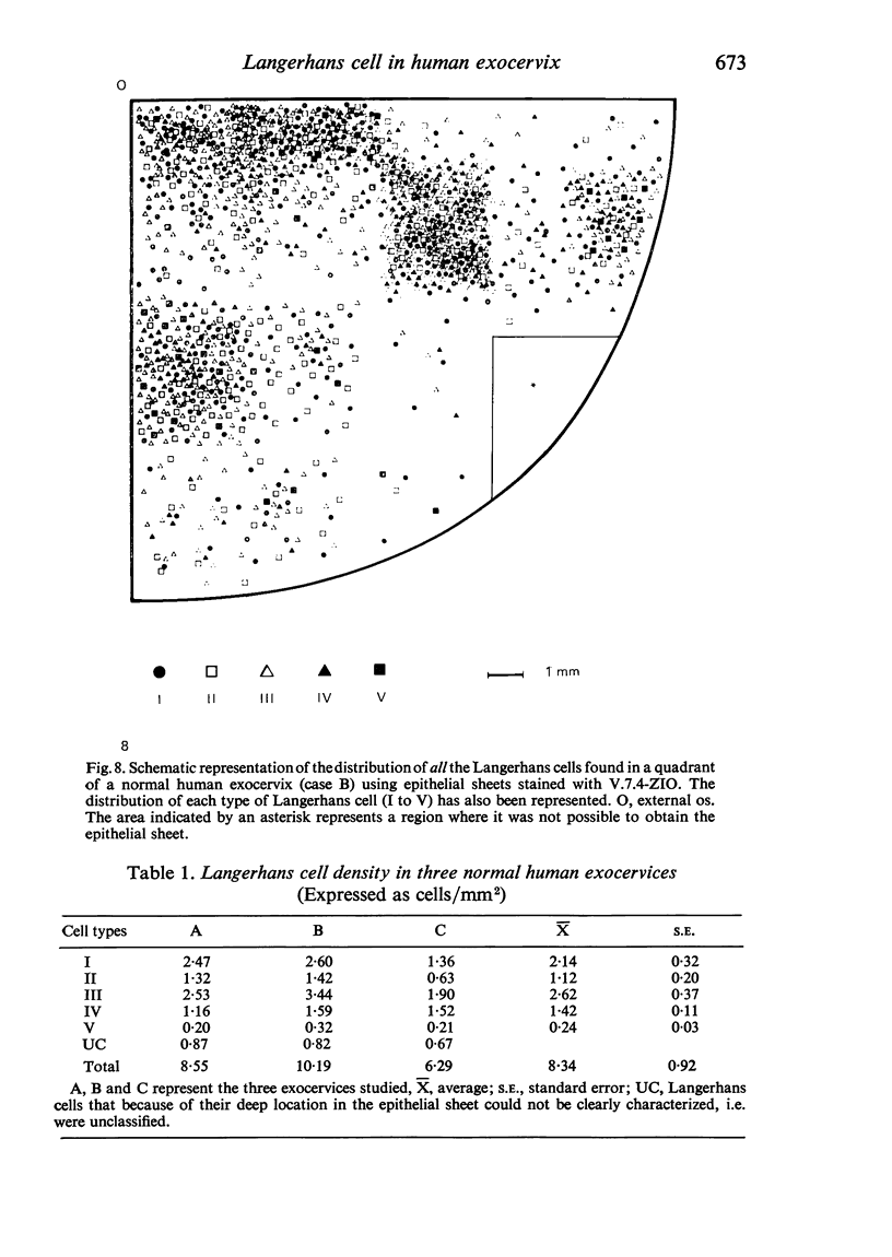
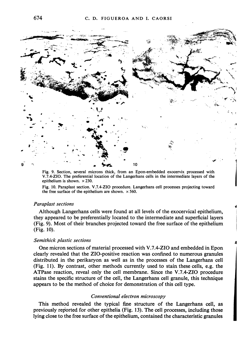
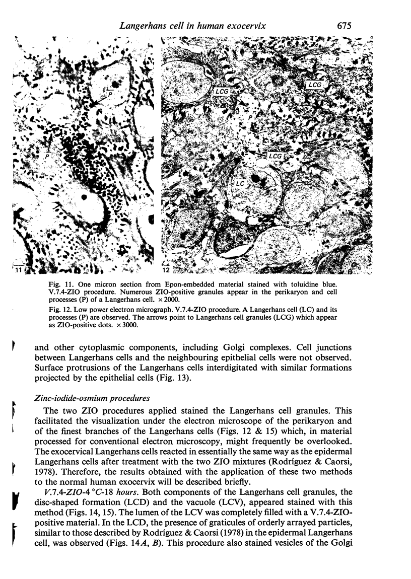
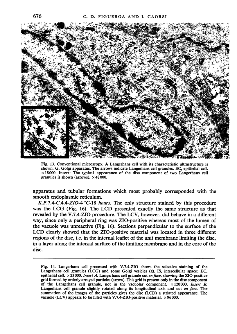
The gross morphology, density, distribution and ultrastructure of the Langerhans cell of the normal human exocervix were investigated. Two standardized zinc-iodide-osmium (ZIO) procedures were applied to epithelial sheets as well as to tissue samples processed for both light and electron microscopy. Conventional electron microscopical techniques were also used. The epithelial sheet preparation allowed the visualization of the whole profile of the Langerhans cell. It was found that the number of cell processes and the degree of their branching varied greatly from one cell to another. The cells were tentatively grouped into five types and it is suggested that they represent different degrees of cell activity. The cells appeared unevenly distributed, but with a preferential location around the external os. In the three cases studied the cell density averaged 8.3 LC/mm2. The ultrastructural study revealed the classical fine structure of the Langerhans cell. The cell processes and their branches contained all the organelles found in the perikaryon, including Golgi complexes and Langerhans cell granules. The two ZIO procedures revealed that the complex inner organization of the granule does not differ from that in the epidermal Langerhans cell. It is concluded that the Langerhans cell is a constant component of the normal human exocervix.
Full text
PDF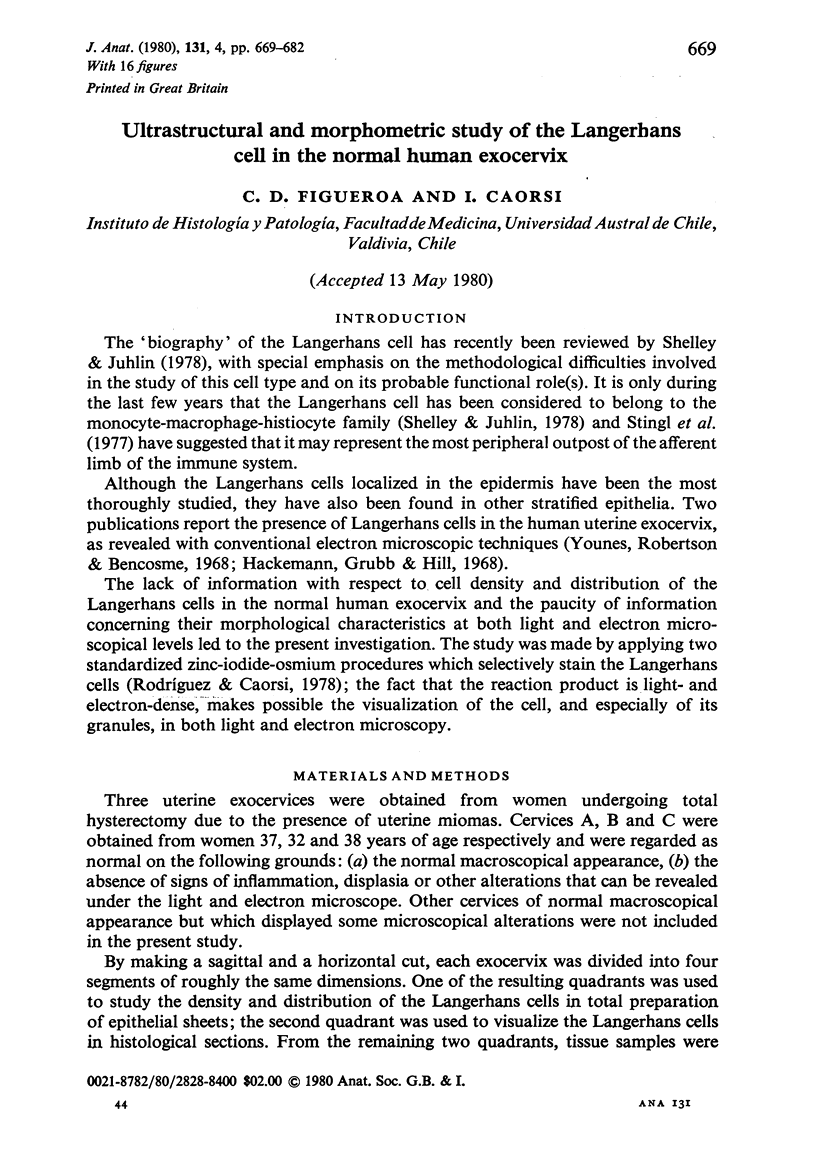


Images in this article
Selected References
These references are in PubMed. This may not be the complete list of references from this article.
- Brown J., Winklemann R. K., Wolff K. Langerhans cells in vitiligo: a qualitative study. J Invest Dermatol. 1967 Oct;49(4):386–390. [PubMed] [Google Scholar]
- Hackemann M., Grubb C., Hill K. R. The ultrastructure of normal squamous epithelium of the human cervix uteri. J Ultrastruct Res. 1968 Mar;22(5):443–457. doi: 10.1016/s0022-5320(68)90033-6. [DOI] [PubMed] [Google Scholar]
- Katz S. I., Tamaki K., Sachs D. H. Epidermal Langerhans cells are derived from cells originating in bone marrow. Nature. 1979 Nov 15;282(5736):324–326. doi: 10.1038/282324a0. [DOI] [PubMed] [Google Scholar]
- Klareskog L., Tjernlund U., Forsum U., Peterson P. A. Epidermal Langerhans cells express Ia antigens. Nature. 1977 Jul 21;268(5617):248–250. doi: 10.1038/268248a0. [DOI] [PubMed] [Google Scholar]
- RICHARDSON K. C., JARETT L., FINKE E. H. Embedding in epoxy resins for ultrathin sectioning in electron microscopy. Stain Technol. 1960 Nov;35:313–323. doi: 10.3109/10520296009114754. [DOI] [PubMed] [Google Scholar]
- Rodríguez E. M., Caorsi I. A second look at the ultrastructure of the Langerhans cell of the human epidermis. J Ultrastruct Res. 1978 Dec;65(3):279–295. doi: 10.1016/s0022-5320(78)80065-3. [DOI] [PubMed] [Google Scholar]
- Rodríguez E. M. Fixation of the central nervous system by perfusion of the cerebral ventricles with a threefold aldehyde mixture. Brain Res. 1969 Oct;15(2):395–412. doi: 10.1016/0006-8993(69)90163-2. [DOI] [PubMed] [Google Scholar]
- Rowden G., Lewis M. G., Sullivan A. K. Ia antigen expression on human epidermal Langerhans cells. Nature. 1977 Jul 21;268(5617):247–248. doi: 10.1038/268247a0. [DOI] [PubMed] [Google Scholar]
- Scaletta L. J., MacCallum D. K. A fine structural study of divalent cation-mediated epithelial union with connective tissue in human oral mucosa. Am J Anat. 1972 Apr;133(4):431–453. doi: 10.1002/aja.1001330406. [DOI] [PubMed] [Google Scholar]
- Shelley W. B., Juhlin L. The Langerhans cell: its origin, nature, and function. Acta Derm Venereol Suppl (Stockh) 1978;58(79):7–22. [PubMed] [Google Scholar]
- Stingl G., Katz S. I., Shevach E. M., Rosenthal A. S., Green I. Analogous functions of macrophages and Langerhans cells in the initiation in the immune response. J Invest Dermatol. 1978 Jul;71(1):59–64. doi: 10.1111/1523-1747.ep12544055. [DOI] [PubMed] [Google Scholar]
- Stingl G., Katz S. I., Shevach E. M., Wolff-Schreiner E., Green I. Detection of Ia antigens on Langerhans cells in guinea pig skin. J Immunol. 1978 Feb;120(2):570–578. [PubMed] [Google Scholar]
- Stingl G., Wolff-Schreiner E. C., Pichler W. J., Gschnait F., Knapp W., Wolff K. Epidermal Langerhans cells bear Fc and C3 receptors. Nature. 1977 Jul 21;268(5617):245–246. doi: 10.1038/268245a0. [DOI] [PubMed] [Google Scholar]
- Younes M. S., Robertson E. M., Bencosme S. A. Electron microscope observations on Langerhans cells in the cervix. Am J Obstet Gynecol. 1968 Oct 1;102(3):397–403. doi: 10.1016/0002-9378(68)90012-4. [DOI] [PubMed] [Google Scholar]