Abstract
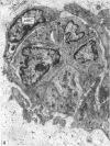
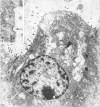
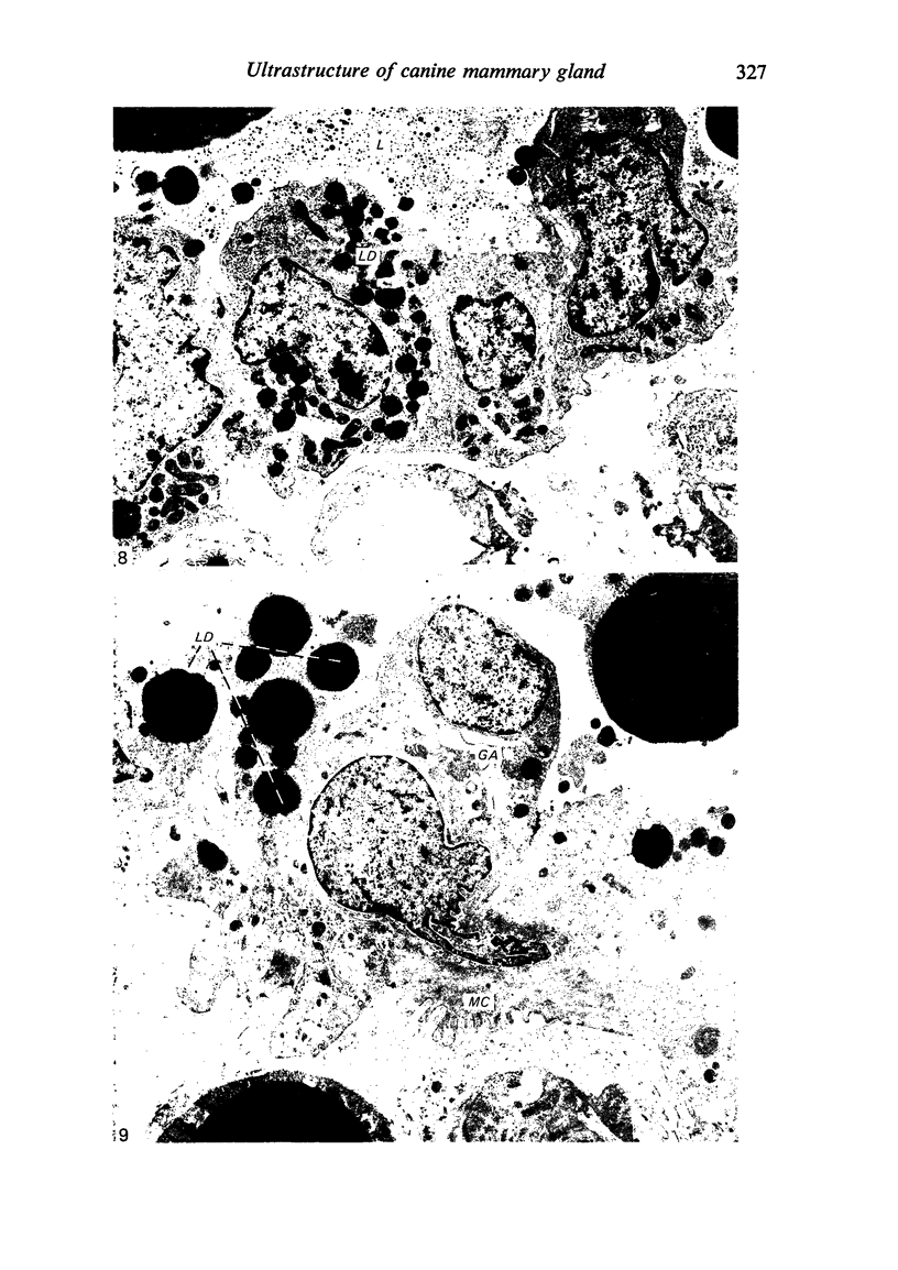
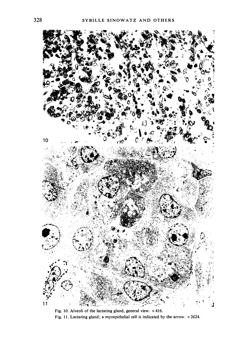
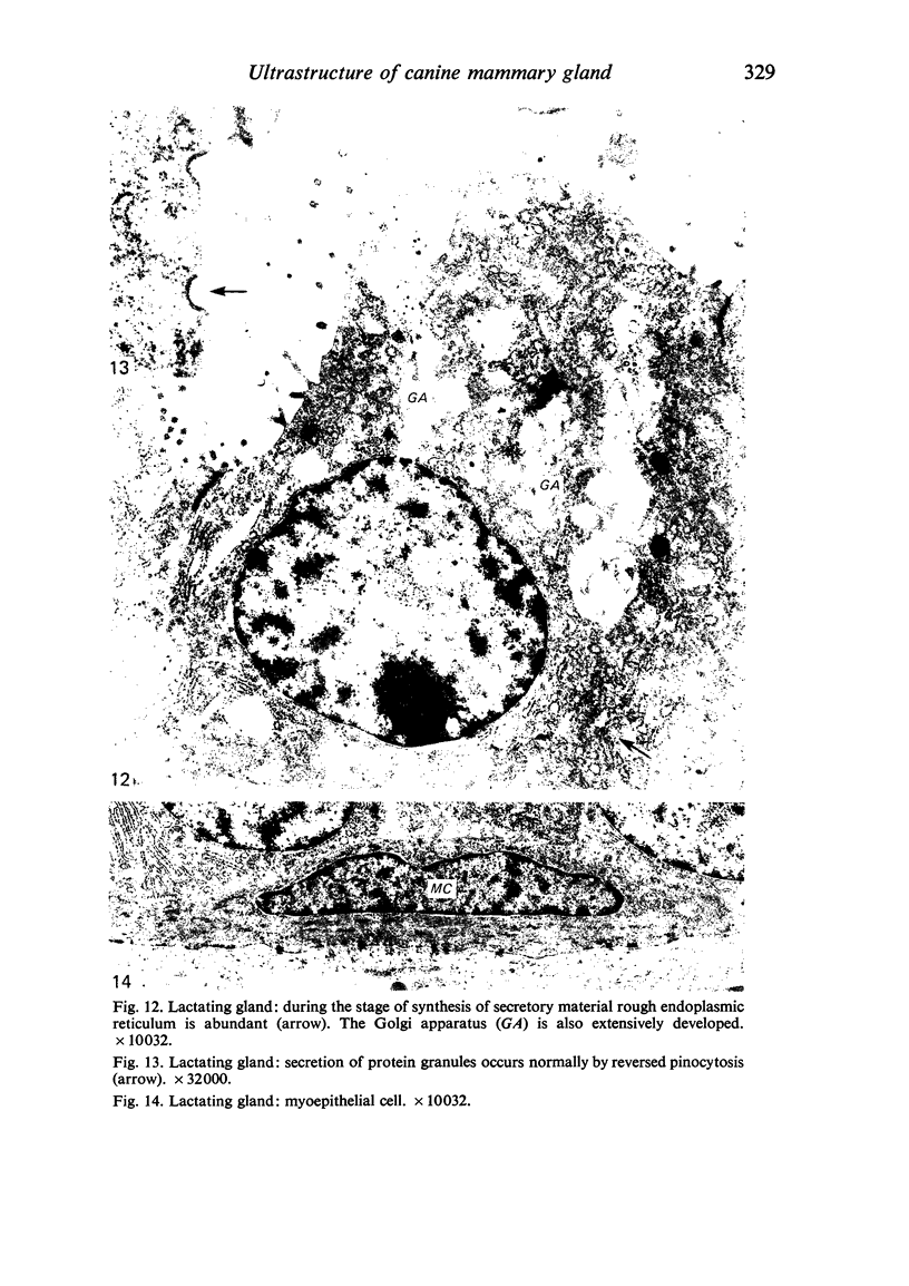
During pregnancy and lactation marked changes are observed in the fine structure of the secretory cells in the Beagle mammary gland: especially pronounced are differences in cellular height, shape and size of the nuclei and distribution of mitochondria. In later stages of pregnancy a proceeding development of those cellular organelles involved in synthesis and extrusion of secretory material (i.e. rough endoplasmic reticulum, Golgi apparatus) can be observed. Myoepithelial cells which can be first discerned from secretory cells by ultrastructural features from day 40 on show only minor variations of their ultrastructure during pregnancy and lactation.
Full text
PDF
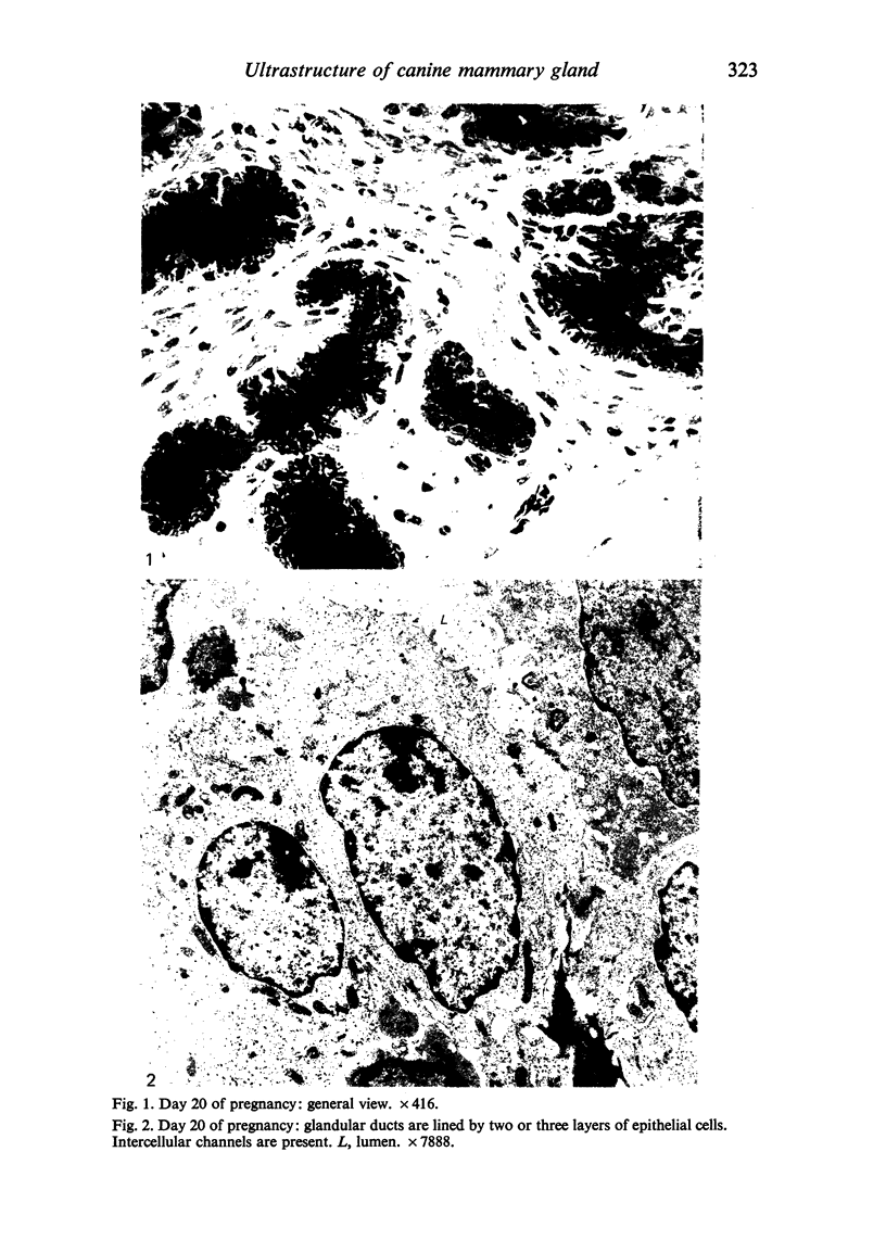
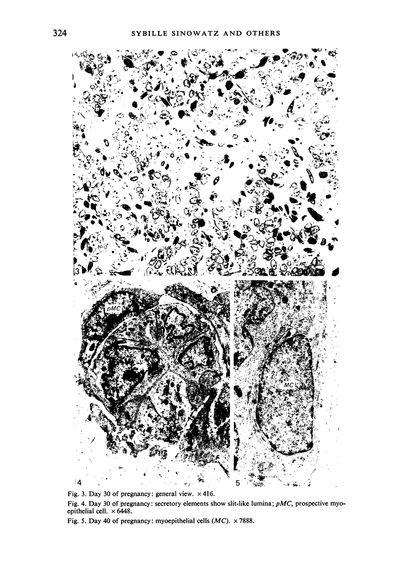
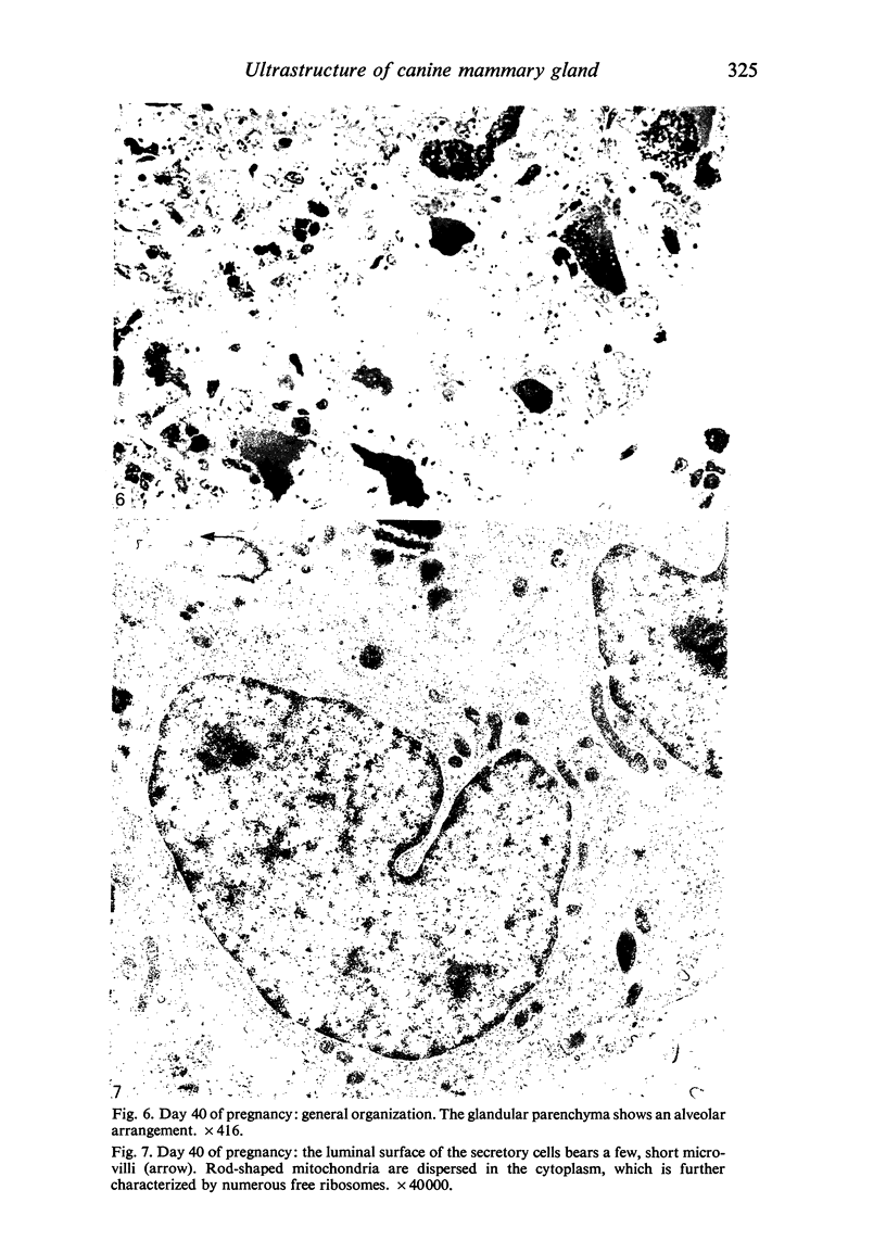
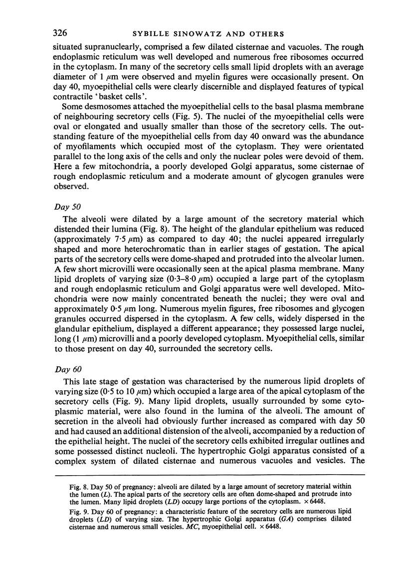
Images in this article
Selected References
These references are in PubMed. This may not be the complete list of references from this article.
- Adamiker D., Glawischnig E. Elektronenmikroskopische Untersuchungen an der Schweinemilchdrüse. II. Diskussion der Befunde. Wien Tierarztl Monatsschr. 1967 Sep;54(9):575–583. [PubMed] [Google Scholar]
- Adamiker D., Glawischnig E. Elektronenmikroskopische Unterushunge an der Schweinemilchdrüse. I. Befunde an Drüsen virgineller, gravider und Iakterender Tiere. Wien Tierarztl Monatsschr. 1967 Aug;54(8):507–contd. [PubMed] [Google Scholar]
- BARGMANN W., KNOOP A. Uber die Morphologie der Milchsekretion; lichtund elektronenmikroskopische Studien an der Milchdrüse Ratte. Z Zellforsch Mikrosk Anat. 1959;49(3):344–388. [PubMed] [Google Scholar]
- Carter D., Yardley J. H., Shelley W. M. Lobular carcinoma of the breast an ultrastructural comparison with certain duct carcinomas and benign lesions. Johns Hopkins Med J. 1969 Jul;125(1):25–32. [PubMed] [Google Scholar]
- FELDMAN J. D. Fine structure of the cow's udder during gestation and lactation. Lab Invest. 1961 Mar-Apr;10:238–255. [PubMed] [Google Scholar]
- Frank D. W., Kirton K. T., Murchison T. E., Quinlan W. J., Coleman M. E., Gilbertson T. J., Feenstra E. S., Kimball F. A. Mammary tumors and serum hormones in the bitch treated with medroxyprogesterone acetate or progesterone for four years. Fertil Steril. 1979 Mar;31(3):340–346. doi: 10.1016/s0015-0282(16)43886-0. [DOI] [PubMed] [Google Scholar]
- Gros C. M., Girardie J. Ultrastructure de la glande mammaire humaine non cancéreuse. Experientia. 1970 Dec 15;26(12):1345–1347. doi: 10.1007/BF02113021. [DOI] [PubMed] [Google Scholar]
- LANGER E., HUHN S. Der submikroskopische Bau der Myoepithelzelle. Z Zellforsch Mikrosk Anat. 1958;47(5):507–516. [PubMed] [Google Scholar]
- Murad T. M., Von Haam E. Ultrastructure of myoepithelial cells in human mammary gland tumors. Cancer. 1968 Jun;21(6):1137–1149. doi: 10.1002/1097-0142(196806)21:6<1137::aid-cncr2820210615>3.0.co;2-u. [DOI] [PubMed] [Google Scholar]
- Pulley L. T. Ultrastructural and histochemical demonstration of myoepithelium in the normal canine mammary gland. Am J Vet Res. 1973 Dec;34(12):1505–1512. [PubMed] [Google Scholar]
- Radnor C. J. Myoepithelium in the prelactating and lactating mammary glands of the rat. J Anat. 1972 Sep;112(Pt 3):337–353. [PMC free article] [PubMed] [Google Scholar]
- Stirling J. W., Chandler J. A. The fine structure of the normal, resting terminal ductal-lobular unit of the female breast. Virchows Arch A Pathol Anat Histol. 1976 Dec 27;372(3):205–226. doi: 10.1007/BF00433280. [DOI] [PubMed] [Google Scholar]
- Tannenbaum M., Weiss M., Marx A. J. Ultrastructure of the human mammary ductule. Cancer. 1969 Apr;23(4):958–978. doi: 10.1002/1097-0142(196904)23:4<958::aid-cncr2820230435>3.0.co;2-h. [DOI] [PubMed] [Google Scholar]
- Tateyama S., Nosaka D., Ashizawa H. Detection of actin, myosin and intermediate filaments in the myoepithelial cells in the mammary glands of lactating mice. J Electron Microsc (Tokyo) 1979;28(1):46–48. [PubMed] [Google Scholar]
- Toker C. Observations on the ultrastructure of a mammary ductule. J Ultrastruct Res. 1967 Nov;21(1):9–25. doi: 10.1016/s0022-5320(67)80003-0. [DOI] [PubMed] [Google Scholar]
- WAUGH D., VAN DER HOEVEN E. Fine structure of the human adult female breast. Lab Invest. 1962 Mar;11:220–228. [PubMed] [Google Scholar]
- Wrobel K. H., El Etreby M., Günzel P. Histochemische und histologische Untersuchungen an der Milchdrüse der Beagle-Hündin während verschiedener Funktionszustände. Acta Histochem. 1974;51(1):61–78. [PubMed] [Google Scholar]
- von Bomhard D., Kappes H. Ultrastrukturelle und ultrahistochemische Untersuchungen an der Milchdrüse des Hundes. I. Ultrastrukturelle Untersuchungen. Zentralbl Veterinarmed C. 1976 Mar;5(1):68–78. [PubMed] [Google Scholar]


