Abstract
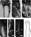
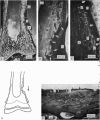
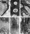
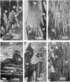
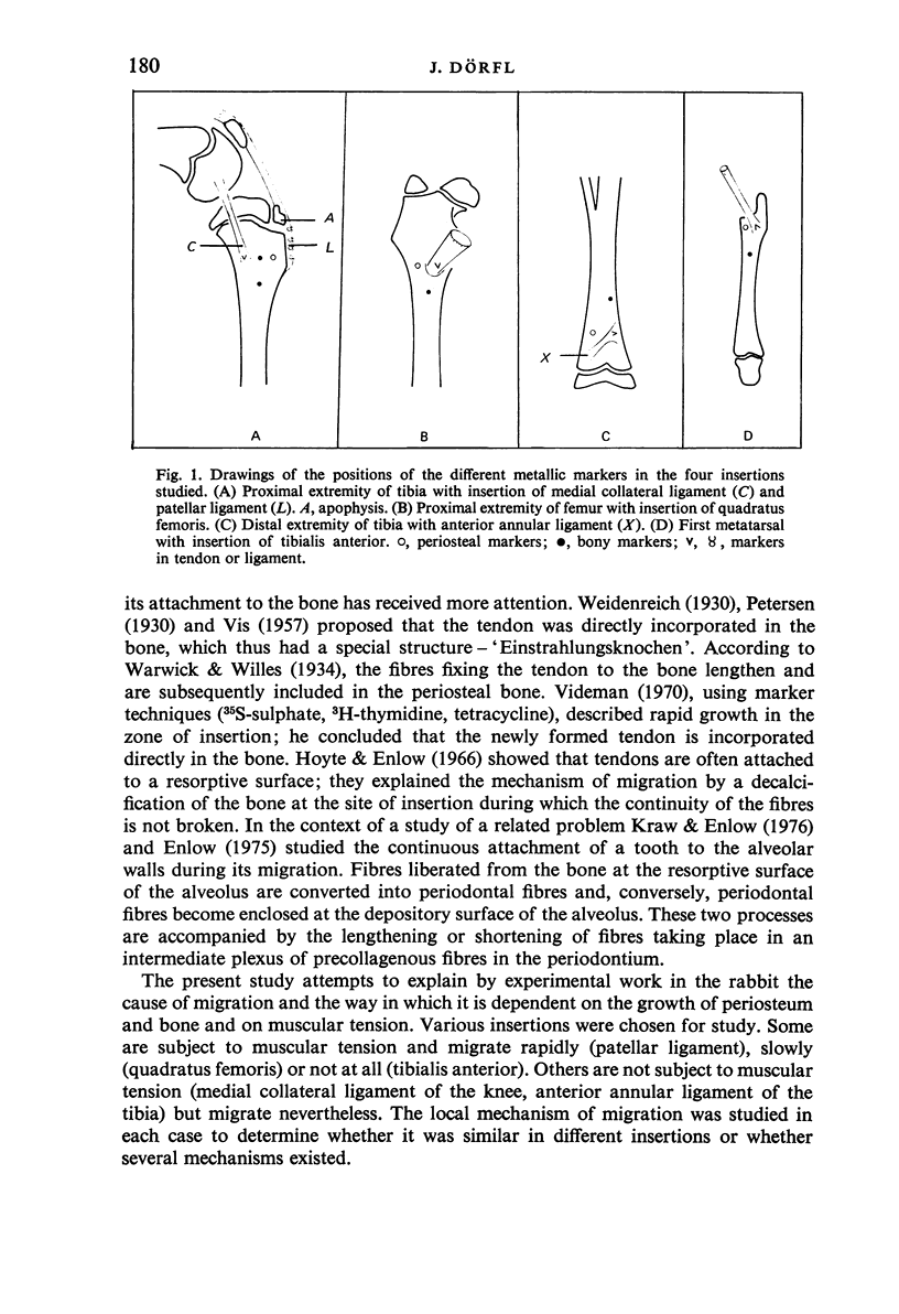
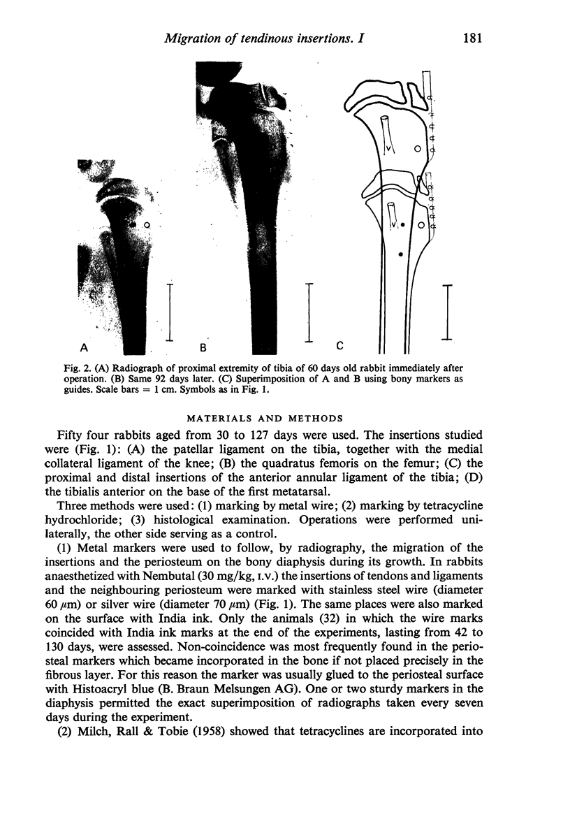
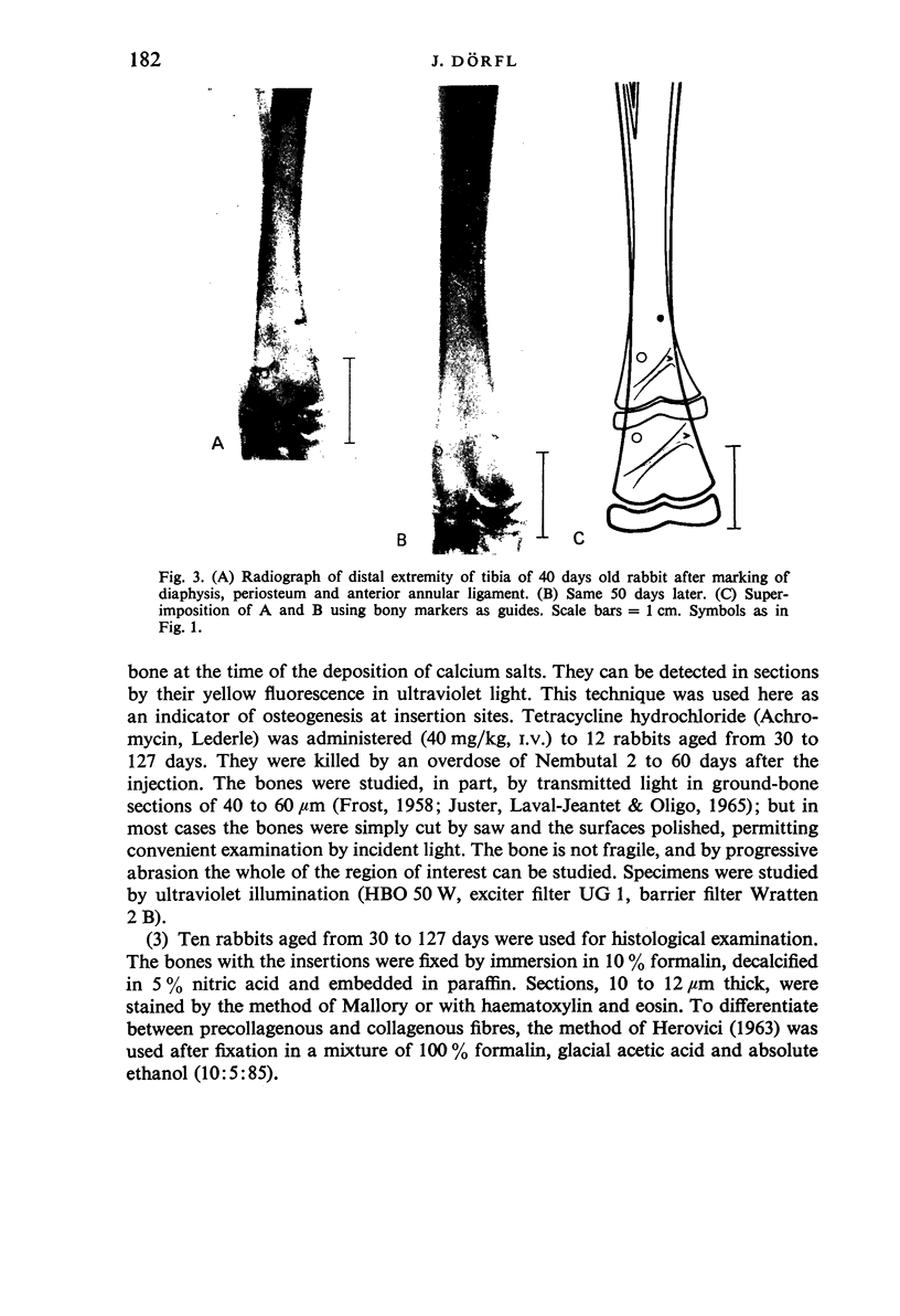
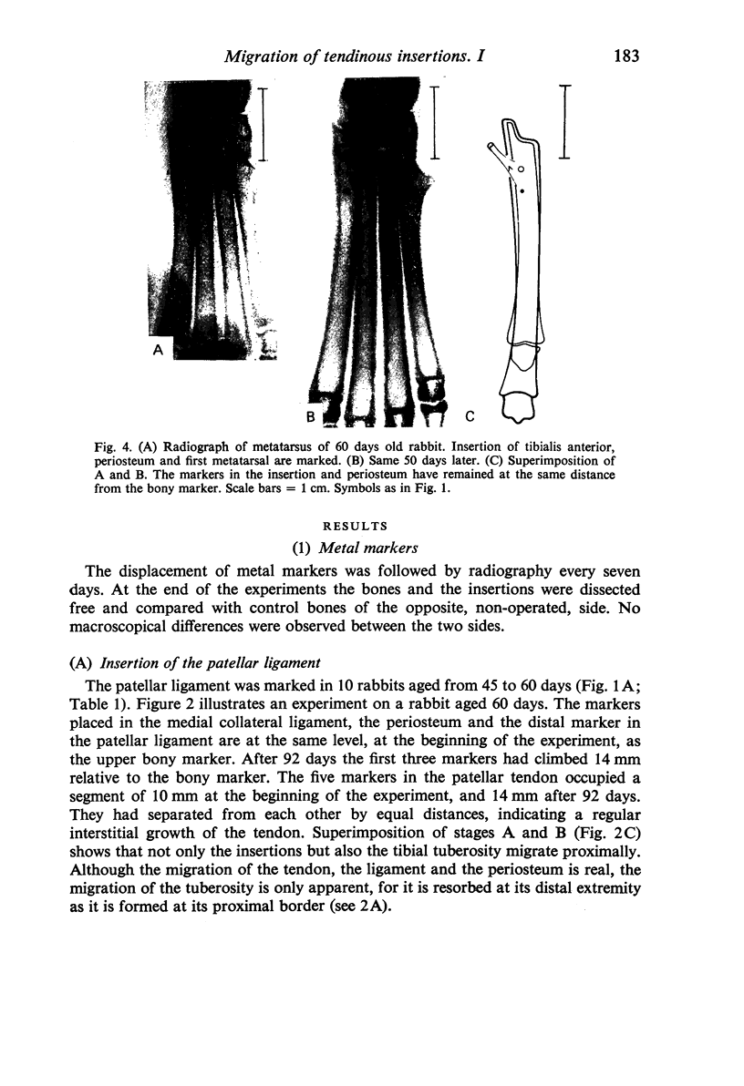
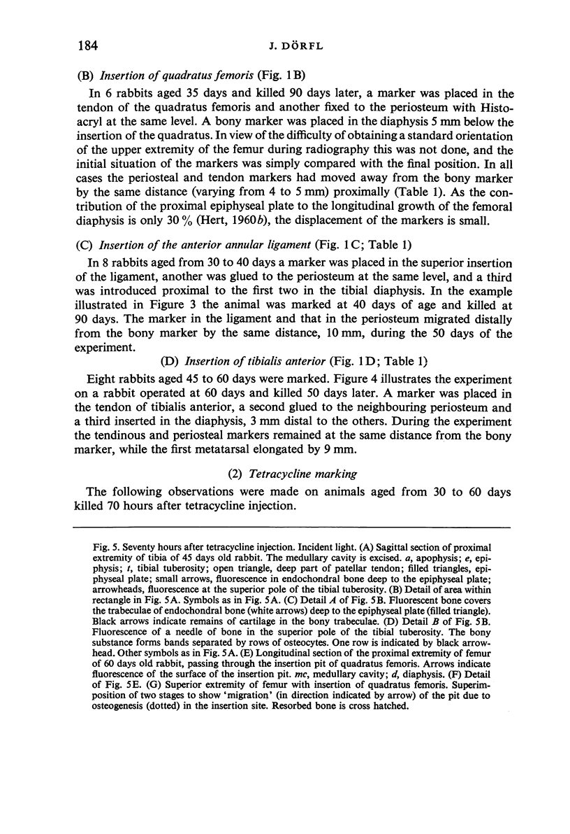
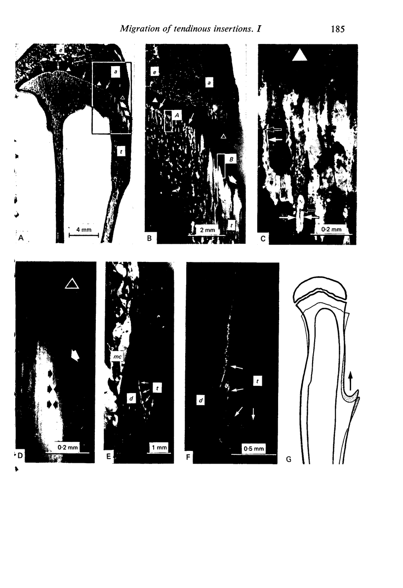
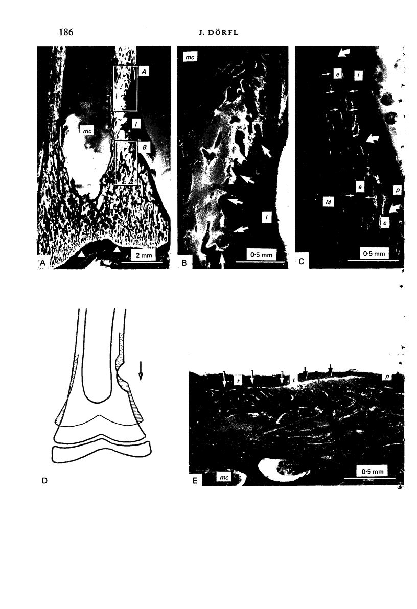
The cause and mechanisms of the migration of tendons and ligaments were studied in young rabbits. Three techniques were used: (1) Marking of insertions, the neighbouring periosteum and the diaphysis with metallic markers. (2) Marking of insertion sites by tetracycline as an indicator of osteogenesis. (3) Histological examination. The insertions used in the study were of three different characters: (1) Insertions subject to muscular traction (patellar ligament, quadratus femoris muscle, tibialis anterior muscle). (2) The distal insertions of the medial collateral ligament of the knee, stretched by the activity of the proximal epiphyseal cartilage of the tibia. (3) The proximal and distal insertions of the anterior annular ligament of the tibia, inserted solely in bone and periosteum. The cause of migration is the growth of periosteum dragging the insertions during its stretching, caused itself by the activity of the epiphyseal plates. The local mechanism governing migration while ensuring a continuous connexion with the bone is not the same in all sites. It depends upon the character of the bony surface at the insertion and of the function of the insertion zone, which can be osteogenic, resorptive or both. A plexus of precollagenous fibres is present at all resorptive insertion sites, and at some of the osteogenic sites.
Full text
PDF
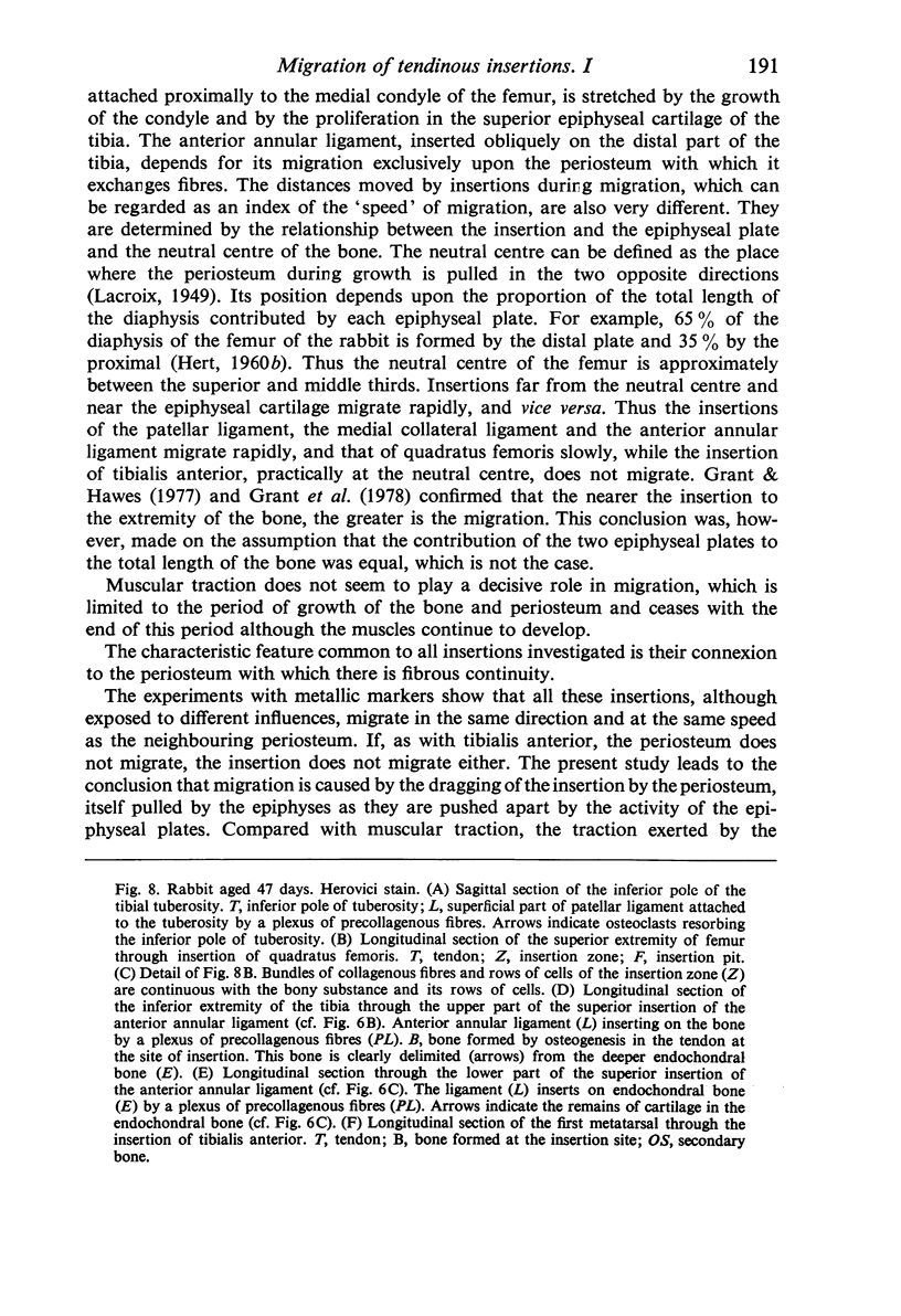
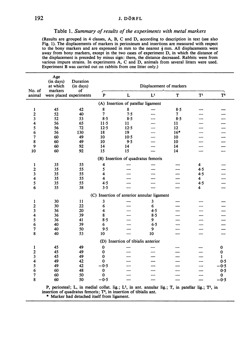
Images in this article
Selected References
These references are in PubMed. This may not be the complete list of references from this article.
- BIERMANN H. Die Knochenbildung im Bereich periostaler-diaphysärer Sehnenund Bandanstäze. Z Zellforsch Mikrosk Anat. 1957;46(6):635–671. [PubMed] [Google Scholar]
- FRIEND W. G. A polychrome stainfor differentiating precollagen from collagen. Stain Technol. 1963 May;38:204–206. [PubMed] [Google Scholar]
- FROST H. M. Staining of fresh, undecalcified, thin bone sections. Stain Technol. 1959 May;34(3):135–146. doi: 10.3109/10520295909114665. [DOI] [PubMed] [Google Scholar]
- Grant P. G., Buschang P. H., Drolet D. W. Positional relationships of structures attached to long bones during growth. Cross-sectional studies. Acta Anat (Basel) 1978;102(4):378–384. doi: 10.1159/000145661. [DOI] [PubMed] [Google Scholar]
- Grant P. G., Hawes M. R. Experimental modification of muscle migration in the rabbit. J Anat. 1977 Apr;123(Pt 2):361–367. [PMC free article] [PubMed] [Google Scholar]
- Hoyte D. A., Enlow D. H. Wolff's law and the problem of muscle attachment on resorptive surfaces of bone. Am J Phys Anthropol. 1966 Mar;24(2):205–213. doi: 10.1002/ajpa.1330240209. [DOI] [PubMed] [Google Scholar]
- MILCH R. A., RALL D. P., TOBIE J. E. Fluorescence of tetracycline antibiotics in bone. J Bone Joint Surg Am. 1958 Jul;40-A(4):897–910. [PubMed] [Google Scholar]
- Videman T. An experimental study of the effects of growth on the relationship of tendons and ligaments to bone at the site of diaphyseal insertion. Acta Orthop Scand Suppl. 1970:1–22. doi: 10.3109/ort.1970.41.suppl-131.01. [DOI] [PubMed] [Google Scholar]


