

Abstract
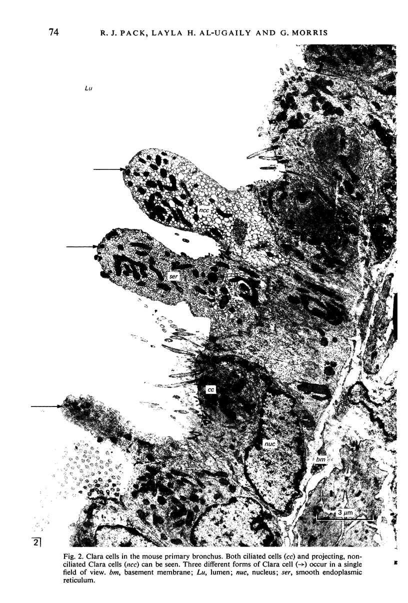
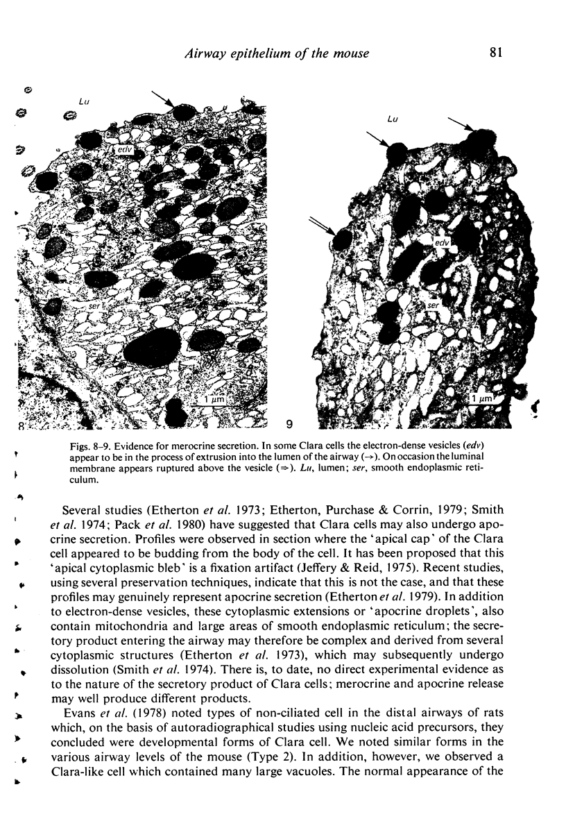
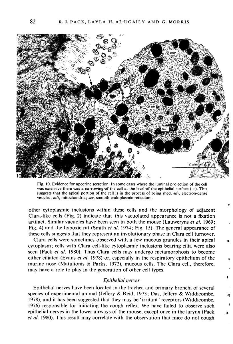
The epithelium of the conducting airways of the mouse consists of a single layer of cells. The number, type and form of these cells have been investigated at five airway levels from the trachea to the distal conducting bronchi with both light and electron microscopes. Contrary to what is found in other species, the majority (50-60%) of cells in the murine airway epithelium are Clara cells. Mucus-producing tissue was infrequent throughout the airways, though epithelial mucous cells occurred in increased numbers at the carina and in the primary bronchus. No mucous or serous cells or submucosal glands were seen in intralobular airways. On a morphological basis, three distinct forms of Clara cell were recognized. On occasion, cells were observed which were apparently transitional types between these and also between Clara cells and mucous or ciliated cells. It is suggested that the 'transforming' cells may indicate a role for the Clara cell as a developmental cell involved in the epithelial cell turnover. Evidence is also provided that Clara cells may undergo both apocrine and merocrine secretion and, it is argued that the latter may be of a PAS + ve material. Free nerve endings were not seen in the epithelium. This may be related to athe restricted ability of mice to cough. It is suggested that the lack of mucus-producing tissue and of cough reflex may be due to the small diameter of the mouse airways.
Full text
PDF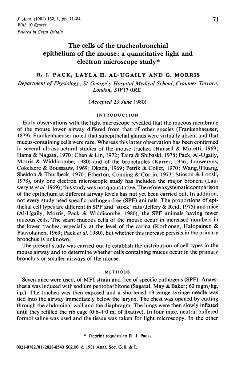

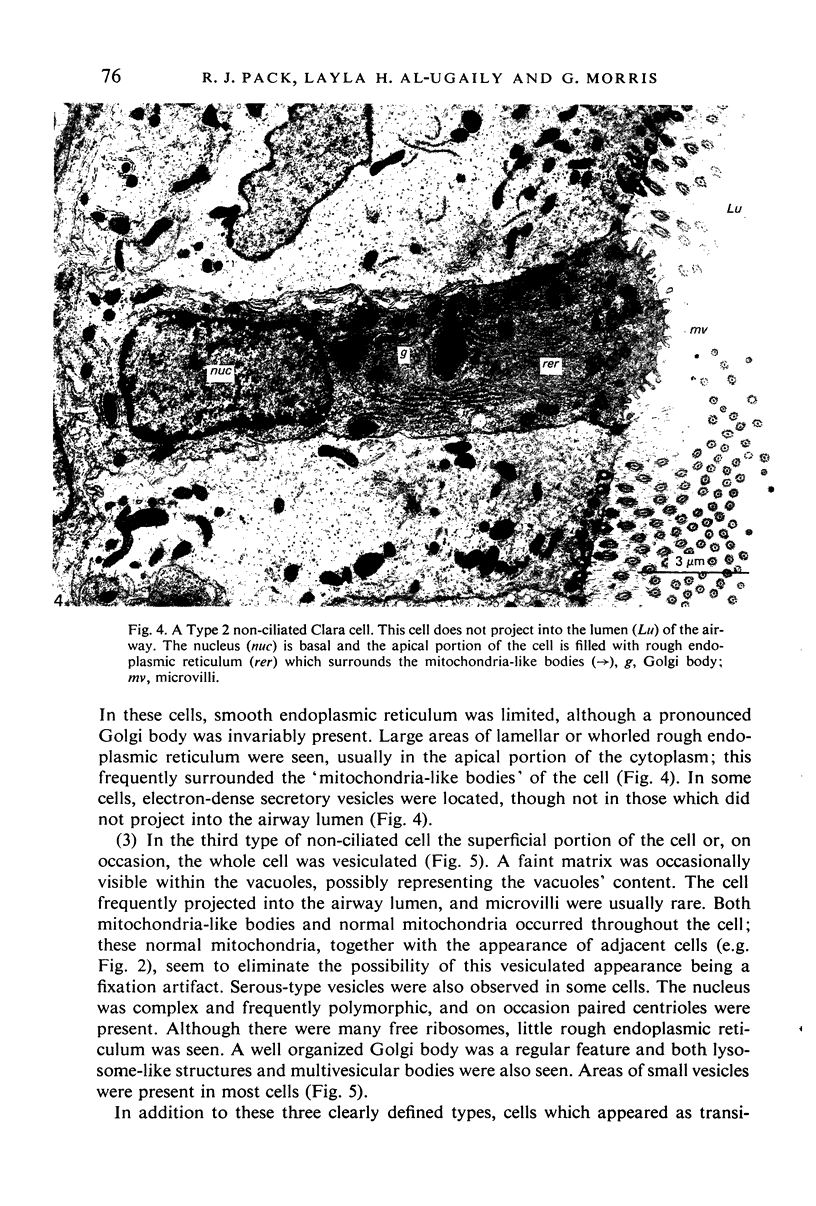
Images in this article
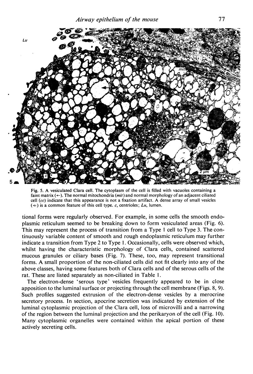
Selected References
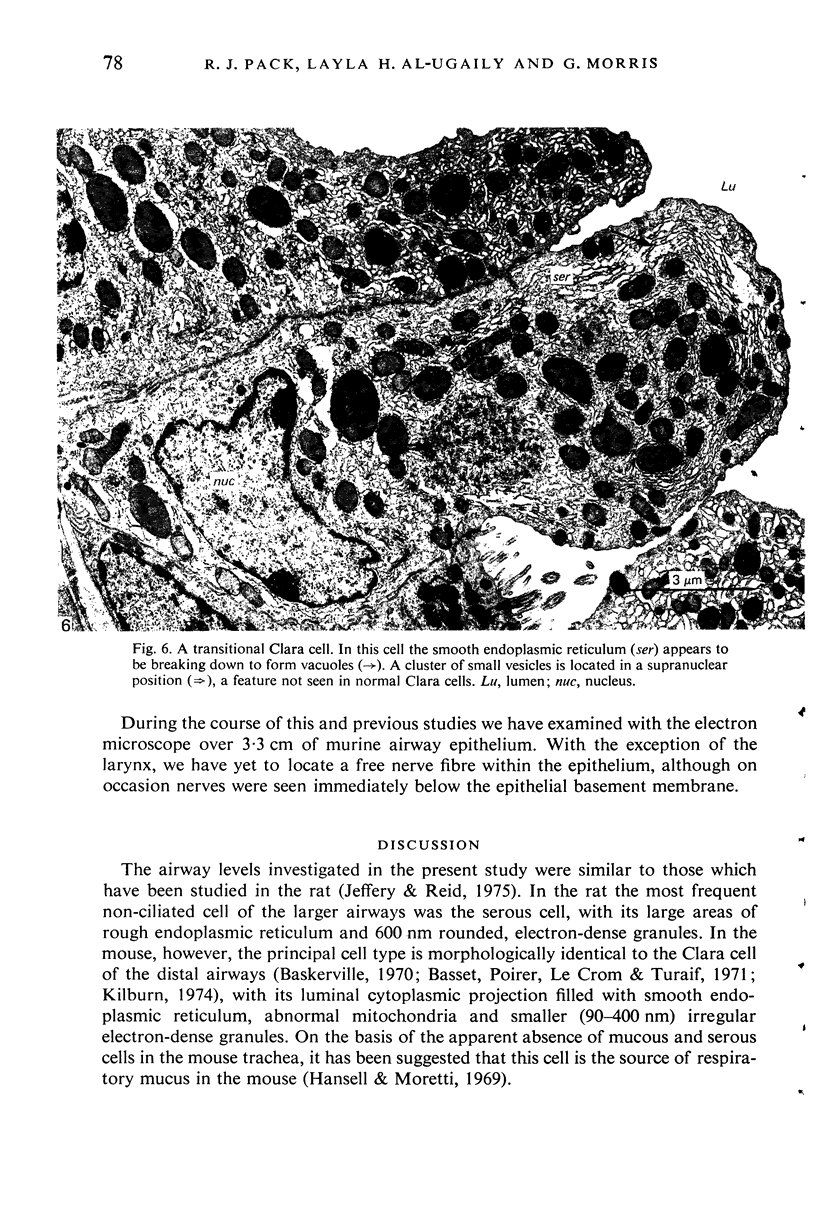
These references are in PubMed. This may not be the complete list of references from this article.
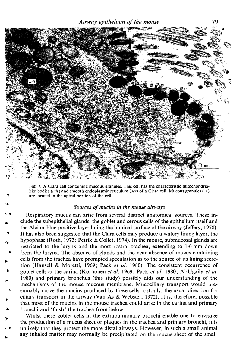
- Azzopardi A., Thurlbeck W. M. The histochemistry of the nonciliated bronchiolar epithelial cell. Am Rev Respir Dis. 1969 Apr;99(4):516–525. doi: 10.1164/arrd.1969.99.4P1.516. [DOI] [PubMed] [Google Scholar]
- Basset F., Poirier J., Le Crom M., Turiaf J. Etude ultrastructurale de l'épithélium bronchiolaire humain. Z Zellforsch Mikrosk Anat. 1971;116(3):425–442. [PubMed] [Google Scholar]
- Breeze R. G., Wheeldon E. B. The cells of the pulmonary airways. Am Rev Respir Dis. 1977 Oct;116(4):705–777. doi: 10.1164/arrd.1977.116.4.705. [DOI] [PubMed] [Google Scholar]
- Cutz E., Conen P. E. Ultrastructure and cytochemistry of Clara cells. Am J Pathol. 1971 Jan;62(1):127–141. [PMC free article] [PubMed] [Google Scholar]
- Das R. M., Jeffrey P. K., Widdicombe J. G. The epithelial innervation of the lower respiratory tract of the cat. J Anat. 1978 May;126(Pt 1):123–131. [PMC free article] [PubMed] [Google Scholar]
- Etherton J. E., Conning D. M., Corrin B. Autoradiographical and morphological evidence for apocrine secretion of dipalmitoyl lecithin in the terminal bronchiole of mouse lung. Am J Anat. 1973 Sep;138(1):11–35. doi: 10.1002/aja.1001380103. [DOI] [PubMed] [Google Scholar]
- Etherton J. E., Purchase I. F., Corrin B. Apocrine secretion in the terminal bronchiole of mouse lung. J Anat. 1979 Sep;129(Pt 2):305–322. [PMC free article] [PubMed] [Google Scholar]
- Evans M. J., Cabral-Anderson L. J., Freeman G. Role of the Clara cell in renewal of the bronchiolar epithelium. Lab Invest. 1978 Jun;38(6):648–653. [PubMed] [Google Scholar]
- Hama K., Nagata F. A stereoscope observation of tracheal epithelium of mouse by means of the high voltage electron microscope. J Cell Biol. 1970 Jun;45(3):654–659. doi: 10.1083/jcb.45.3.654. [DOI] [PMC free article] [PubMed] [Google Scholar]
- Hansell M. M., Moretti R. L. Ultrastructure of the mouse tracheal epithelium. J Morphol. 1969 Jun;128(2):159–169. doi: 10.1002/jmor.1051280203. [DOI] [PubMed] [Google Scholar]
- Jeffery P. K., Reid L. New observations of rat airway epithelium: a quantitative and electron microscopic study. J Anat. 1975 Nov;120(Pt 2):295–320. [PMC free article] [PubMed] [Google Scholar]
- Jeffery P., Reid L. Intra-epithelial nerves in normal rat airways: a quantitative electron microscopic study. J Anat. 1973 Jan;114(Pt 1):35–45. [PMC free article] [PubMed] [Google Scholar]
- KARRER H. E. Electron microscopic study of bronchiolar epithelium of normal mouse lung; preliminary report. Exp Cell Res. 1956 Feb;10(1):237–241. doi: 10.1016/0014-4827(56)90093-3. [DOI] [PubMed] [Google Scholar]
- Kilburn K. H. Functional morphology of the distal lung. Int Rev Cytol. 1974;37(0):153–270. doi: 10.1016/s0074-7696(08)61359-5. [DOI] [PubMed] [Google Scholar]
- Korhonen L. K., Holopainen E., Paavolainen M. Some histochemical characteristics of tracheobronchial tree and pulmonary neoplasms. Acta Histochem. 1969;32(1):57–73. [PubMed] [Google Scholar]
- Kuhn C., 3rd, Callaway L. A., Askin F. B. The formation of granules in the bronchiolar Clara cells of the rat. 1. Electron microscopy,. J Ultrastruct Res. 1974 Dec;49(3):387–400. doi: 10.1016/s0022-5320(74)90052-5. [DOI] [PubMed] [Google Scholar]
- Niden A. H. Bronchiolar and large alveolar cell in pulmonary phospholipid metabolism. Science. 1967 Dec 8;158(3806):1323–1324. doi: 10.1126/science.158.3806.1323. [DOI] [PubMed] [Google Scholar]
- Okada Y. The ultrastructure of the Clara cell in the bronchiolar epithelium. Bull Chest Dis Res Inst Kyoto Univ. 1969 Sep;3(1):1–10. [PubMed] [Google Scholar]
- Pack R. J., Al-Ugaily L. H., Morris G., Widdicombe J. G. The distribution and structure of cells in the tracheal epithelium of the mouse. Cell Tissue Res. 1980;208(1):65–84. doi: 10.1007/BF00234174. [DOI] [PubMed] [Google Scholar]
- Petrik P., Collet A. J. Infrastructure des cellules bronchiolaires non ciliées chez la souris. Rev Can Biol. 1970 Jun;29(2):141–152. [PubMed] [Google Scholar]
- Petrik P., Collet A. J. Quantitative electron microscopic autoradiography of in vivo incorporation of 3H-choline, 3H-leucine, 3H-acetate and 3H-galactose in non-ciliated bronchiolar (Clara) cells of mice. Am J Anat. 1974 Apr;139(4):519–533. doi: 10.1002/aja.1001390405. [DOI] [PubMed] [Google Scholar]
- REYNOLDS E. S. The use of lead citrate at high pH as an electron-opaque stain in electron microscopy. J Cell Biol. 1963 Apr;17:208–212. doi: 10.1083/jcb.17.1.208. [DOI] [PMC free article] [PubMed] [Google Scholar]
- STEMPAK J. G., WARD R. T. AN IMPROVED STAINING METHOD FOR ELECTRON MICROSCOPY. J Cell Biol. 1964 Sep;22:697–701. doi: 10.1083/jcb.22.3.697. [DOI] [PMC free article] [PubMed] [Google Scholar]
- Smith P., Heath D., Moosavi H. The Clara cell. Thorax. 1974 Mar;29(2):147–163. doi: 10.1136/thx.29.2.147. [DOI] [PMC free article] [PubMed] [Google Scholar]
- Stinson S. F., Loosli C. G. Ultrastructural evidence concerning the mode of secretion of electron-dense granules by Clara cells. J Anat. 1978 Oct;127(Pt 2):291–298. [PMC free article] [PubMed] [Google Scholar]
- Taira K., Shibasaki S. A fine structure study of the non-ciliated cells in the mouse tracheal epithelium with special reference to the relation of "brush cells" and "endocrine cells". Arch Histol Jpn. 1978 Sep;41(4):351–366. doi: 10.1679/aohc1950.41.351. [DOI] [PubMed] [Google Scholar]
- Wang N. S., Huang S. N., Sheldon H., Thurlbeck W. M. Ultrastructural changes of Clara and type II alveolar cells in adrenalin-induced pulmonary edema in mice. Am J Pathol. 1971 Feb;62(2):237–252. [PMC free article] [PubMed] [Google Scholar]
- van As A., Webster I. The organization of ciliary activity and mucus transport in pulmonary airways. S Afr Med J. 1972 Mar 25;46(13):347–350. [PubMed] [Google Scholar]