Abstract
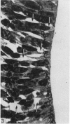
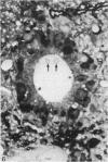
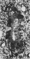
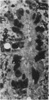
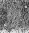
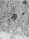
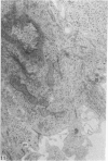
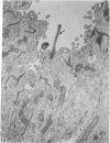
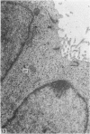
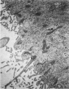
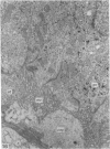
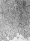
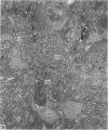
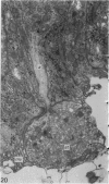
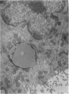
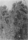
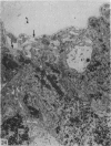
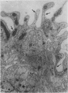
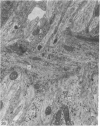
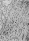
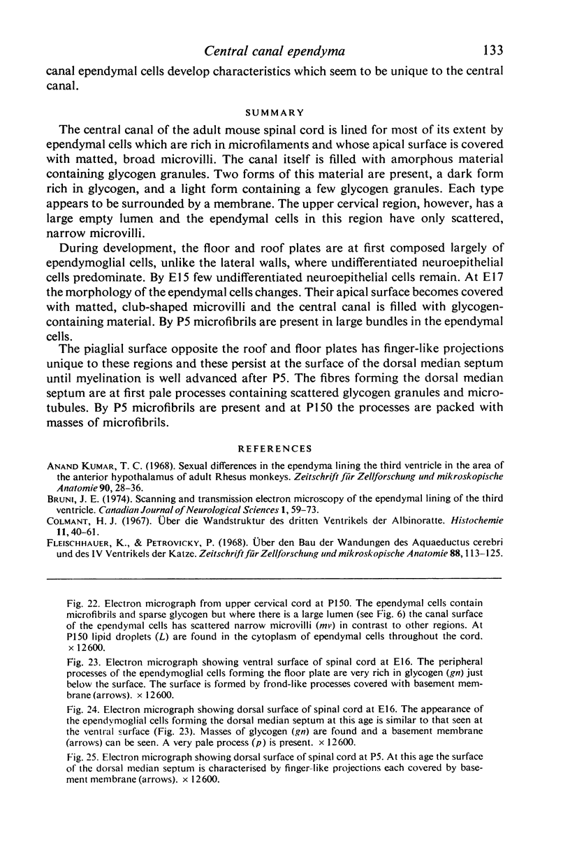
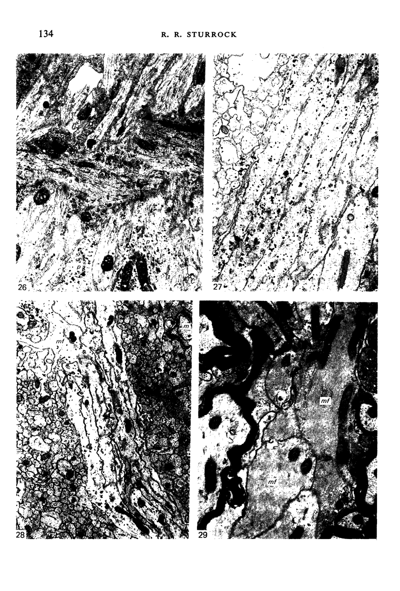
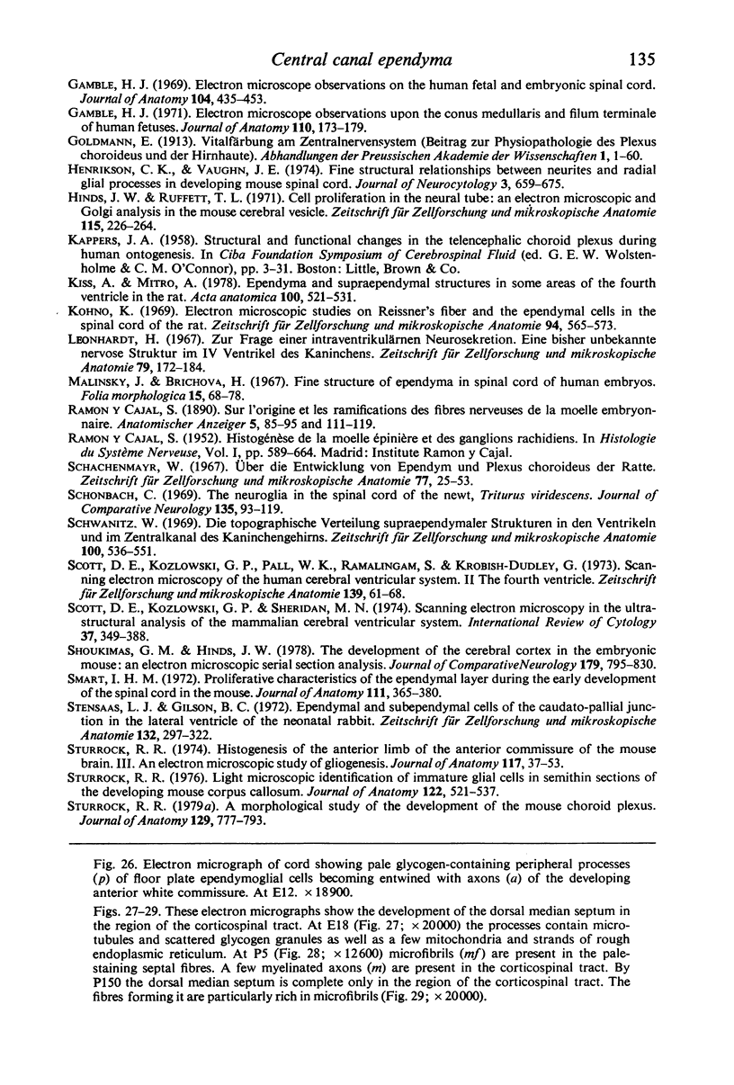
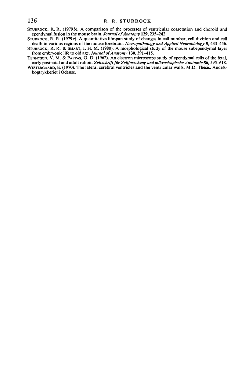
The central canal of the adult mouse spinal cord is lined for most of its extent by ependymal cells which are rich in microfilaments and whose apical surface is covered with matted, broad microvilli. The canal itself is filled with amorphous material containing glycogen granules. Two forms of this material are present, a dark form rich in glycogen, and a light form containing a few glycogen granules. Each type appears to be surrounded by a membrane. The upper cervical region, however, has a large empty lumen and the ependymal cells in this region have only scattered, narrow microvilli. During development, the floor and roof plates are at first composed largely of ependymoglial cells, unlike the lateral walls, where undifferentiated neuroepithelial cells predominate. By E15 few undifferentiated neuroepithelial cells remain. At E17 the morphology of the ependymal cells changes. Their apical surface becomes covered with matted, club-shaped microvilli and the central canal is filled with glycogen-containing material. By P5 microfibrils are present in large bundles in the ependymal cells. The piaglial surface opposite the roof and floor plates has finger-like projections unique to these regions and these persist at the surface of the dorsal median septum until myelination is well advanced after P5. The fibres forming the dorsal median septum are at first pale processes containing scattered glycogen granules and microtubules. By P5 microfibrils are present and at P150 the processes are packed with masses of microfibrils.
Full text
PDF
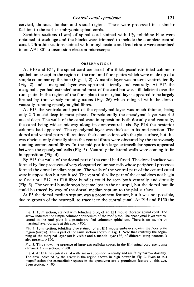
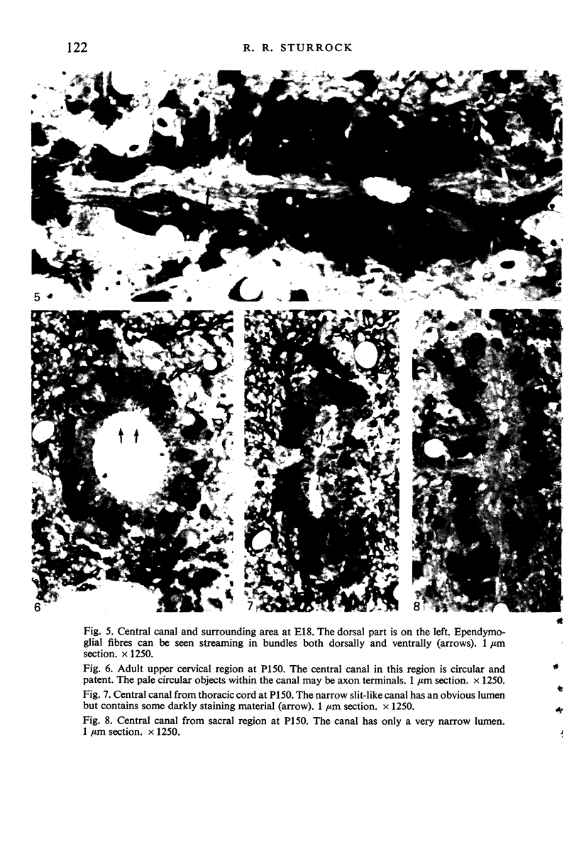
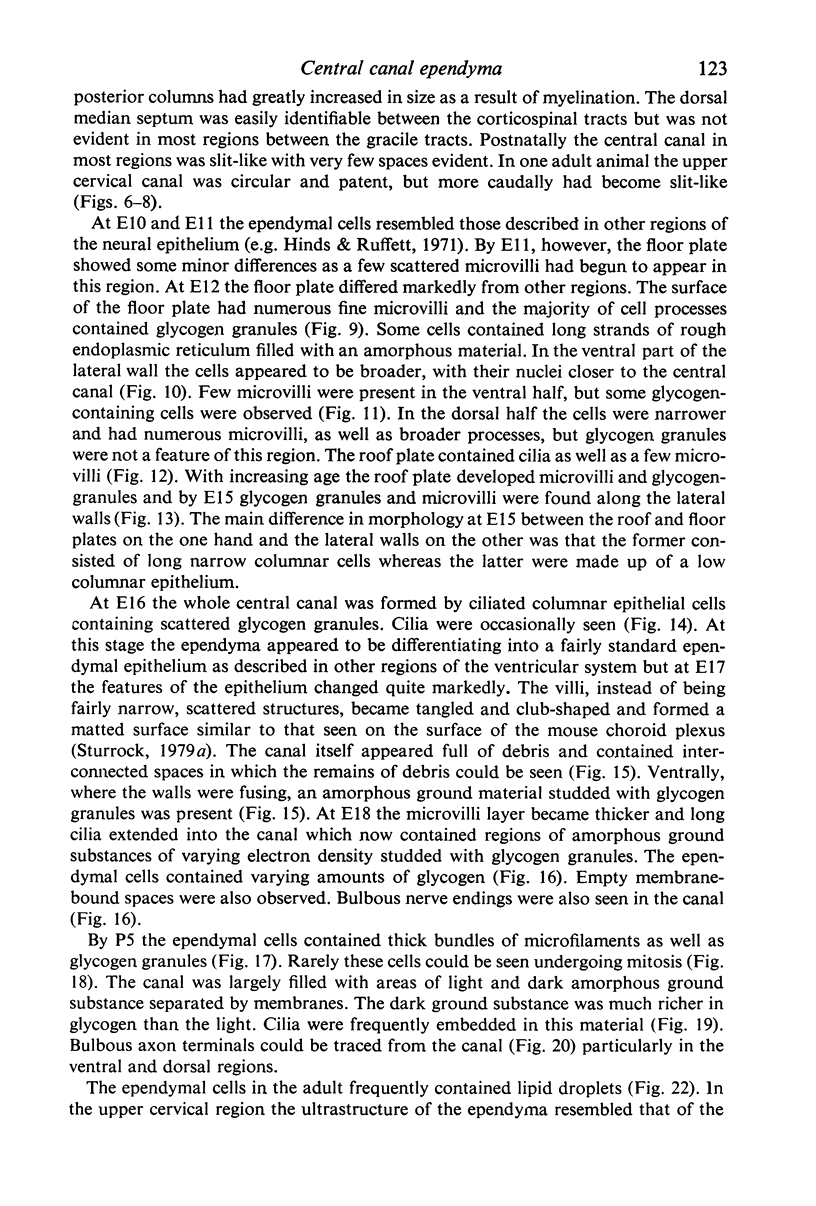
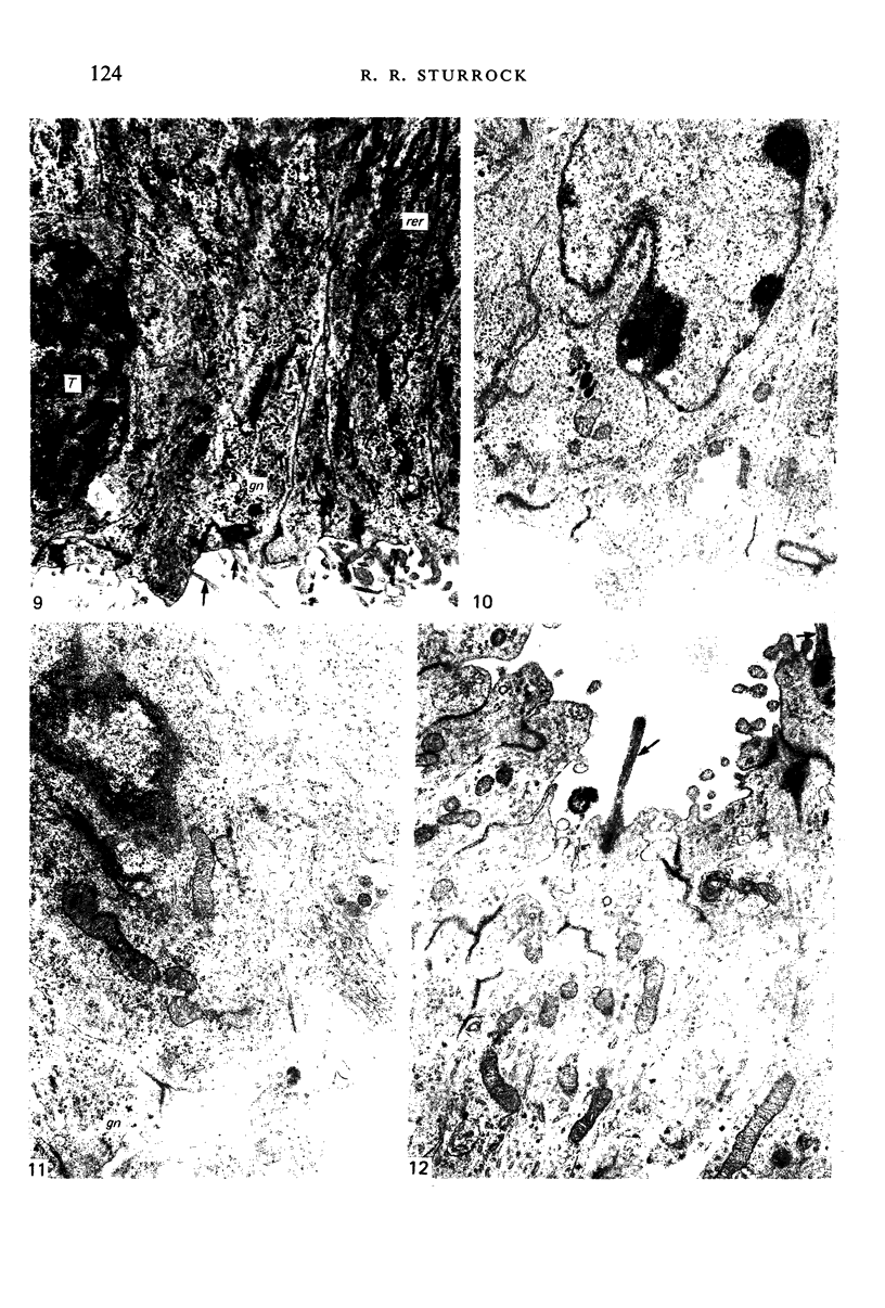
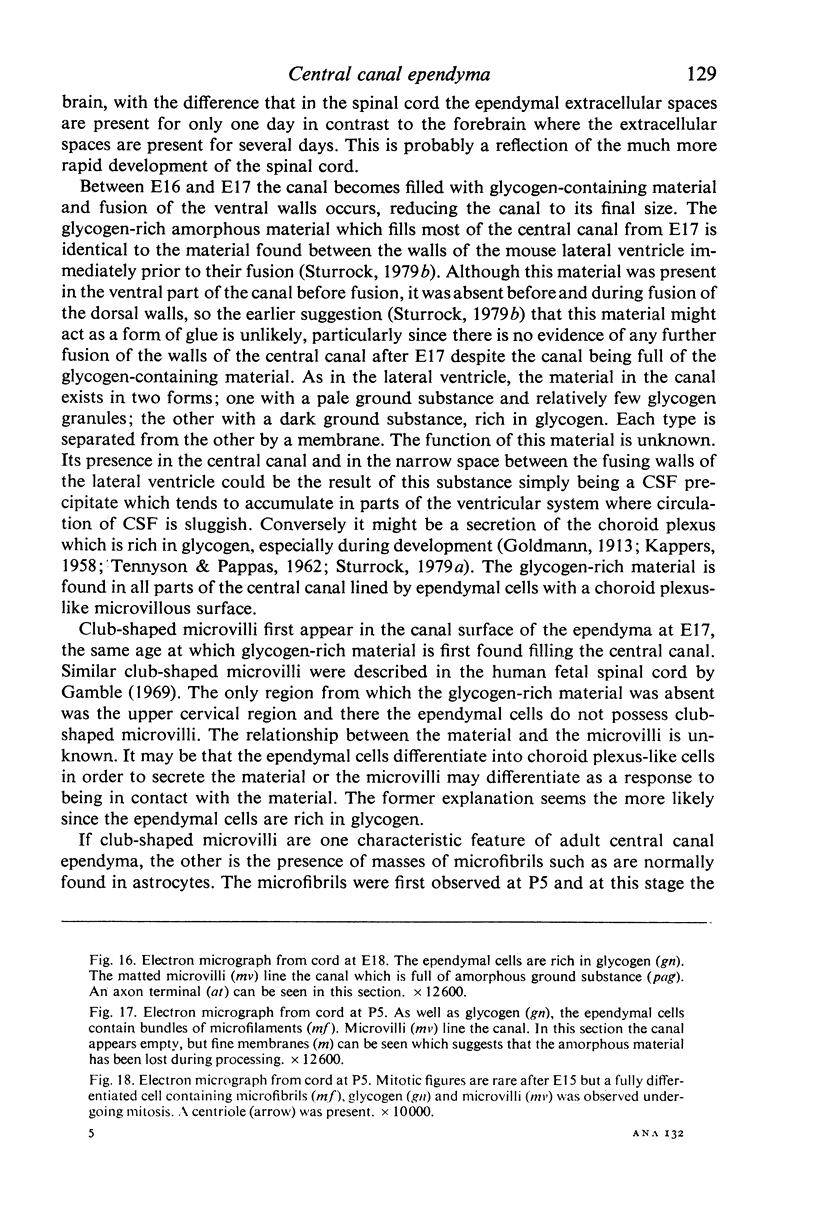
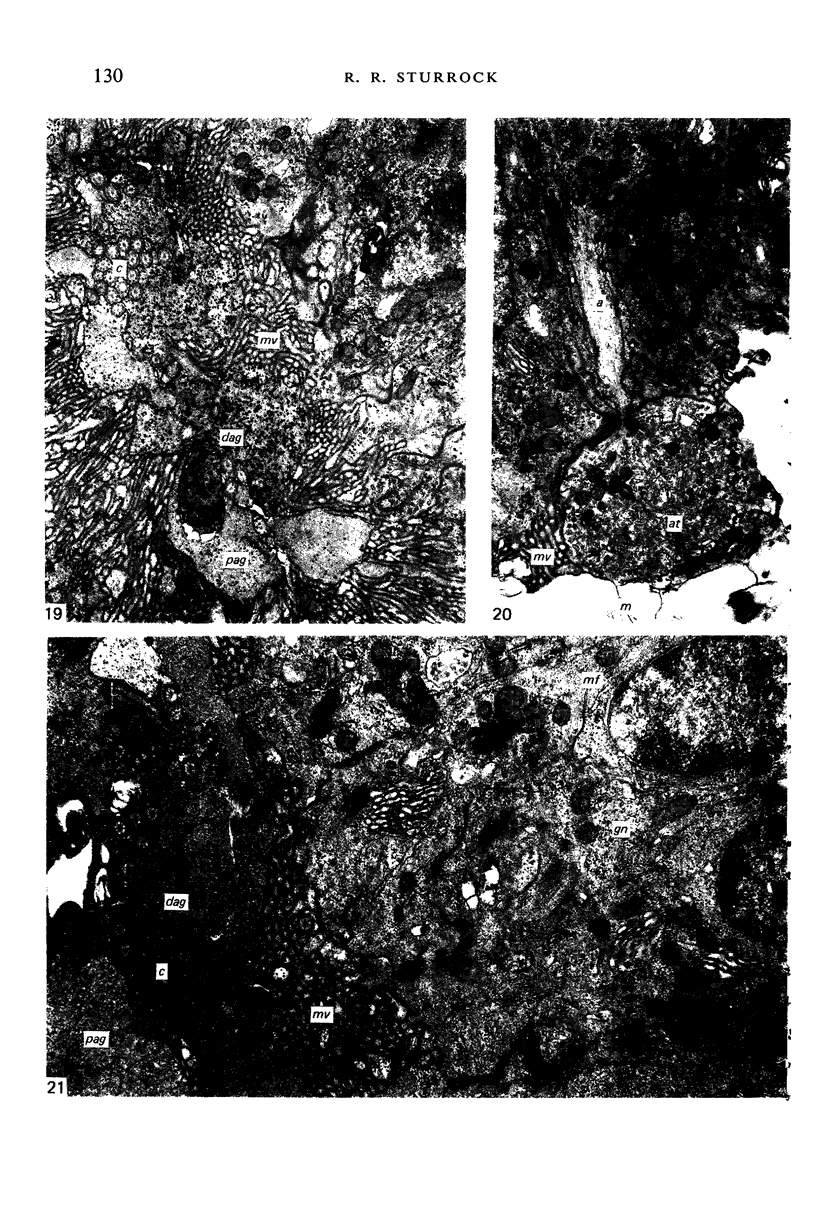
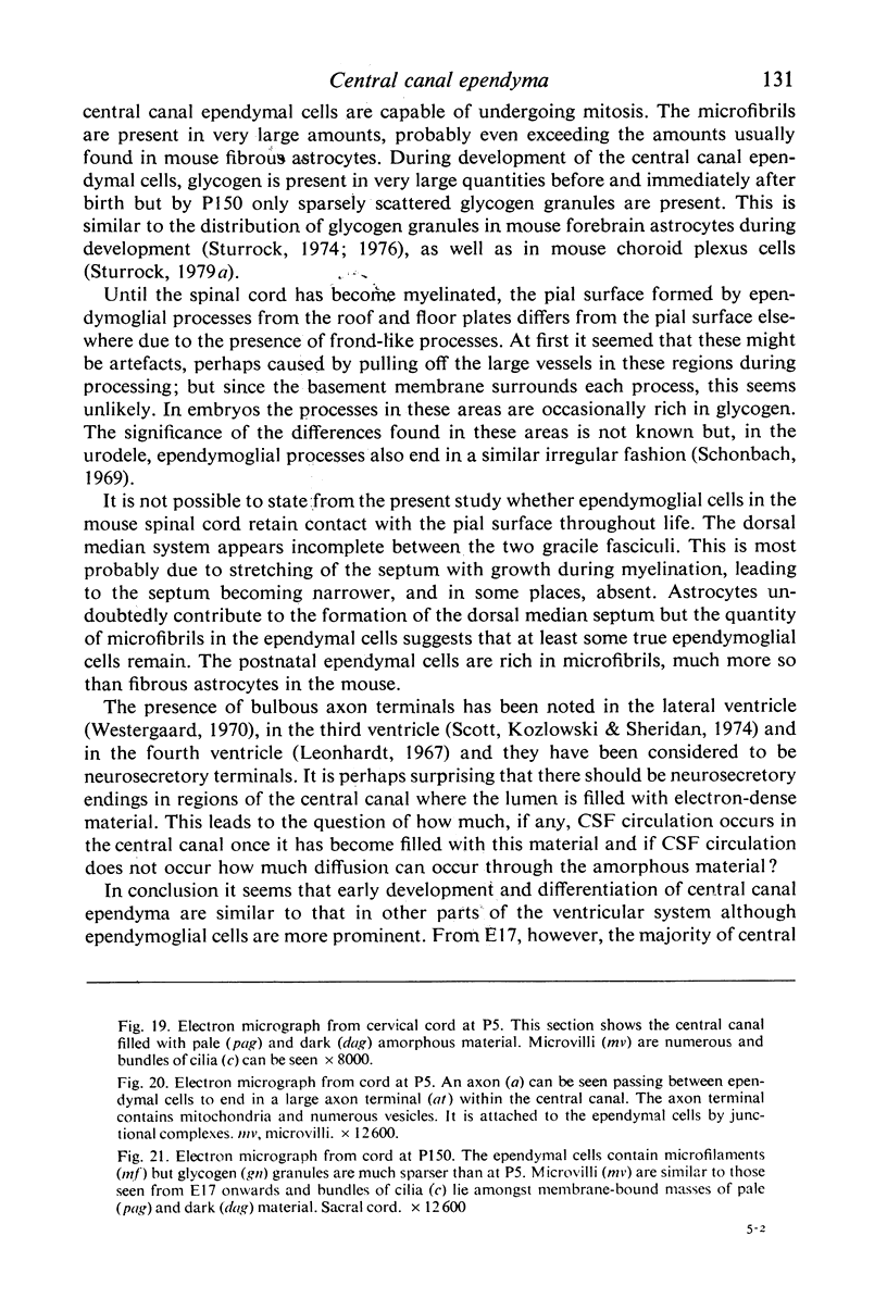
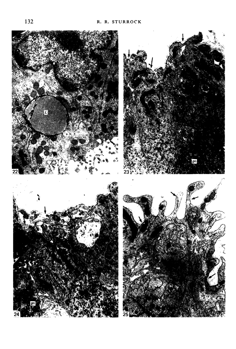
Images in this article
Selected References
These references are in PubMed. This may not be the complete list of references from this article.
- Bruni J. E. Scanning and transmission electron microscopy of the ependymal lining of the third ventricle. Can J Neurol Sci. 1974 Feb;1(1):59–73. [PubMed] [Google Scholar]
- Colmant H. J. Uber die Wandstruktur des dritten Ventrikels der Albinoratte. Histochemie. 1967;11(1):40–61. doi: 10.1007/BF00326611. [DOI] [PubMed] [Google Scholar]
- Gamble H. J. Electron microscope observations on the human foetal and embryonic spinal cord. J Anat. 1969 May;104(Pt 3):435–453. [PMC free article] [PubMed] [Google Scholar]
- Gamble H. J. Electron microscope observations upon the conus medullaris and filum terminale of human fetuses. J Anat. 1971 Nov;110(Pt 2):173–179. [PMC free article] [PubMed] [Google Scholar]
- Henrikson C. K., Vaughn J. E. Fine structural relationships between neurites and radial glial processes in developing mouse spinal cord. J Neurocytol. 1974 Dec;3(6):659–675. doi: 10.1007/BF01097190. [DOI] [PubMed] [Google Scholar]
- Hinds J. W., Ruffett T. L. Cell proliferation in the neural tube: an electron microscopic and golgi analysis in the mouse cerebral vesicle. Z Zellforsch Mikrosk Anat. 1971;115(2):226–264. doi: 10.1007/BF00391127. [DOI] [PubMed] [Google Scholar]
- Kiss A., Mitro A. Ependyma and supraependymal structures in some areas of the fourth ventricle in the rat. Acta Anat (Basel) 1978;100(4):521–531. doi: 10.1159/000144936. [DOI] [PubMed] [Google Scholar]
- Kohno K. Electron microscopic studies on Reissner's fiber and the ependymal cells in the spinal cord of the rat. Z Zellforsch Mikrosk Anat. 1969;94(4):565–573. doi: 10.1007/BF00936062. [DOI] [PubMed] [Google Scholar]
- Kumar T. C. Sexual differences in the ependyma LINING THE THIRD VENTRICLE IN THE AREA OF THE ANTERIOR HYPOTHALAMUS OF ADULT RHESUS MONKEYS. Z Zellforsch Mikrosk Anat. 1968;90(1):28–36. doi: 10.1007/BF00496700. [DOI] [PubMed] [Google Scholar]
- Leonhardt H. Zur Frage einer intraventrikulären Neurosekretion. Eine bisher unbekannte nervse Struktur im IV. Ventrikel des Kaninchens. Z Zellforsch Mikrosk Anat. 1967;79(2):172–184. [PubMed] [Google Scholar]
- Malinský J., Brichová H. Fine structure of ependyma in spinal cord of human embryos. Folia Morphol (Praha) 1967;15(1):68–78. [PubMed] [Google Scholar]
- Schonbach C. The neuroglia in the spinal cord of the newt, Triturus viridescens. J Comp Neurol. 1969 Jan;135(1):93–120. doi: 10.1002/cne.901350107. [DOI] [PubMed] [Google Scholar]
- Schwanitz W. Die topographische Verteilung supraependymaler Strukturen in den Ventrikeln und im Zentralkanal des Kaninchengehirns. Z Zellforsch Mikrosk Anat. 1969 Sep 22;100(4):536–551. [PubMed] [Google Scholar]
- Scott D. E., Kozlowski G. P., Paull W. K., Ramalingam S., Krobisch-Dudley G. Scanning electron microscopy of the human cerebral ventricular system. II. The fourth ventricle. Z Zellforsch Mikrosk Anat. 1973 May 11;139(1):61–68. doi: 10.1007/BF00307461. [DOI] [PubMed] [Google Scholar]
- Scott D. E., Kozlowski G. P., Sheridan M. N. Scanning electron microscopy in the ultrastructural analysis of the mammalian cerebral ventricular system. Int Rev Cytol. 1974;37(0):349–388. doi: 10.1016/s0074-7696(08)61362-5. [DOI] [PubMed] [Google Scholar]
- Shoukimas G. M., Hinds J. W. The development of the cerebral cortex in the embryonic mouse: an electron microscopic serial section analysis. J Comp Neurol. 1978 Jun 15;179(4):795–830. doi: 10.1002/cne.901790407. [DOI] [PubMed] [Google Scholar]
- Smart I. H. Proliferative characteristics of the ependymal layer during the early development of the spinal cord in the mouse. J Anat. 1972 Apr;111(Pt 3):365–380. [PMC free article] [PubMed] [Google Scholar]
- Stensaas L. J., Gilson B. C. Ependymal and subependymal cells of the caudato-pallial junction in the lateral ventricle of the neonatal rabbit. Z Zellforsch Mikrosk Anat. 1972;132(3):297–322. doi: 10.1007/BF02450711. [DOI] [PubMed] [Google Scholar]
- Sturrock R. R. A comparison of the processes of ventricular coarctation and choroid and ependymal fusion in the mouse brain. J Anat. 1979 Sep;129(Pt 2):235–242. [PMC free article] [PubMed] [Google Scholar]
- Sturrock R. R. A morphological study of the development of the mouse choroid plexus. J Anat. 1979 Dec;129(Pt 4):777–793. [PMC free article] [PubMed] [Google Scholar]
- Sturrock R. R. A quantitative lifespan study of changes in cell number, cell division and cell death in various regions of the mouse forebrain. Neuropathol Appl Neurobiol. 1979 Nov-Dec;5(6):433–456. doi: 10.1111/j.1365-2990.1979.tb00642.x. [DOI] [PubMed] [Google Scholar]
- Sturrock R. R. Histogenesis of the anterior limb of the anterior commissure of the mouse brain. 3. An electron microscopic study of gliogenesis. J Anat. 1974 Feb;117(Pt 1):37–53. [PMC free article] [PubMed] [Google Scholar]
- Sturrock R. R. Light microscopic identification of immature glial cells in semithin sections of the developing mouse corpus callosum. J Anat. 1976 Dec;122(Pt 3):521–537. [PMC free article] [PubMed] [Google Scholar]
- Sturrock R. R., Smart I. H. A morphological study of the mouse subependymal layer from embryonic life to old age. J Anat. 1980 Mar;130(Pt 2):391–415. [PMC free article] [PubMed] [Google Scholar]
- TENNYSON V. M., PAPPAS G. D. An electron microscope study of ependymal cells of the fetal, early postnatal and adult rabbit. Z Zellforsch Mikrosk Anat. 1962;56:595–618. doi: 10.1007/BF00540584. [DOI] [PubMed] [Google Scholar]


