Abstract
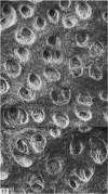
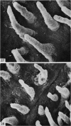
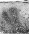
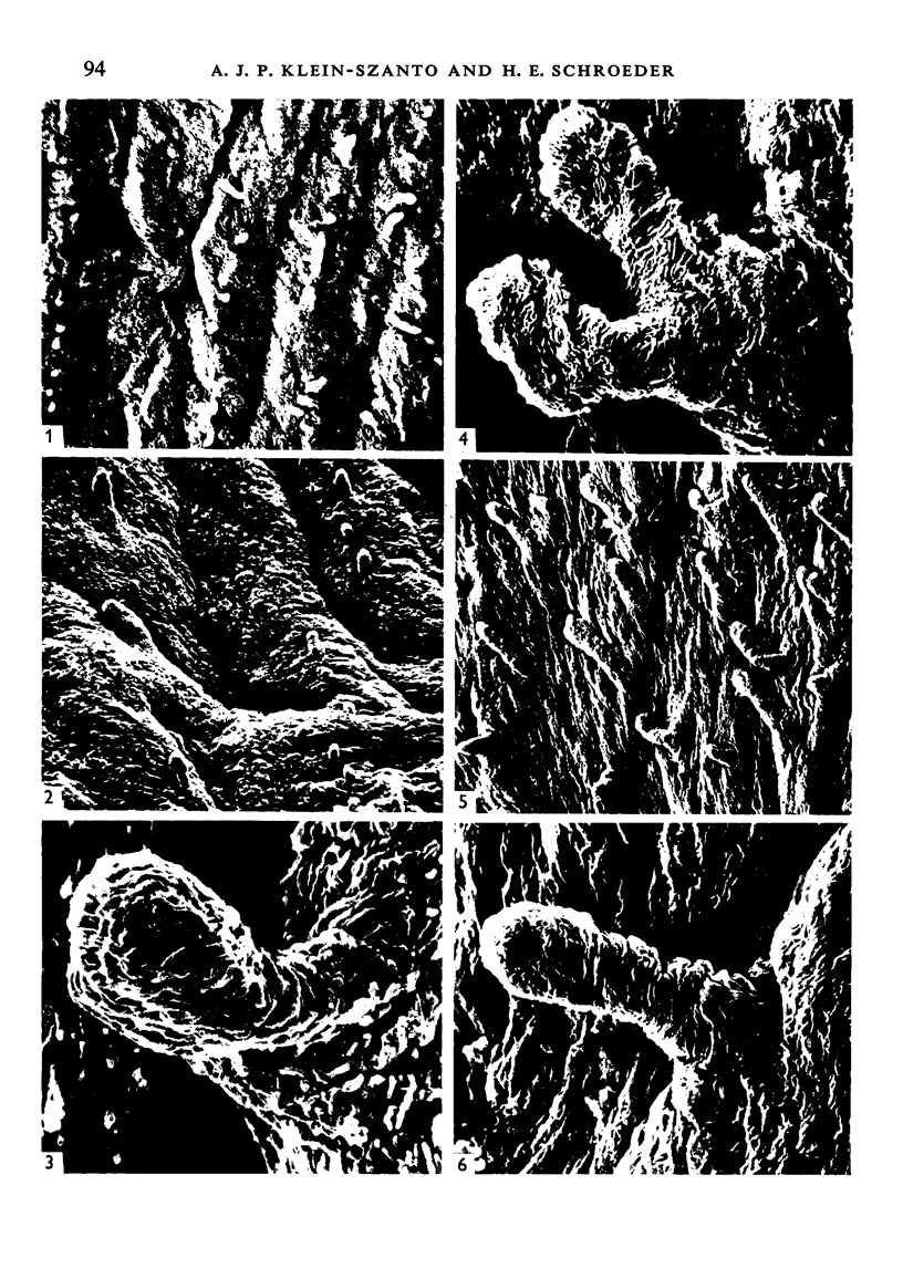
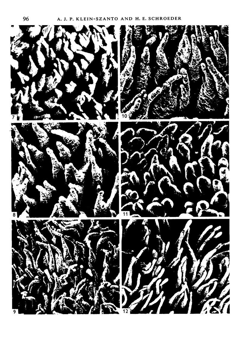
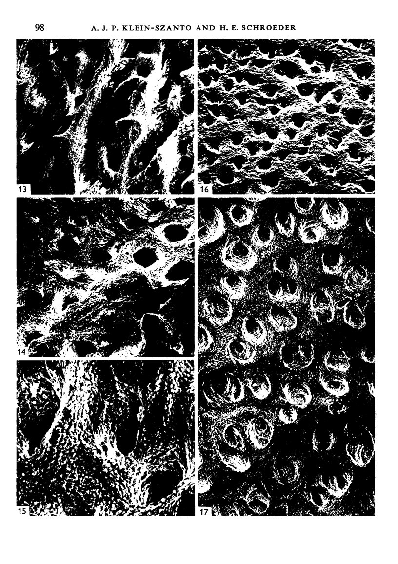
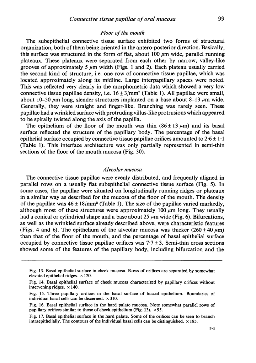
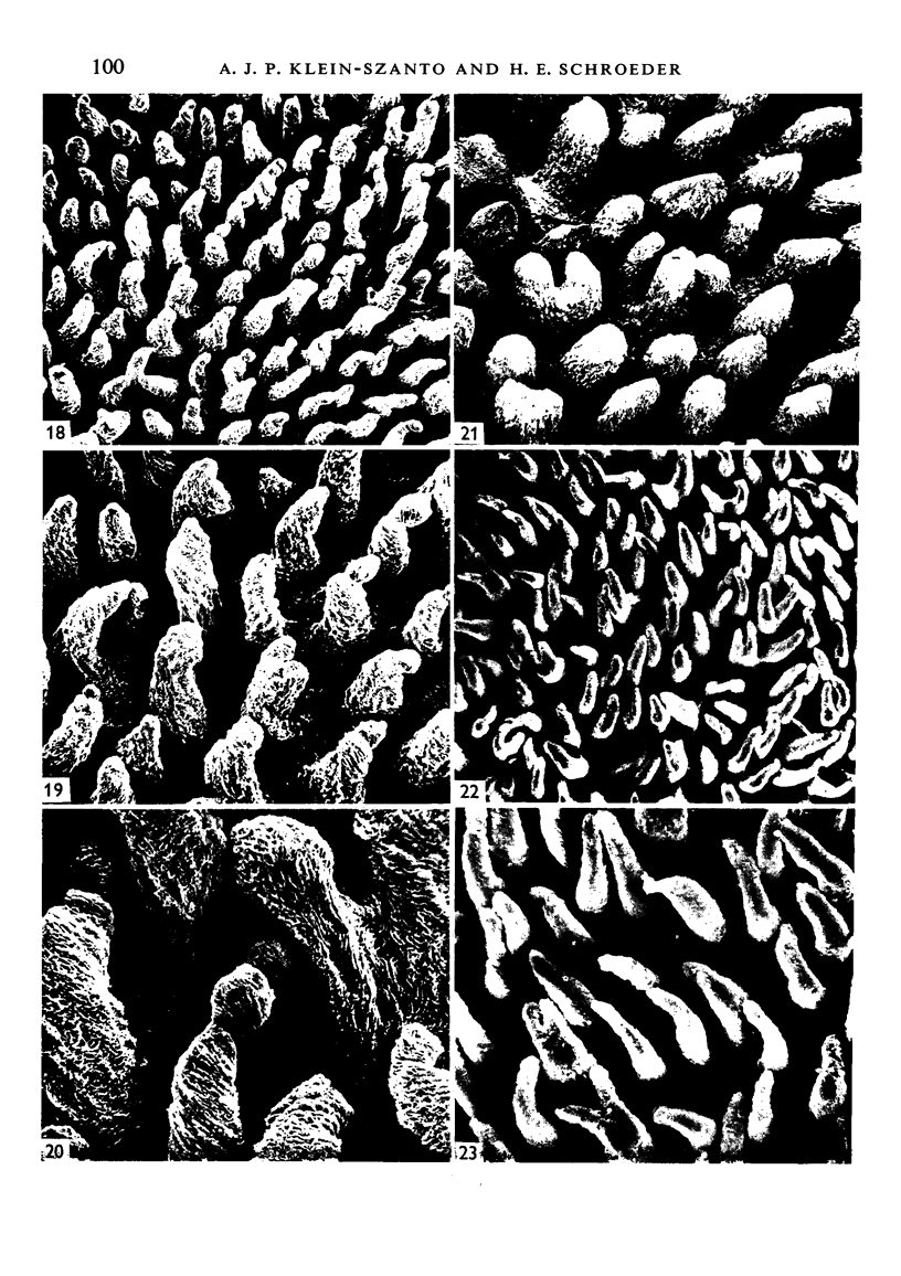
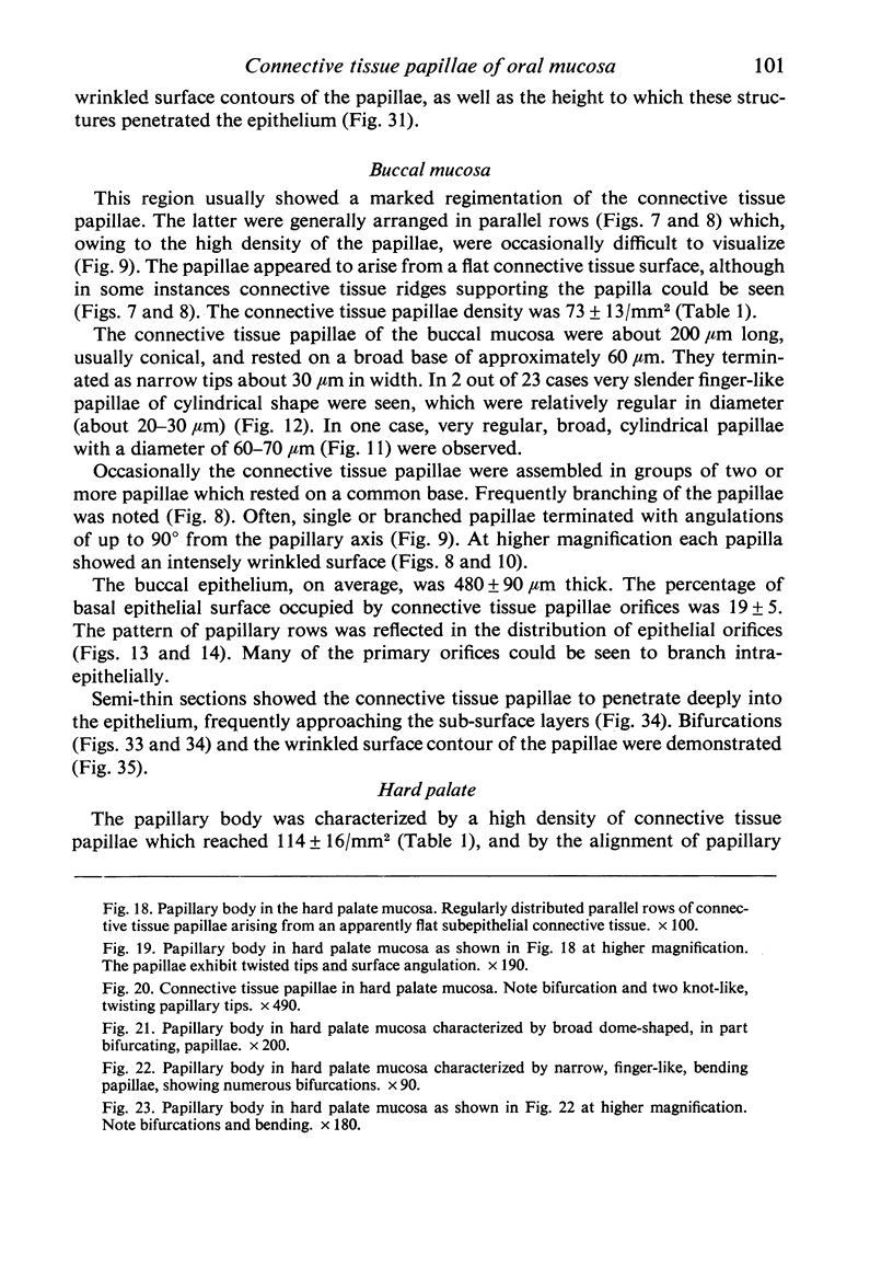
The papillary body of the human oral mucosa was studied at six different sites. Biopsy and autopsy material from 57 individuals, 11-81 years of age, was split chemically along the basal lamina and the epithelium-connective tissue interface examined by light and scanning electron microscopy. Morphometric techniques were employed in order to determine: epithelial thickness, height and density of connective tissue papillae and the percentage of basal epithelial surfaces occupied by them. In the majority of sites, connective tissue plateaux or ridges carrying a variable number of single or grouped papillae were found to be the basic structural units of the papillary body. Three regions with diferent characteristics of the epithelium-connective tissue interface could be identified: (1) floor of the mouth, (2) lip and cheek, (3) gingiva and hard palate. The floor of the mouth showed the lowest connective tissue papillae density, the smallest papillae, and connective tissue plateaux separated by narrow grooves. Lip and cheek mucosae revealed an intermediate density, the papillae were frequently bifurcated and angulated. Gingiva and hard palate were characterized by the highest papillary density and by papillae which were cylindrical, slender and erect. The alveolar mucosa exhibited intermediate features between those of the floor of the mouth and those of the cheek mucosa.
Full text
PDF
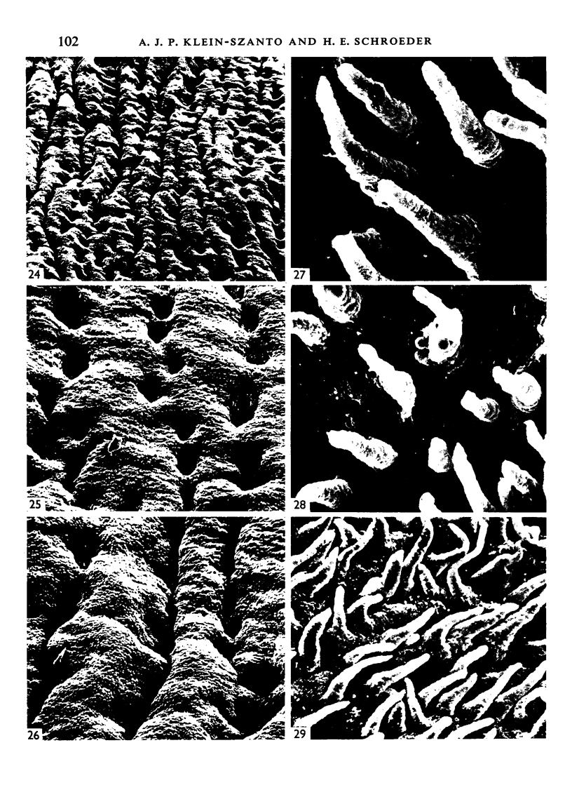
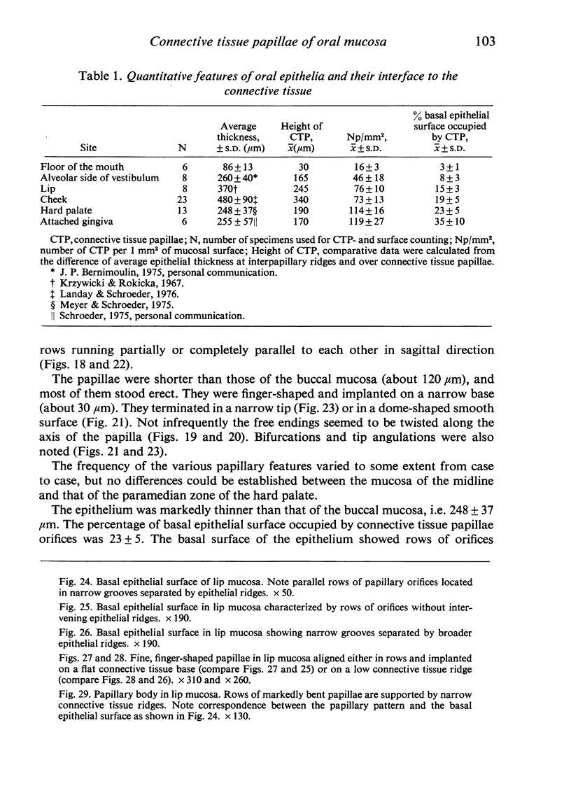
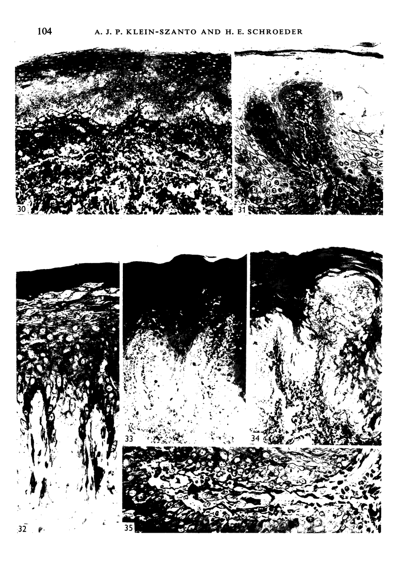
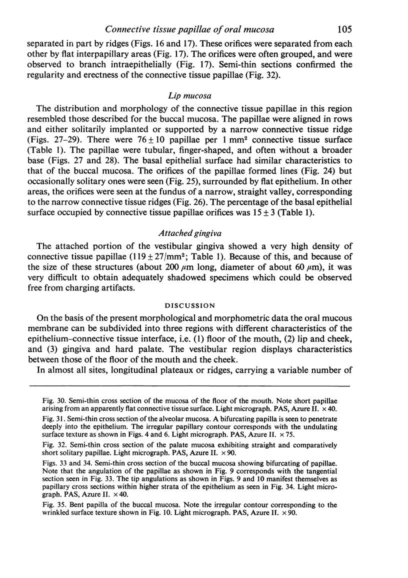
Images in this article
Selected References
These references are in PubMed. This may not be the complete list of references from this article.
- Bollinger K., Riethe P. Zur Morphologie der menschlichen Gingiva. Das dreidimensionale Reliefbild an den Grenzflächen zwischen Epithel und Lamina propria der "attached Gingiva". Dtsch Zahnarztl Z. 1973 Dec;28(12):1212–1219. [PubMed] [Google Scholar]
- Frenkel G., Abraham R. Untersuchungen am epithelo-cutanen Grenzflächenrelief bei chronisch entzündlich veränderter Mundschleimhaut. Dtsch Zahnarztl Z. 1968 Mar;23(3):274–278. [PubMed] [Google Scholar]
- Frenkel G., Abraham R. Untersuchungen am epithelo-cutanen Grenzflächenrelief bei pathologisch veränderter Mundschleimhaut, insbesondere bei Neoplasmen. Dtsch Zahnarztl Z. 1969 Apr;24(4):312–316. [PubMed] [Google Scholar]
- HALE A. R. Morphogenesis of volar skin in the human fetus. Am J Anat. 1952 Jul;91(1):147–181. doi: 10.1002/aja.1000910105. [DOI] [PubMed] [Google Scholar]
- HORSTMANN E. Morphologie und Morphogenese des Papillarkörpers der Schleimhäute in der Mundhöhle des Menschen. Z Zellforsch Mikrosk Anat. 1954;39(5):479–514. [PubMed] [Google Scholar]
- Karring T., Lang N. P., Löe H. The role of gingival connective tissue in determining epithelial differentiation. J Periodontal Res. 1975 Feb;10(1):1–11. doi: 10.1111/j.1600-0765.1975.tb00001.x. [DOI] [PubMed] [Google Scholar]
- Karring T. Mitotic activity in the oral epithelium. J Periodontal Res Suppl. 1973;13:1–47. [PubMed] [Google Scholar]
- Karring T., Ostergaard E., Löe H. Conservation of tissue specificity after heterotopic transplantation of gingiva and alveolar mucosa. J Periodontal Res. 1971;6(4):282–293. doi: 10.1111/j.1600-0765.1971.tb00619.x. [DOI] [PubMed] [Google Scholar]
- Krzywicki J., Rokicka A. Morphological picture of the oral mucosa. Planimetric studies. Pol Med J. 1967;6(2):520–527. [PubMed] [Google Scholar]
- Löe H., Karring T. The three-dimensional morphology of the epithelium-connective tissue interface of the gingiva as related to age and sex. Scand J Dent Res. 1971;79(5):315–326. doi: 10.1111/j.1600-0722.1971.tb02017.x. [DOI] [PubMed] [Google Scholar]
- Meller S. M., Coppe M. R., Ito S., Waterman R. E. Transmission electron microscopy of critical point dried tissue after observation in the scanning electron microscope. Anat Rec. 1973 Jun;176(2):245–252. doi: 10.1002/ar.1091760210. [DOI] [PubMed] [Google Scholar]
- Meyer M., Schroeder H. E. A quantitative electron microscopic analysis of the keratinizing epithelium of noral human hard palate. Cell Tissue Res. 1975;158(2):177–203. doi: 10.1007/BF00219960. [DOI] [PubMed] [Google Scholar]
- Papa C. M., Farber B. Direct scanning electron microscopy of human skin. Arch Dermatol. 1971 Sep;104(3):262–270. [PubMed] [Google Scholar]
- Plagmann H. C., Lange D. E., Bernimoulin J. P., Howe H. Experimentelle Studie über die Epithelneubildung bei heterotopischen Bindegewebstransplantaten. Dtsch Zahnarztl Z. 1974 May;29(5):497–503. [PubMed] [Google Scholar]
- Scaletta L. J., MacCallum D. K. A fine structural study of divalent cation-mediated epithelial union with connective tissue in human oral mucosa. Am J Anat. 1972 Apr;133(4):431–453. doi: 10.1002/aja.1001330406. [DOI] [PubMed] [Google Scholar]
- Schenk P., Wersäll J. Die Ultrastruktur der Papillae filiformes der menschlichen Zunge. Arch Dermatol Forsch. 1975;252(2):91–109. [PubMed] [Google Scholar]
- Schroeder H. E. Transmigration and infiltration of leucocytes in human junctional epithelium. Helv Odontol Acta. 1973 Apr;17(1):6–18. [PubMed] [Google Scholar]
- Shklar G. The effects of aging upon oral mucosa. J Invest Dermatol. 1966 Aug;47(2):115–120. [PubMed] [Google Scholar]
- Weibel E. R. Stereological principles for morphometry in electron microscopic cytology. Int Rev Cytol. 1969;26:235–302. doi: 10.1016/s0074-7696(08)61637-x. [DOI] [PubMed] [Google Scholar]


