Abstract
The cellular changes in the ileum and duodenum and in the mesenteric and prefemoral lymph nodes of gnotobiotic piglets were observed following feeding with a live culture of a non-pathogenic E. coli. There was a rapid and intense reaction in the lower ileum, and follicles were formed; germinal centres were formed in the mesenteric lymph node after a short delay. Germinal centres were not seen in the prefemoral lymph node, though there were pyroninophilic cells in the cortex of this node. Plasma cells were not detected in the medulla of either the mesenteric or the prefemoral lymph nodes, but pyroninophilic cells and plasma cells were found in the lamina propria of the duodenum from 7 days after infection oneards. These observations demonstrate the requirement of an intestinal flora for the development of normal Peyer's patch architecture and indicate that the Peyer's patch response secondarily affects the mesenteric lymph node. The observations also suggest that there is a haematogenous dissemination of pyroninophilic cells from gut-associated lymphoid tissue to, amongst other sites, the duodenum; this may be of importance in both natural and artificial immunization by the oral route.
Full text
PDF
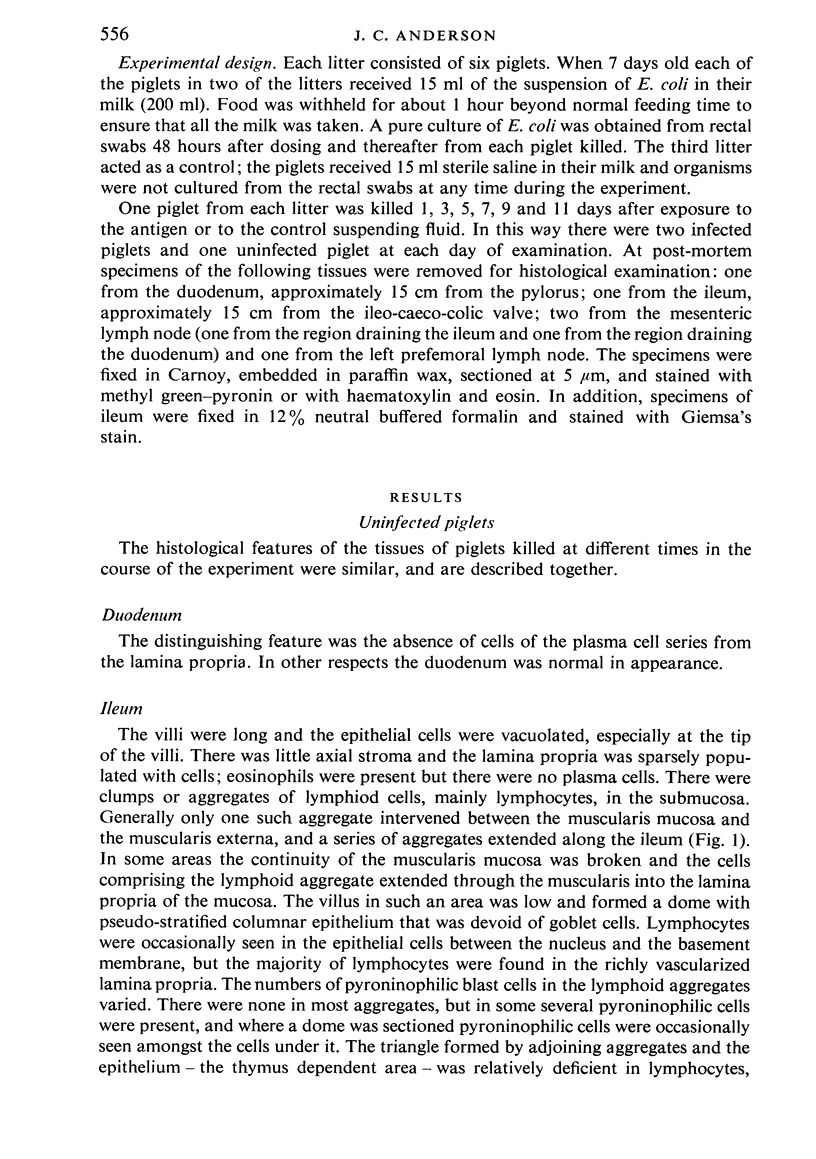
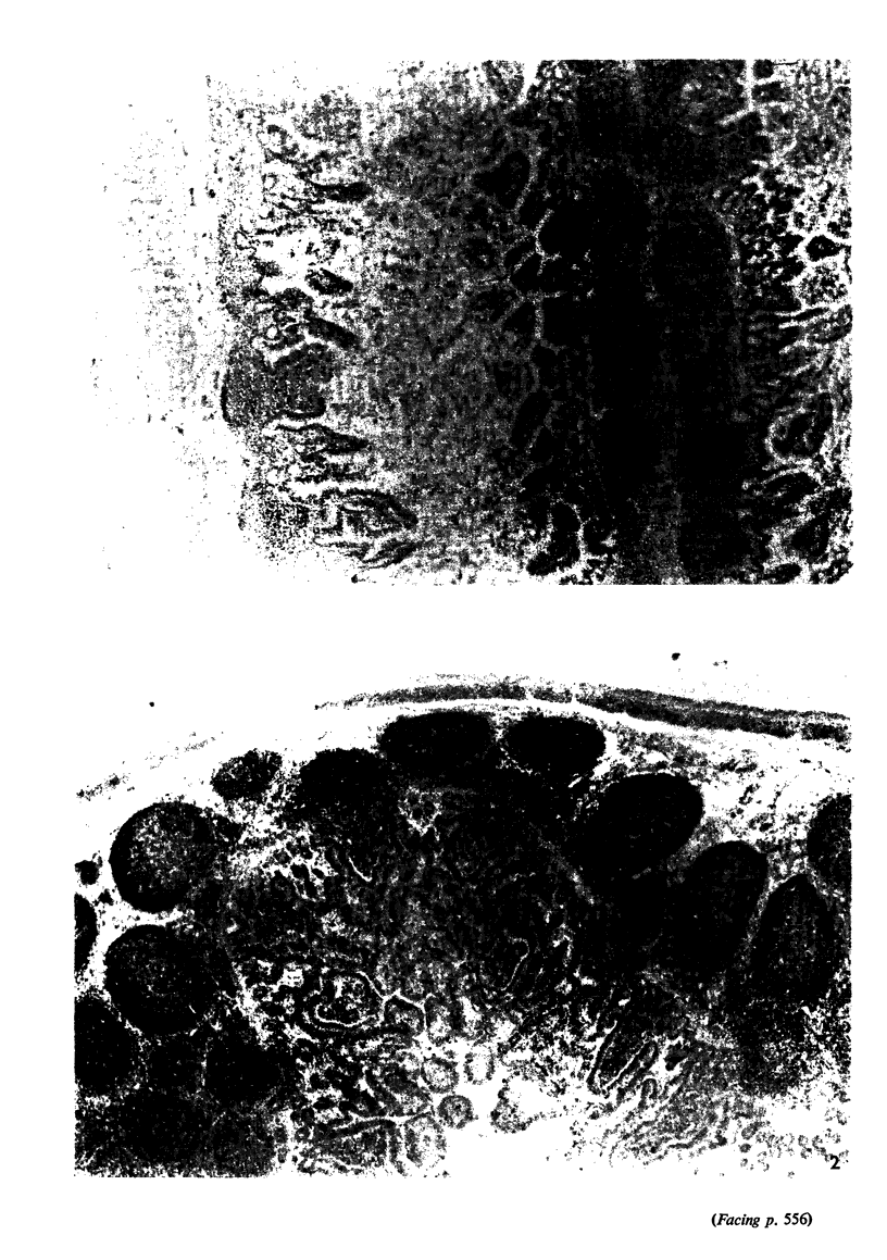
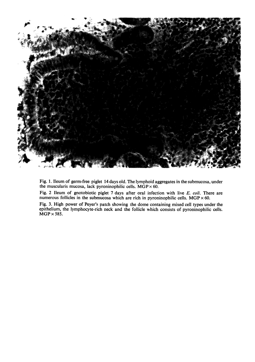
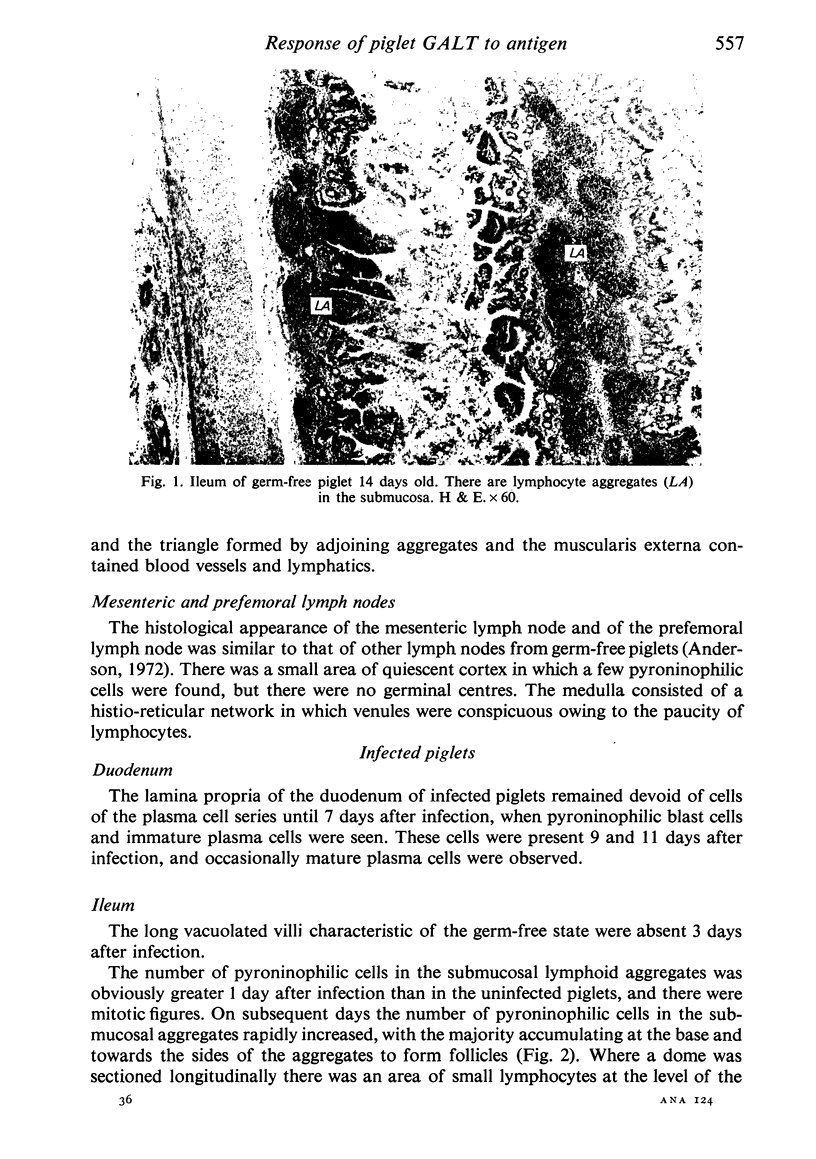
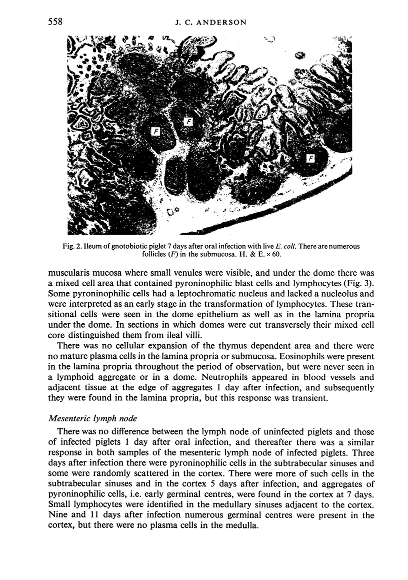
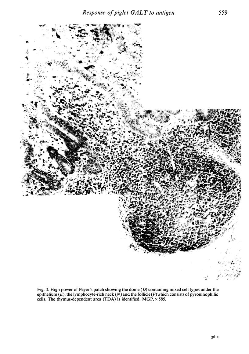
Images in this article
Selected References
These references are in PubMed. This may not be the complete list of references from this article.
- Allen W. D., Porter P. The relative distribution of IgM and IgA cells in intestinal mucosa and lymphoid tissues of the young unweaned pig and their significance in ontogenesis of secretory immunity. Immunology. 1973 Mar;24(3):493–501. [PMC free article] [PubMed] [Google Scholar]
- Anderson J. C. The micro-anatomy of the lymph node of germ-free piglets. Br J Exp Pathol. 1972 Feb;53(1):37–39. [PMC free article] [PubMed] [Google Scholar]
- Anderson J. C. The response of the tonsil and associated lymph nodes of gnotobiotic piglets to the presence of bacterial antigen in the oral cavity. J Anat. 1974 Feb;117(Pt 1):191–198. [PMC free article] [PubMed] [Google Scholar]
- Anderson J. C. The use of germ-free piglets in the study of lymphoid tissue and germinal centre formation. Adv Exp Med Biol. 1973;29(0):643–649. doi: 10.1007/978-1-4615-9017-0_93. [DOI] [PubMed] [Google Scholar]
- Atkins A. M., Schofield G. C., Reeders T. Studies on the structure and distribution of immunoglobulin A-containing cells in the gut of the pig. J Anat. 1971 Sep;109(Pt 3):385–395. [PMC free article] [PubMed] [Google Scholar]
- Bockman D. E., Cooper M. D. Pinocytosis by epithelium associated with lymphoid follicles in the bursa of Fabricius, appendix, and Peyer's patches. An electron microscopic study. Am J Anat. 1973 Apr;136(4):455–477. doi: 10.1002/aja.1001360406. [DOI] [PubMed] [Google Scholar]
- Bradley P. A., Bourne F. J., Brown P. J. The respiratory tract immune system in the pig. I. Distribution of immunoglobulin-containing cells in the respiratory tract mucosa. Vet Pathol. 1976;13(2):81–89. doi: 10.1177/030098587601300201. [DOI] [PubMed] [Google Scholar]
- Brown P. J., Bourne F. J. Distributions of immunoglobulin-containing cells in alimentary tract, spleen, and mesenteric lymph node of the pig demonstrated by peroxidase-conjugated antiserums to porcine immunoglobulins G, A, and M. Am J Vet Res. 1976 Jan;37(1):9–13. [PubMed] [Google Scholar]
- Brown P., Bourne J., Steel M. Immunoperoxidase and immunofluorescence techniques in pig tissues. Histochemistry. 1974;40(4):343–348. doi: 10.1007/BF00495041. [DOI] [PubMed] [Google Scholar]
- Chapman H. A., Johnson J. S., Cooper M. D. Ontogeny of Peyer's patches and immunoglobulin-containing cells in pigs. J Immunol. 1974 Feb;112(2):555–563. [PubMed] [Google Scholar]
- Edwards J. L., Murphy R. C., Cho Y. On the development of the lymphoid follicles of the bursa of Fabricius. Anat Rec. 1975 Apr;181(4):735–753. doi: 10.1002/ar.1091810406. [DOI] [PubMed] [Google Scholar]
- Fichtelius K. E. The mammalian equivalent to bursa Fabricii of birds. Exp Cell Res. 1967 Apr;46(1):231–234. doi: 10.1016/0014-4827(67)90427-2. [DOI] [PubMed] [Google Scholar]
- Kruml J., Kovárů F., Ludvík J., Trebichavský I. The development of lymphoid and haemopoietic tissues in pig fetuses. Folia Microbiol (Praha) 1970;15(1):17–22. doi: 10.1007/BF02867043. [DOI] [PubMed] [Google Scholar]
- Kruml J., Ludvík J., Trebichavský I., Mandel L., Kovárů F. Morphology of germ-free piglets. Folia Microbiol (Praha) 1969;14(5):441–446. doi: 10.1007/BF02872789. [DOI] [PubMed] [Google Scholar]
- Owen R. L., Jones A. L. Epithelial cell specialization within human Peyer's patches: an ultrastructural study of intestinal lymphoid follicles. Gastroenterology. 1974 Feb;66(2):189–203. [PubMed] [Google Scholar]
- Porter P., Kenworthy R., Holme D. W., Horsfield S. Escherichia coli antigens as dietary additives for oral immunisation of pigs: trials with pig creep feeds. Vet Rec. 1973 Jun 16;92(24):630–636. doi: 10.1136/vr.92.24.630. [DOI] [PubMed] [Google Scholar]
- Porter P., Kenworthy R., Noakes D. E., Allen W. D. Intestinal antibody secretion in the young pig in response to oral immunization with Escherichia coli. Immunology. 1974 Nov;27(5):841–853. [PMC free article] [PubMed] [Google Scholar]
- SLOSS M. W. The microscopic anatomy of the digestive tract of Sus scrofa domestica. Am J Vet Res. 1954 Oct;15(57):578–593. [PubMed] [Google Scholar]
- WILBUR R. D., CATRON D. V., QUINN L. Y., SPEER V. C., HAYS V. W. Intestinal flora of the pig as influenced by diet and age. J Nutr. 1960 Jun;71:168–175. doi: 10.1093/jn/71.2.168. [DOI] [PubMed] [Google Scholar]
- Waksman B. H., Ozer H., Blythman H. E. Appendix and M-antibody formation. VI. The functional anatomy of the rabbit appendix. Lab Invest. 1973 May;28(5):614–626. [PubMed] [Google Scholar]


