Abstract
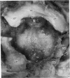
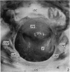
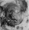
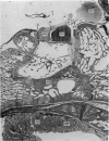
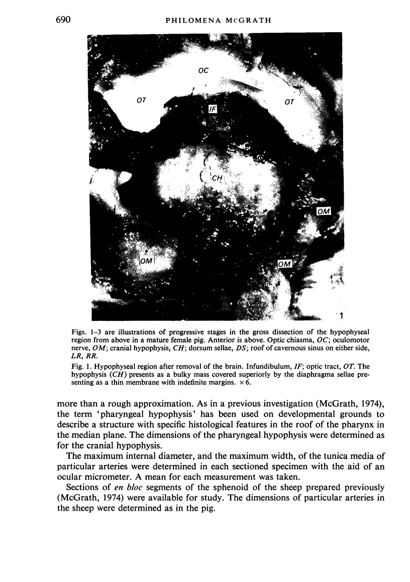
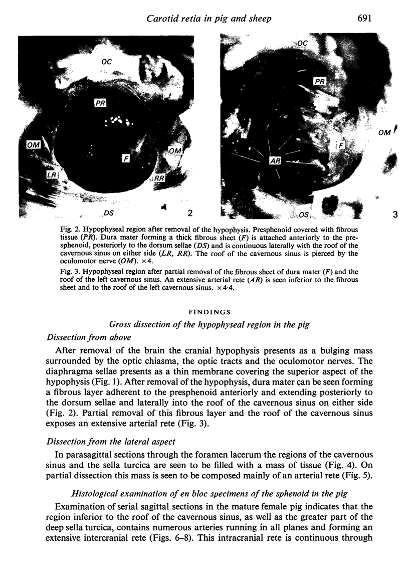
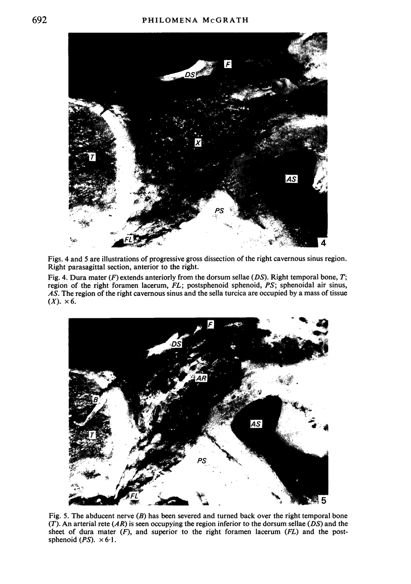
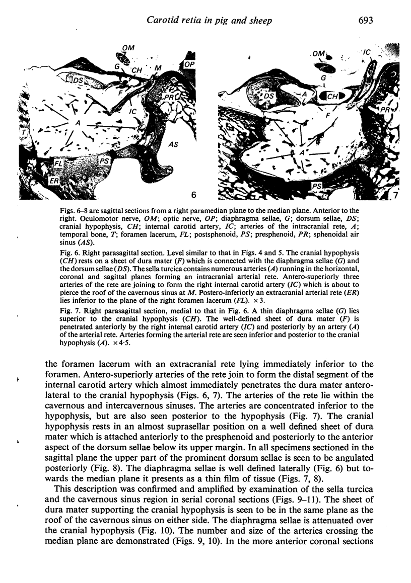
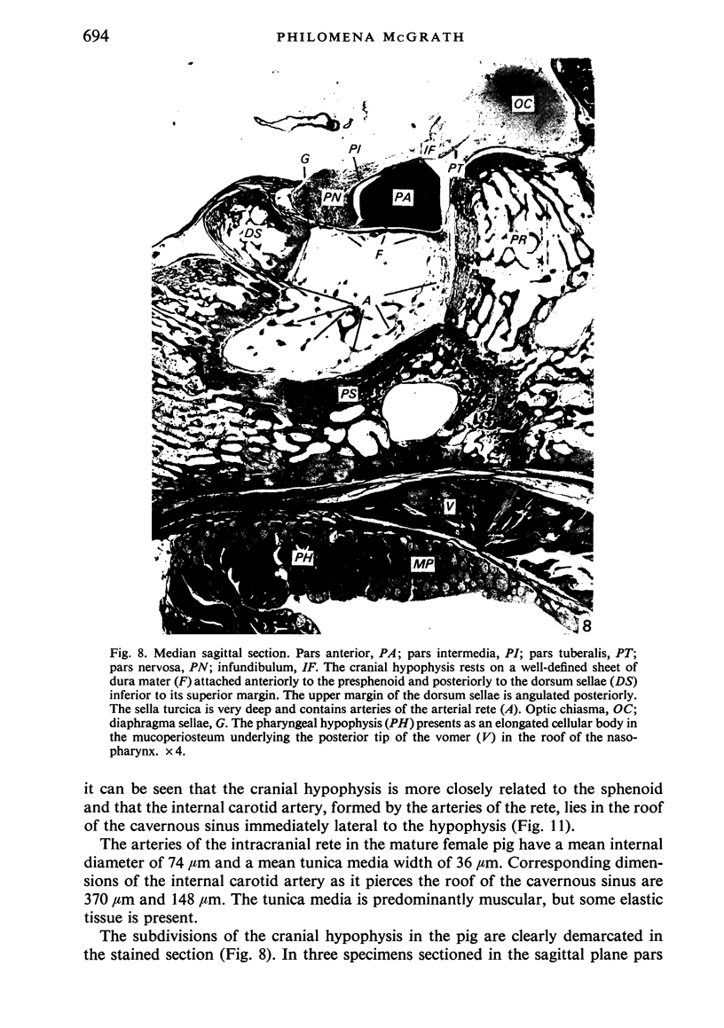
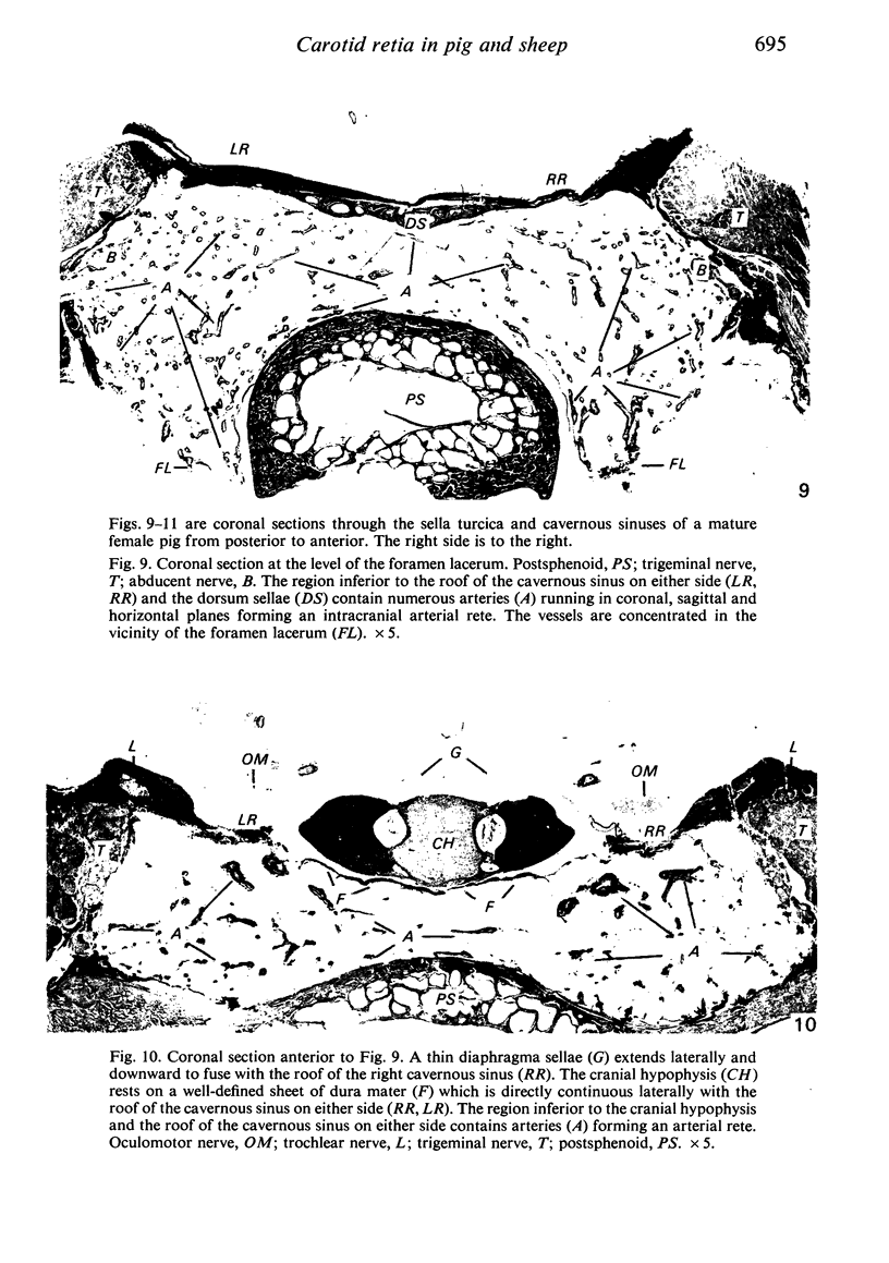
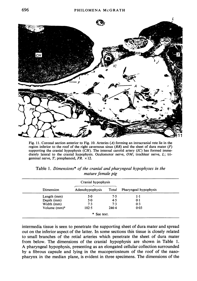
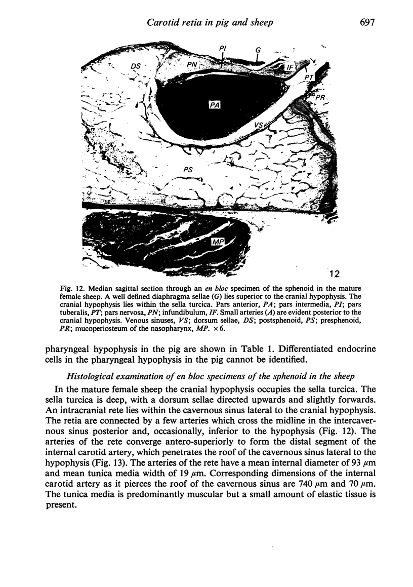
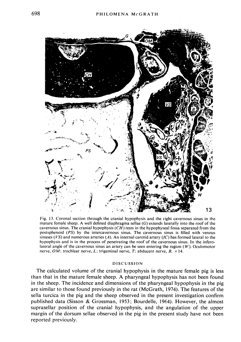
It is accepted that in the pig the intracranial carotid retia are connected across the midline by numerous arteries lying within the intercavernous sinus. The present study has demonstrated that these vascular elements fill the greater part of the very deep sella turcica, the cranial hypophysis occupying an almost suprasellar position. In the sheep the anastomosis between the carotid retia is limited to a few arteries crossing the midline posterior to the hypophysis, and the gland lies wholly within the sella turcica. It is suggested that the position of the cranial hypophysis in the mature female pig results from the inward and upward pressures exerted on the hypophysis by the carotid retia and their extensive interconnexions in this species.
Full text
PDF
Images in this article
Selected References
These references are in PubMed. This may not be the complete list of references from this article.
- BALDWIN B. A. THE ANATOMY OF THE ARTERIAL SUPPLY TO THE CRANIAL REGIONS OF THE SHEEP AND OX. Am J Anat. 1964 Jul;115:101–107. doi: 10.1002/aja.1001150107. [DOI] [PubMed] [Google Scholar]
- Gillan L. A. Blood supply to brains of ungulates with and without a rete mirabile caroticum. J Comp Neurol. 1974 Feb 1;153(3):275–290. doi: 10.1002/cne.901530305. [DOI] [PubMed] [Google Scholar]
- McGrath P. The pharyngeal hypophysis in some laboratory animals. J Anat. 1974 Feb;117(Pt 1):95–115. [PMC free article] [PubMed] [Google Scholar]


