Abstract
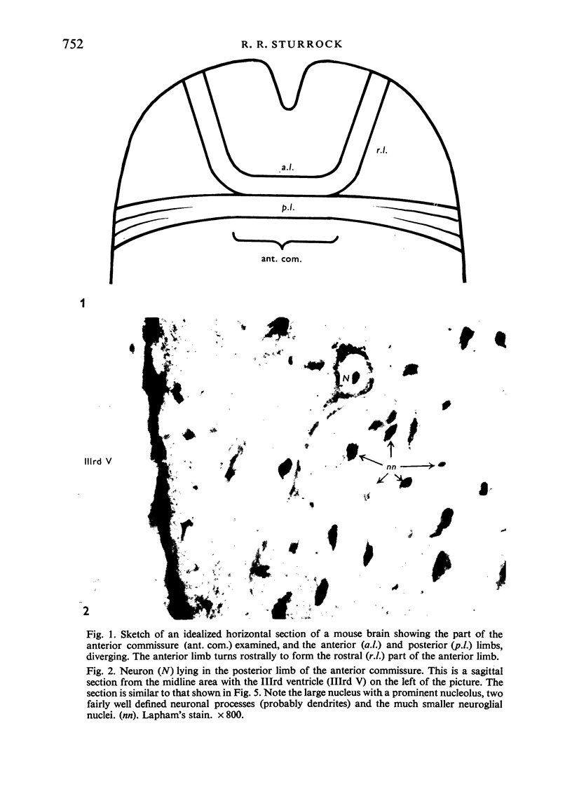
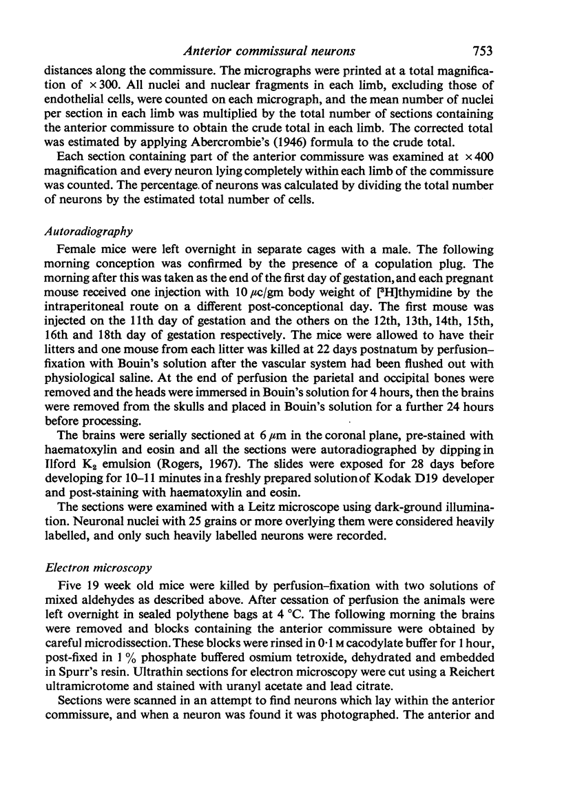
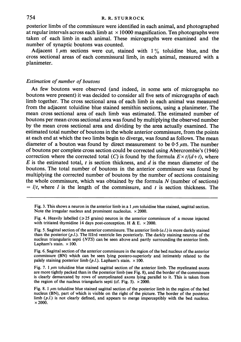
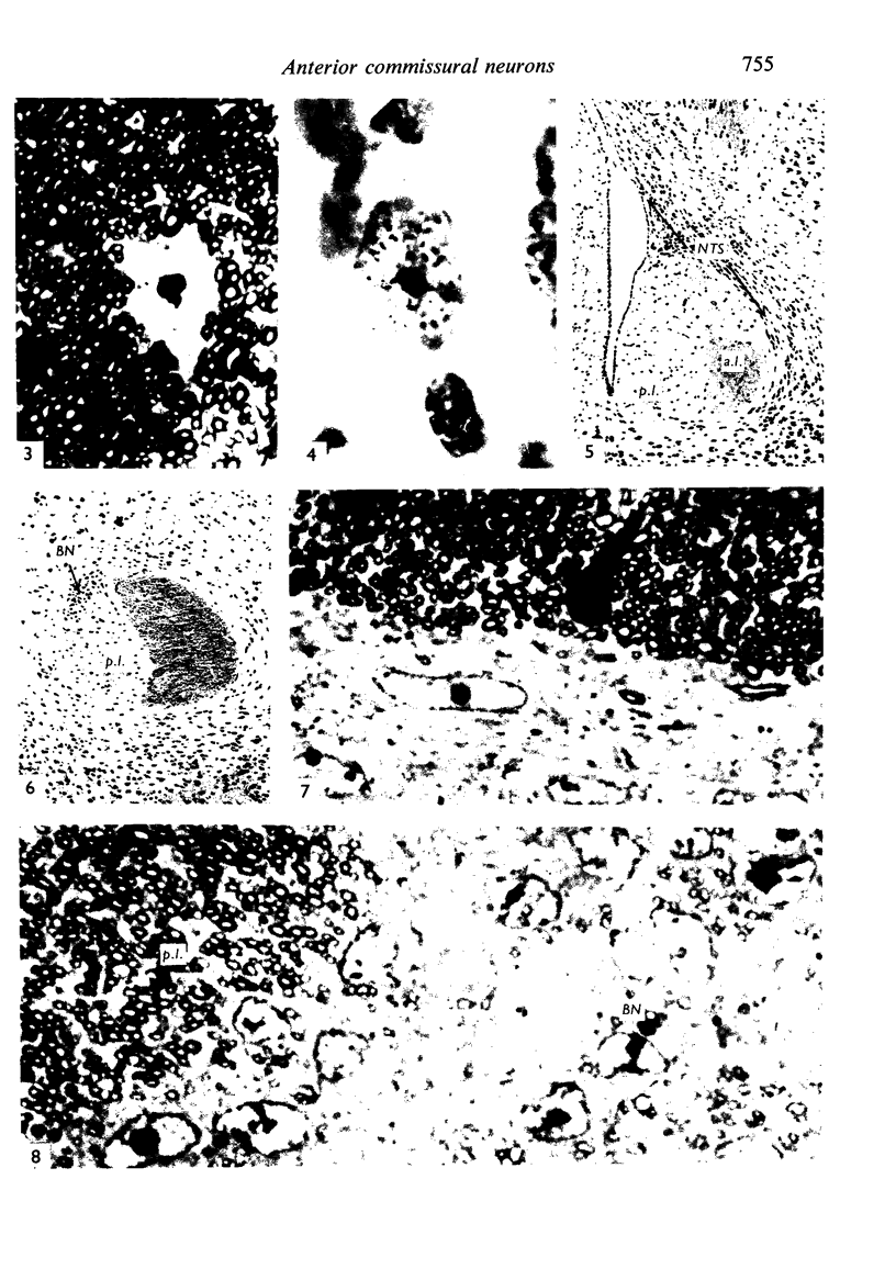
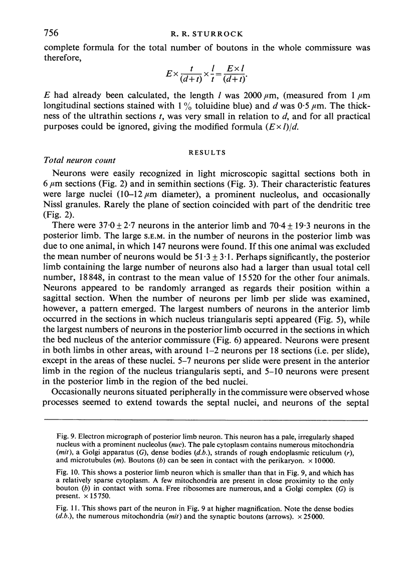
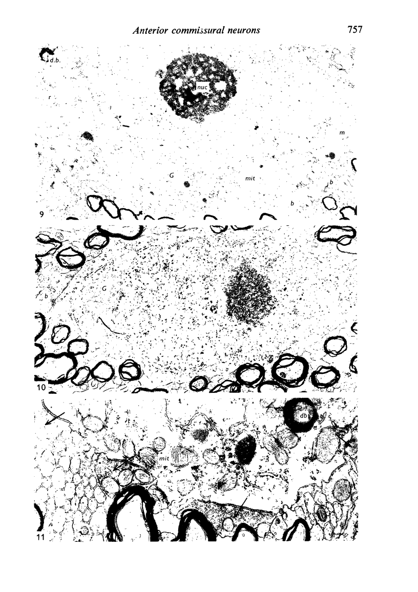
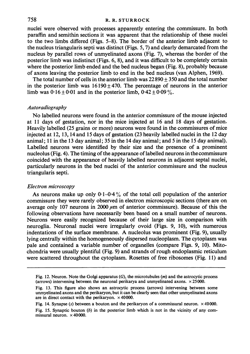
Both limbs of the anterior commissure of the mouse brain were examined to find the number, distribution, times of origin and structure of the neurons present, and also the number of synapses within the commissure. Neurons form between 0-1 and 0-4% of the total cell population and are produced between the twelfth and fourteenth days of gestation. It seems likely that the neurons within the anterior commissure are derived from adjacent septal nuclei, with the bed nuclei of the anterior commissure mainly contributing to the posterior limb and the nucleus triangularis septi mainly contributing to the anterior limb. The neurons are almost certainly functional, and distribute to the nuclei from which they are derived. There are probably also other connexions between these nuclei and both limbs of the anterior commissure through dendrites from the septal nuclei which ramify throughout the commissure. The large number of synapses scattered throughout the anterior commissure suggests that the neurons within the commissure, and dendrites entering it, may contribute substantially to the pathways between the anterior and posterior limbs and the septal nuclei without diminishing the number of axons in the commissure.
Full text
PDF
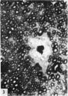
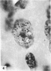
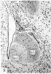
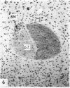
Images in this article
Selected References
These references are in PubMed. This may not be the complete list of references from this article.
- LAPHAM L. W., JOHNSTONE M. A., BRUNDJAR K. H. A NEW PARAFFIN METHOD FOR THE COMBINED STAINING OF MYELIN AND GLIAL FIBERS. J Neuropathol Exp Neurol. 1964 Jan;23:156–160. [PubMed] [Google Scholar]
- Ling E. A., Ahmed M. M. Neuroglia in the corpus callosum of the primate, slow loris (Nycticebus coucang coucang). Tissue Cell. 1974;6(2):361–370. doi: 10.1016/0040-8166(74)90058-5. [DOI] [PubMed] [Google Scholar]
- Sturrock R. R. Development of the mouse anterior commissure. Part I. A comparison of myelination in the anterior and posterior limbs of the anterior commissure of the mouse brain. Zentralbl Veterinarmed C. 1976 Mar;5(1):54–67. doi: 10.1111/j.1439-0264.1976.tb00656.x. [DOI] [PubMed] [Google Scholar]
- Sturrock R. R. Light microscopic identification of immature glial cells in semithin sections of the developing mouse corpus callosum. J Anat. 1976 Dec;122(Pt 3):521–537. [PMC free article] [PubMed] [Google Scholar]
- Van Alphen H. A. The anterior commissure of the rabbit. Acta Anat Suppl (Basel) 1969;57:1–112. [PubMed] [Google Scholar]


