Abstract
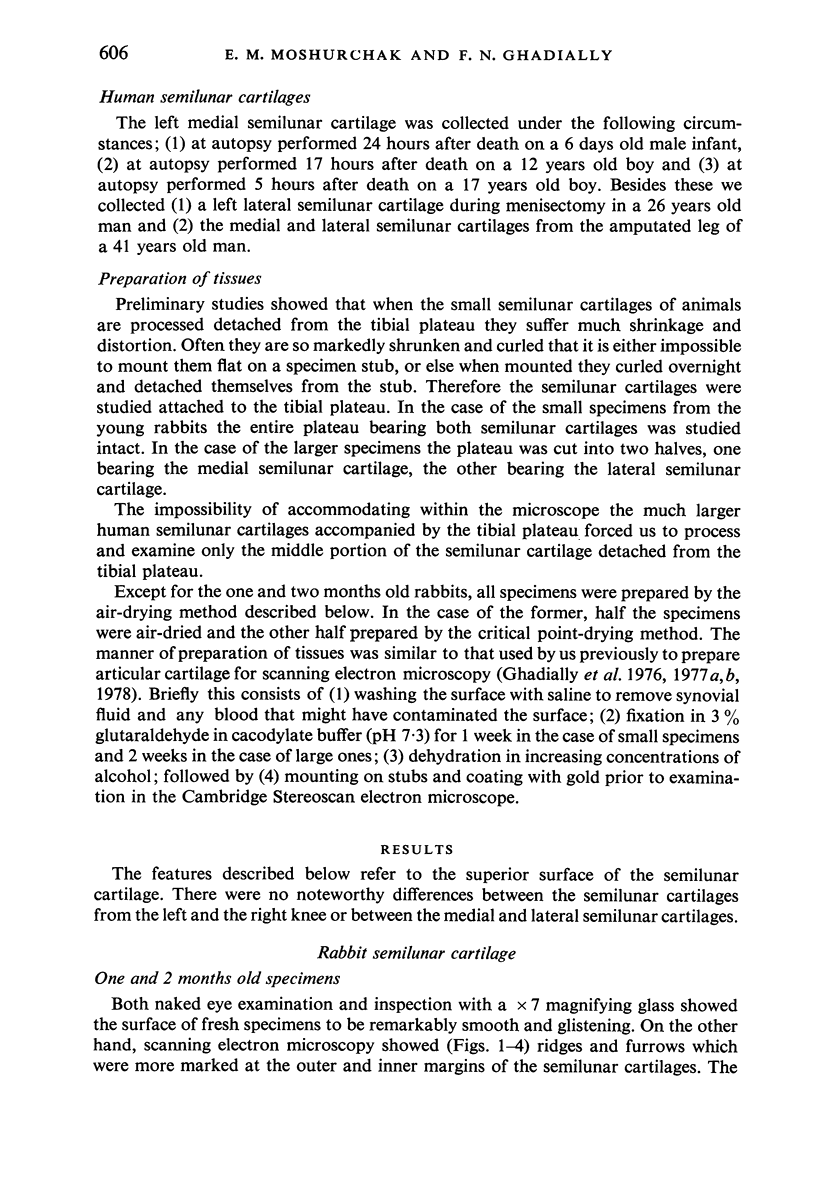
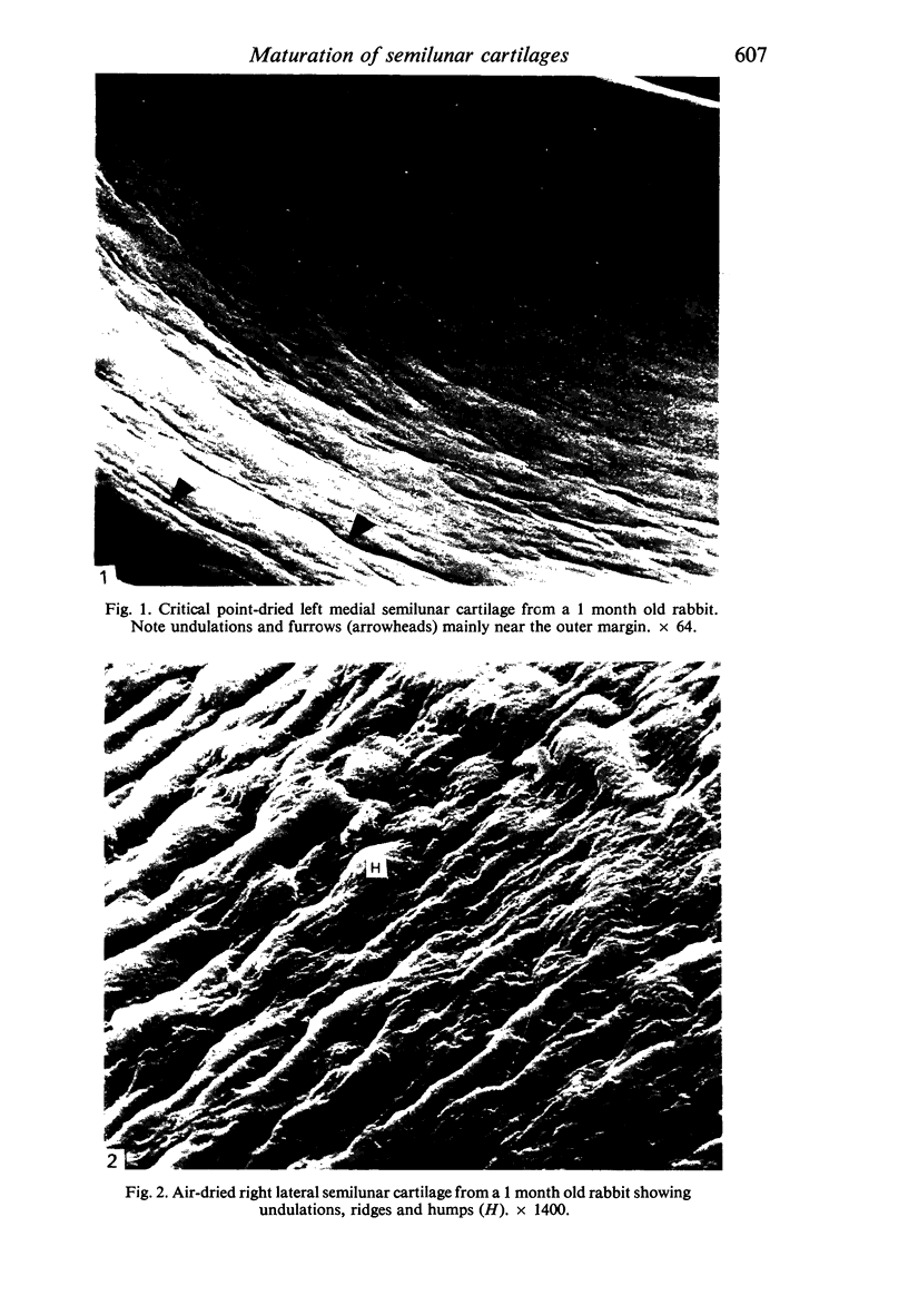
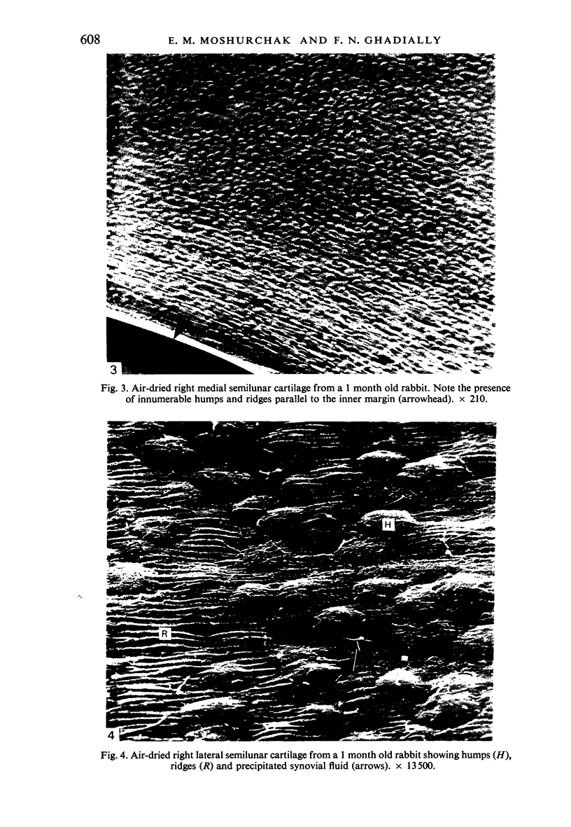
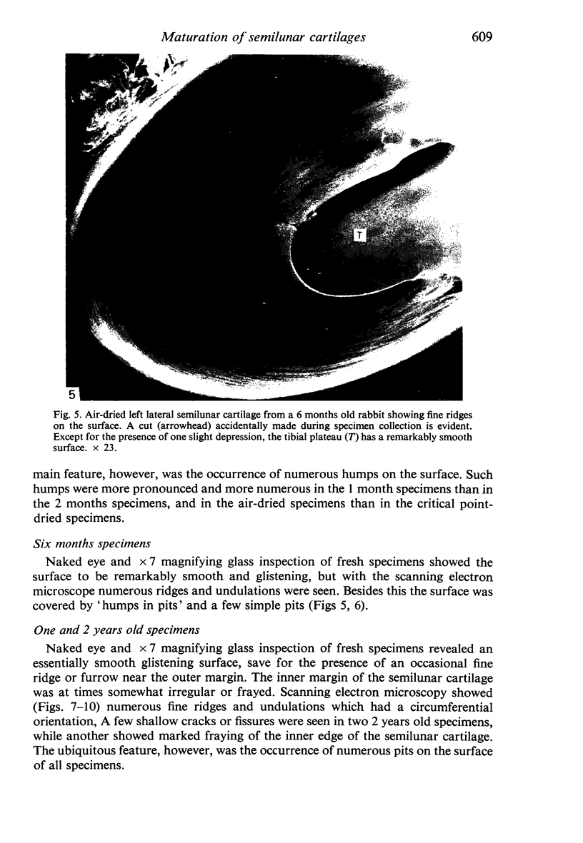
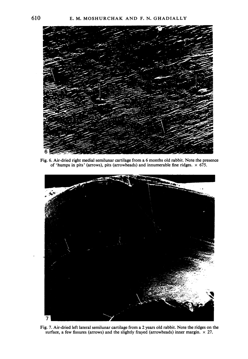
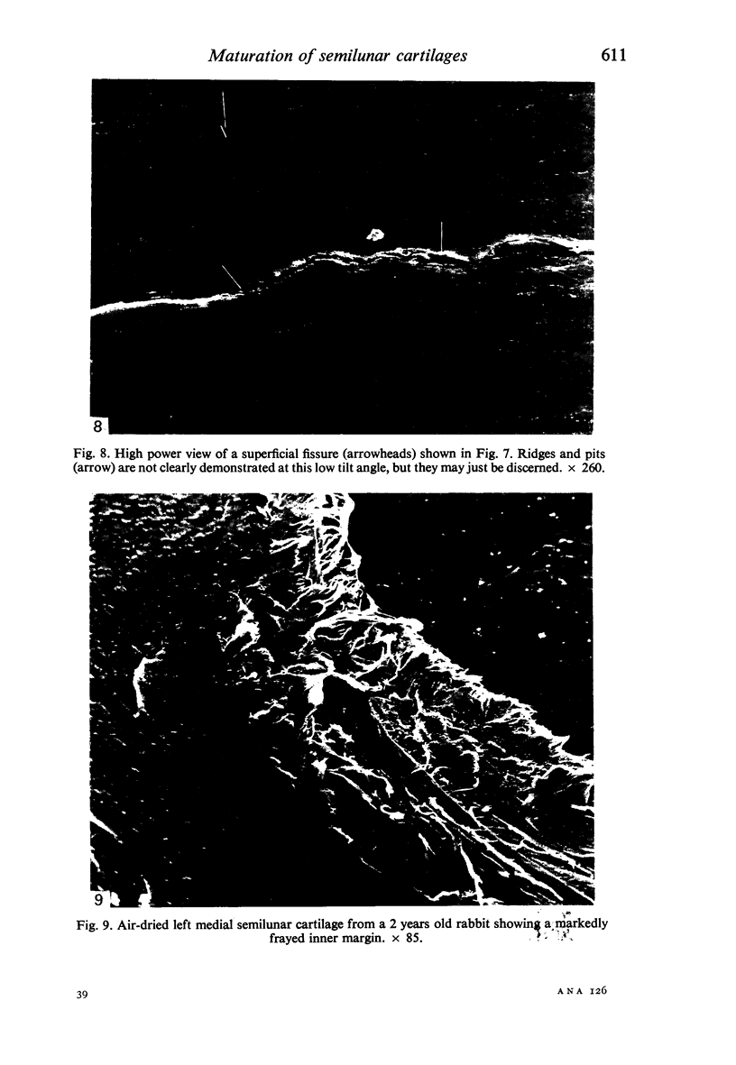
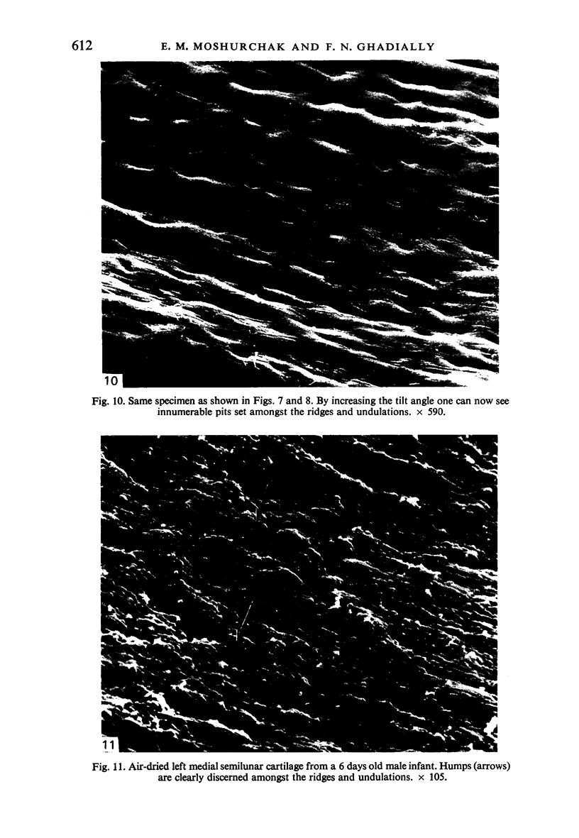
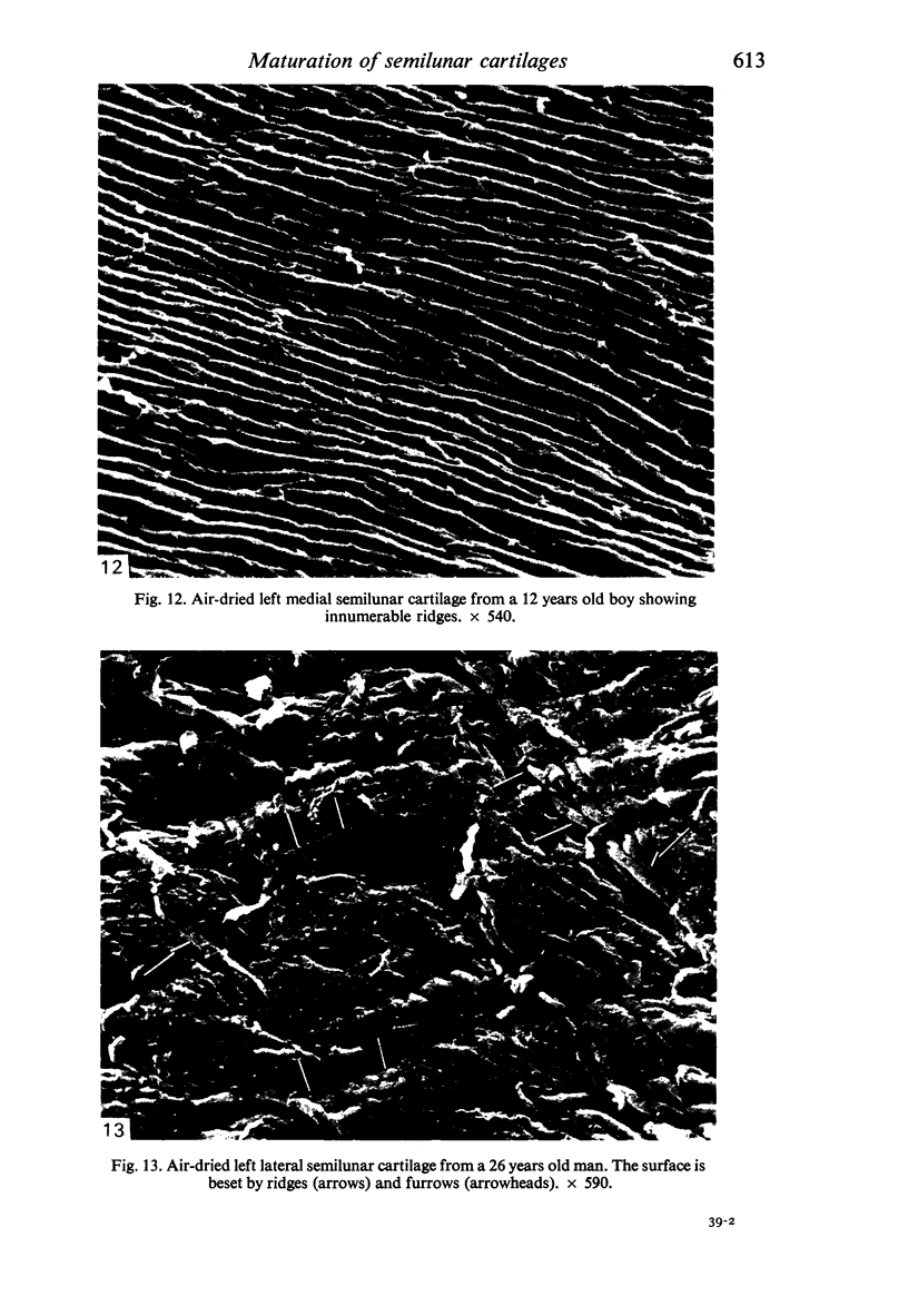
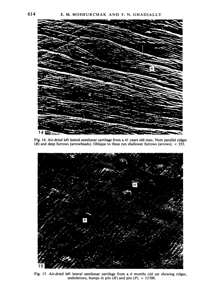
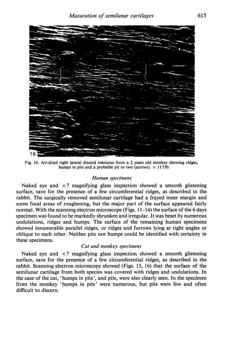
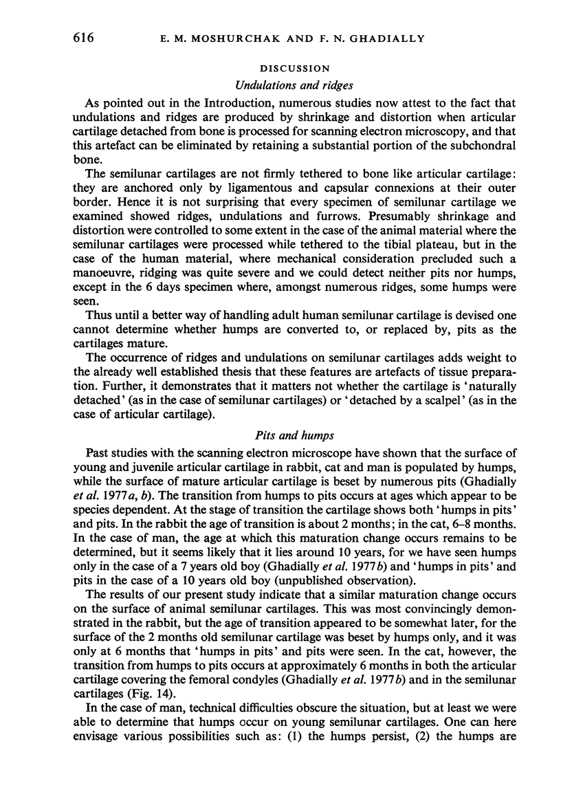
The surface of rabbit, cat, monkey and human semilunar cartilages was examined with the scanning electron microscope. A common feature was the occurrence of numerous ridges, undulations and furrows on the surface, but this was thought to be due to marked shrinkage and distortion of cartilage not firmly attached to bone. Humps were seen on the semilunar cartilages of young animals, but pits occurred in adults. This is thought to reflect a maturation change. Humps were seen in a young human semilunar cartilage, but pits were not seen in adult specimens. It is not clear whether pits are truly absent or just masked by the severe ridging produced during the preparation of large human specimens.
Full text
PDF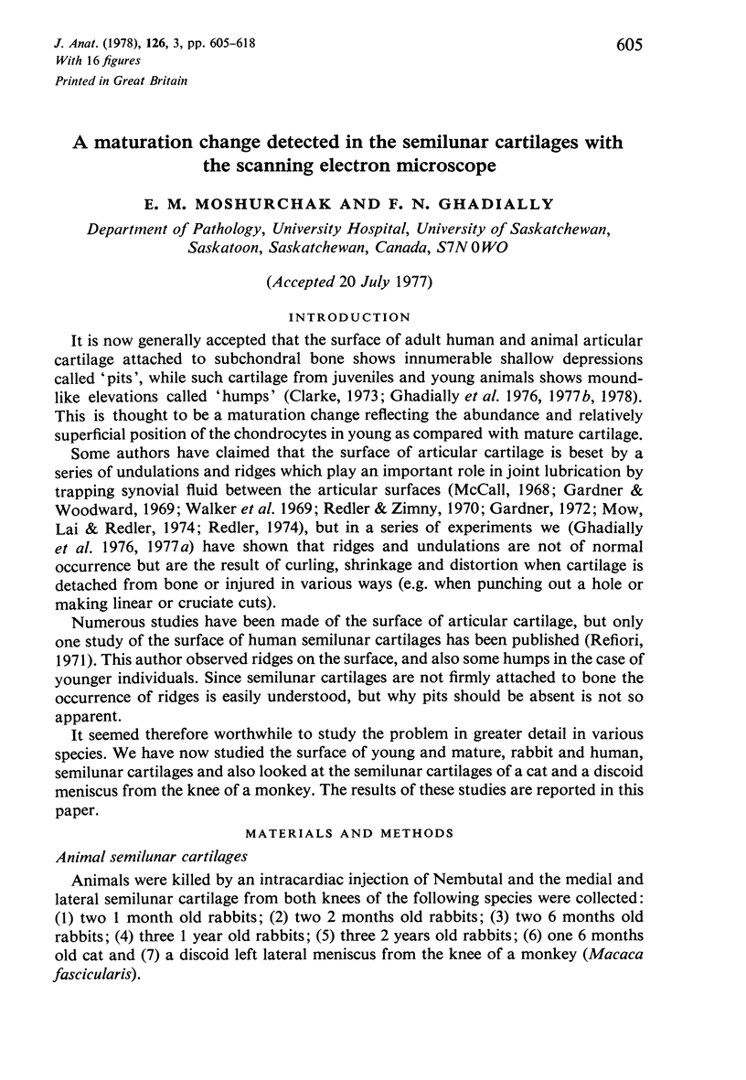
Images in this article
Selected References
These references are in PubMed. This may not be the complete list of references from this article.
- Gardner D. L. The influence of microscopic technology on knowledge of cartilage surface structure. Ann Rheum Dis. 1972 Jul;31(4):235–258. doi: 10.1136/ard.31.4.235. [DOI] [PMC free article] [PubMed] [Google Scholar]
- Gardner D. L., Woodward D. Scanning electron microscopy and replica studies of articular surfaces of guinea-pig synovial joints. Ann Rheum Dis. 1969 Jul;28(4):379–391. doi: 10.1136/ard.28.4.379. [DOI] [PMC free article] [PubMed] [Google Scholar]
- Ghadially F. N., Ghadially J. A., Oryschak A. F., Yong N. K. Experimental production of ridges on rabbit articular cartilage: a scanning electron microscope study. J Anat. 1976 Feb;121(Pt 1):119–132. [PMC free article] [PubMed] [Google Scholar]
- Ghadially F. N., Ghadially J. A., Oryschak A. F., Yong N. K. The surface of dog articular cartilage: a scanning electron microscope study. J Anat. 1977 Apr;123(Pt 2):527–536. [PMC free article] [PubMed] [Google Scholar]
- Ghadially F. N., Moshurchak E. M., Ghadially J. A. A maturation change in the surface of cat articular cartilage detected by the scanning electron microscope. J Anat. 1978 Feb;125(Pt 2):349–360. [PMC free article] [PubMed] [Google Scholar]
- Ghadially F. N., Moshurchak E. M., Thomas I. Humps on young human and rabbit articular cartilage. J Anat. 1977 Nov;124(Pt 2):425–435. [PMC free article] [PubMed] [Google Scholar]
- McCall J. G. Scanning electron microscopy of articular surfaces. Lancet. 1968 Nov 30;2(7579):1194–1194. doi: 10.1016/s0140-6736(68)91680-2. [DOI] [PubMed] [Google Scholar]
- Mow V. C., Lai W. M. Some surface characteristics of articular cartilage. I. A scanning electron microscopy study and a theoretical model for the dynamic interaction of synovial fluid and articular cartilage. J Biomech. 1974 Sep;7(5):449–456. doi: 10.1016/0021-9290(74)90007-4. [DOI] [PubMed] [Google Scholar]
- Redler I. A scanning electron microscopic study of human normal and osteoarthritic articular cartilage. Clin Orthop Relat Res. 1974;(103):262–268. doi: 10.1097/00003086-197409000-00087. [DOI] [PubMed] [Google Scholar]
- Redler I., Zimny M. L. Scanning electron microscopy of normal and abnormal articular cartilage and synovium. J Bone Joint Surg Am. 1970 Oct;52(7):1395–1404. [PubMed] [Google Scholar]
- Refior H. J. Altersabhängige Veränderungen der Meniscusoberfläche--Untersuchungen mit dem Rasterelektronemikroskop. Arch Orthop Unfallchir. 1971;71(4):316–323. [PubMed] [Google Scholar]
- Walker P. S., Sikorski J., Dowson D., Longfield M. D., Wright V., Buckley T. Behaviour of synovial fluid on surfaces of articular cartilage. A scanning electron microscope study. Ann Rheum Dis. 1969 Jan;28(1):1–14. doi: 10.1136/ard.28.1.1. [DOI] [PMC free article] [PubMed] [Google Scholar]


