Abstract
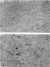
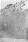
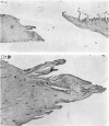
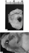
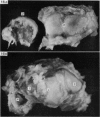
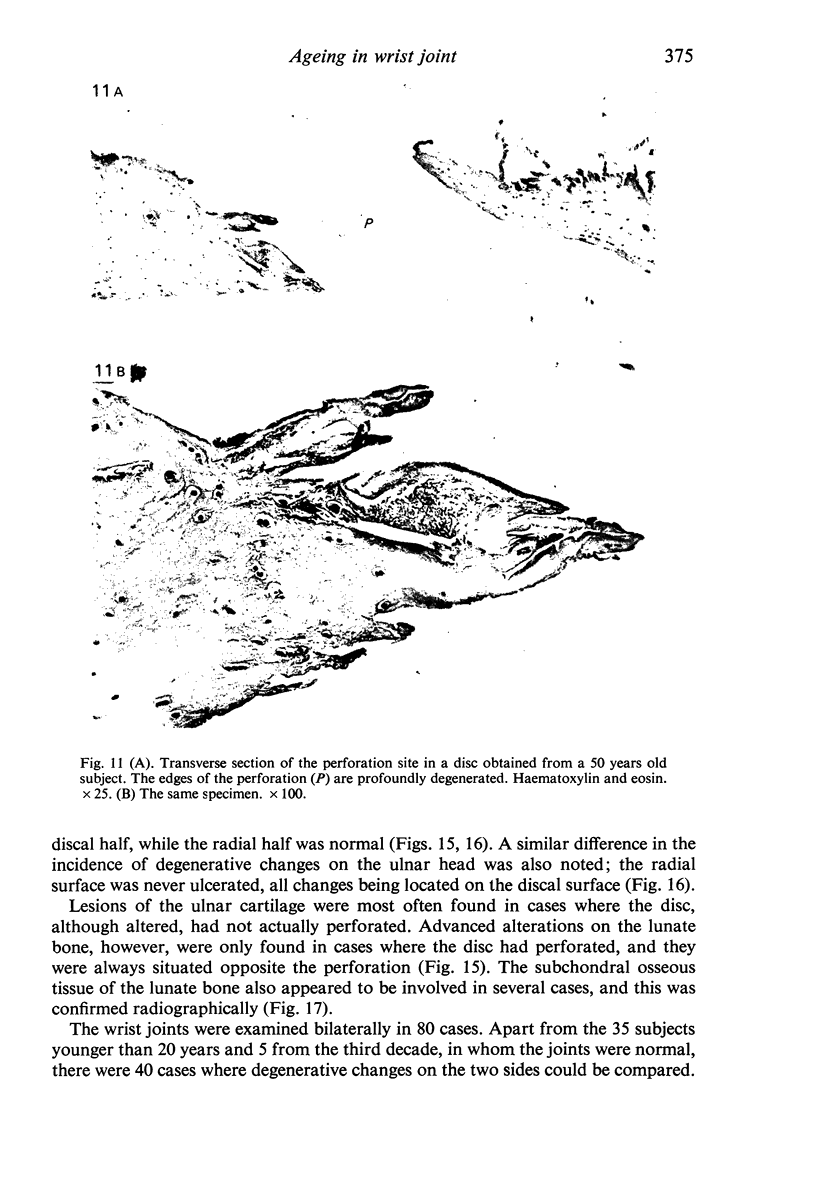
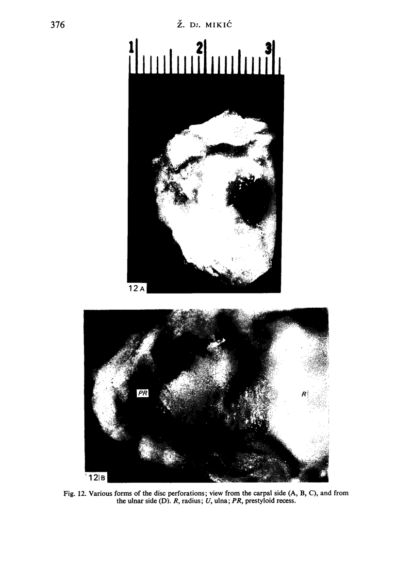
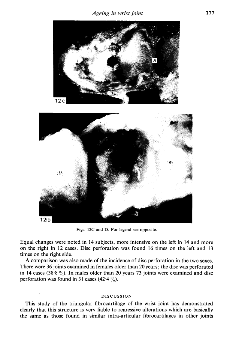
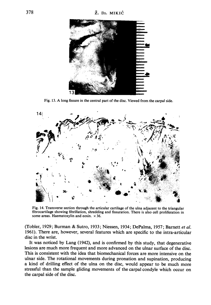
On the basis of a study of 180 wrist joints from 100 fresh cadavers of individuals ranging in age from fetuses to 94 years, it is concluded that the triangular fibro-cartilage is very liable to degenerative alterations associated with ageing. Degeneration begins in the third decade and progressively increases in frequency and severity in subsequent decades. The changes comprise reduced cellularity, loss of elastic fibres, mucoid degeneration of the ground substance, exposure of collagen fibres, fibrillation, erosion, ulceration, abnormal thinning, and, ultimately, disc perforation. The changes are more frequent and more intense on the ulnar surface, and they are always situated in the central part of the disc. It appears that disc perforation is degenerative and age-related: thus there were no perforations in the first two decades of life; in the third there were 7.6%, in the fourth 18.1%, in the fifth 40.0%, in the sixth 42.8%, and in the over sixties 53.1%. There was an associated pattern of degenerative changes in the wrist joint as a whole. The structures adjacent to the articular disc (discal surface of the ulnar head, discal part of the lunate) were much more often involved, and the changes were much more advanced, than on non-discal surfaces. It is argued that this is because of more intensive biomechanical forces, particularly rotational forces, in the disc compartment of the joint.
Full text
PDF
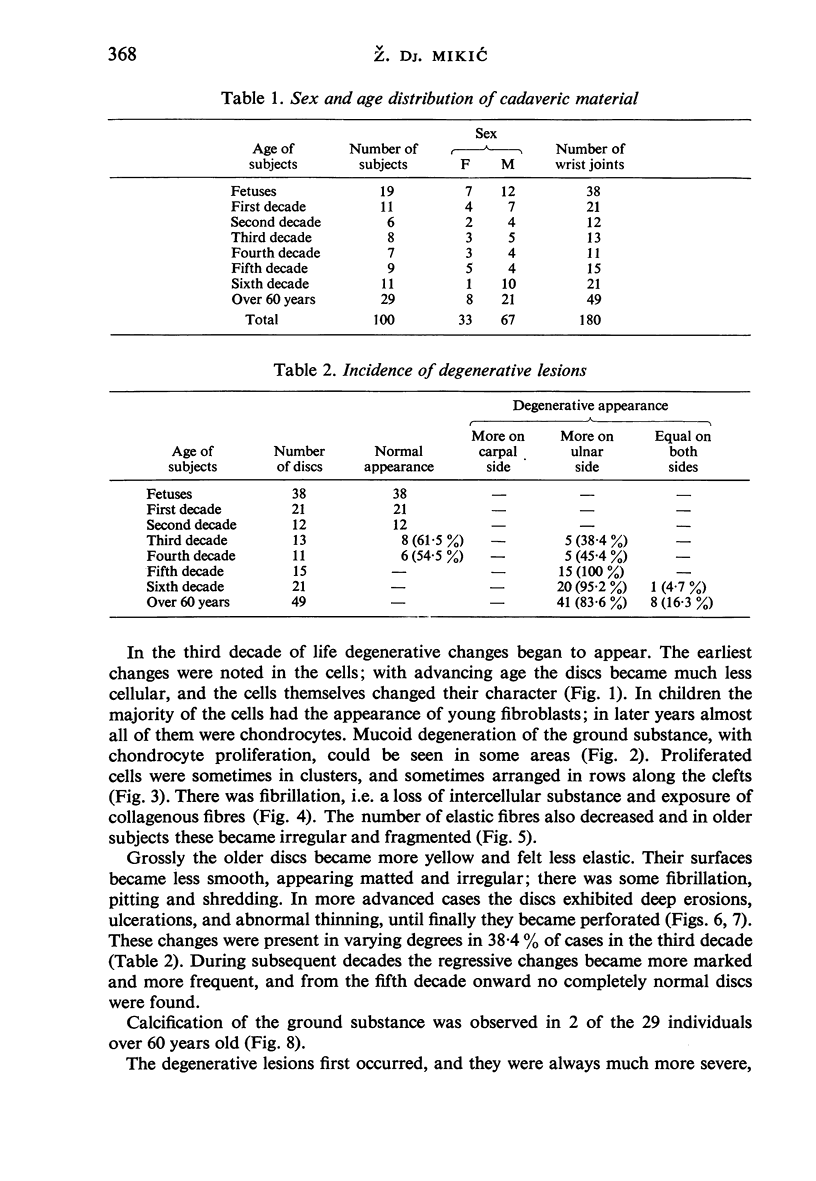
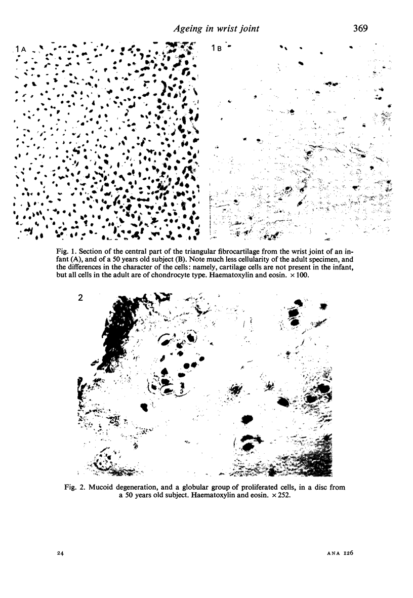
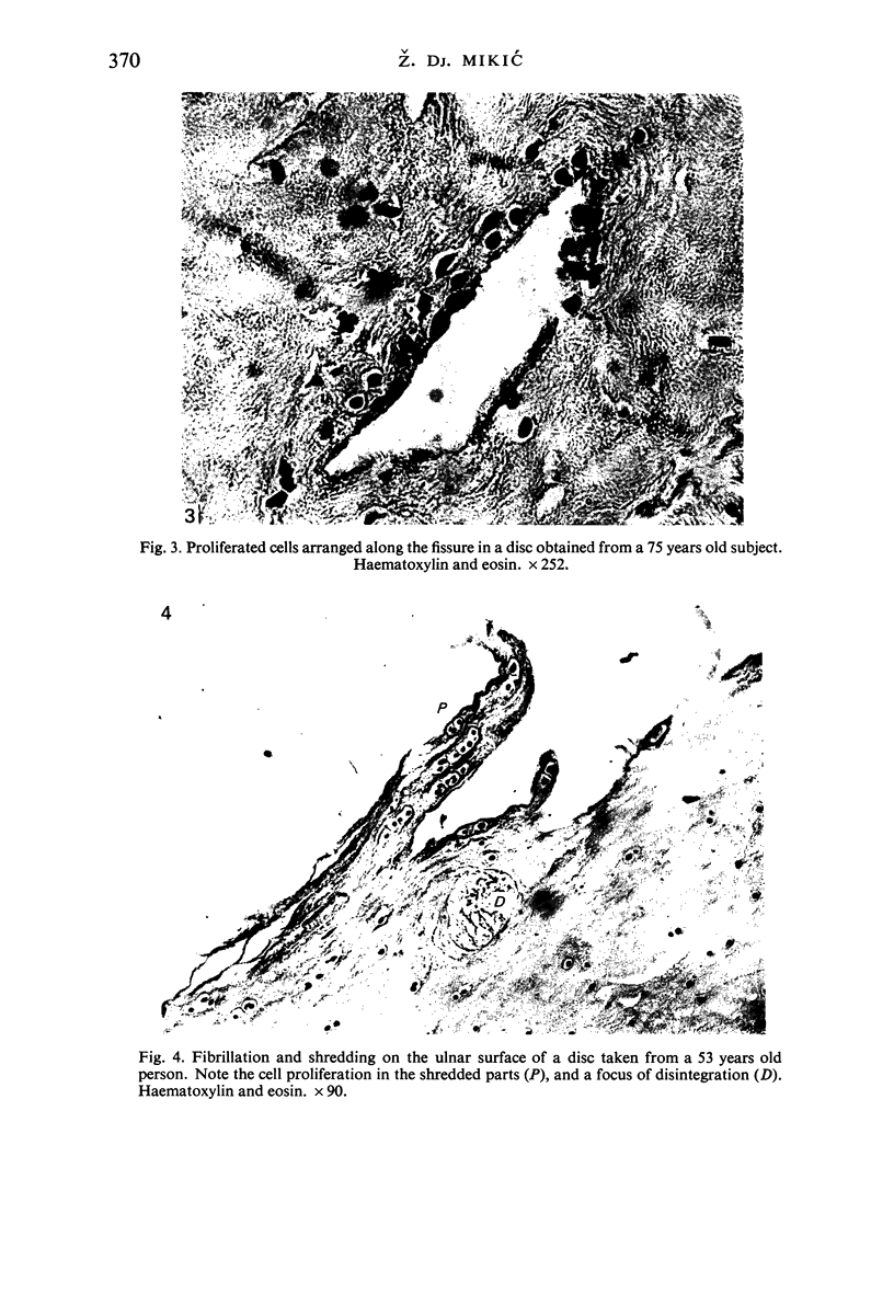
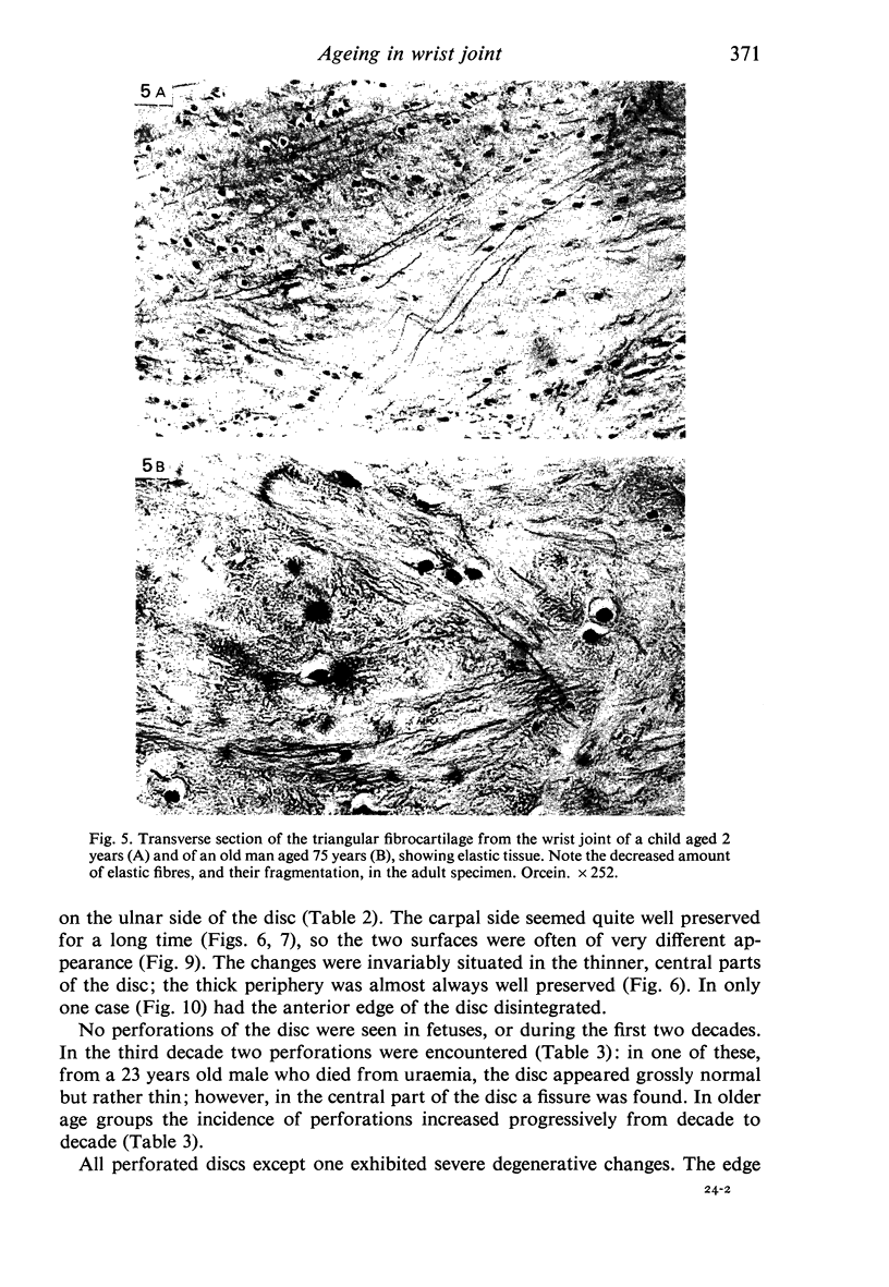

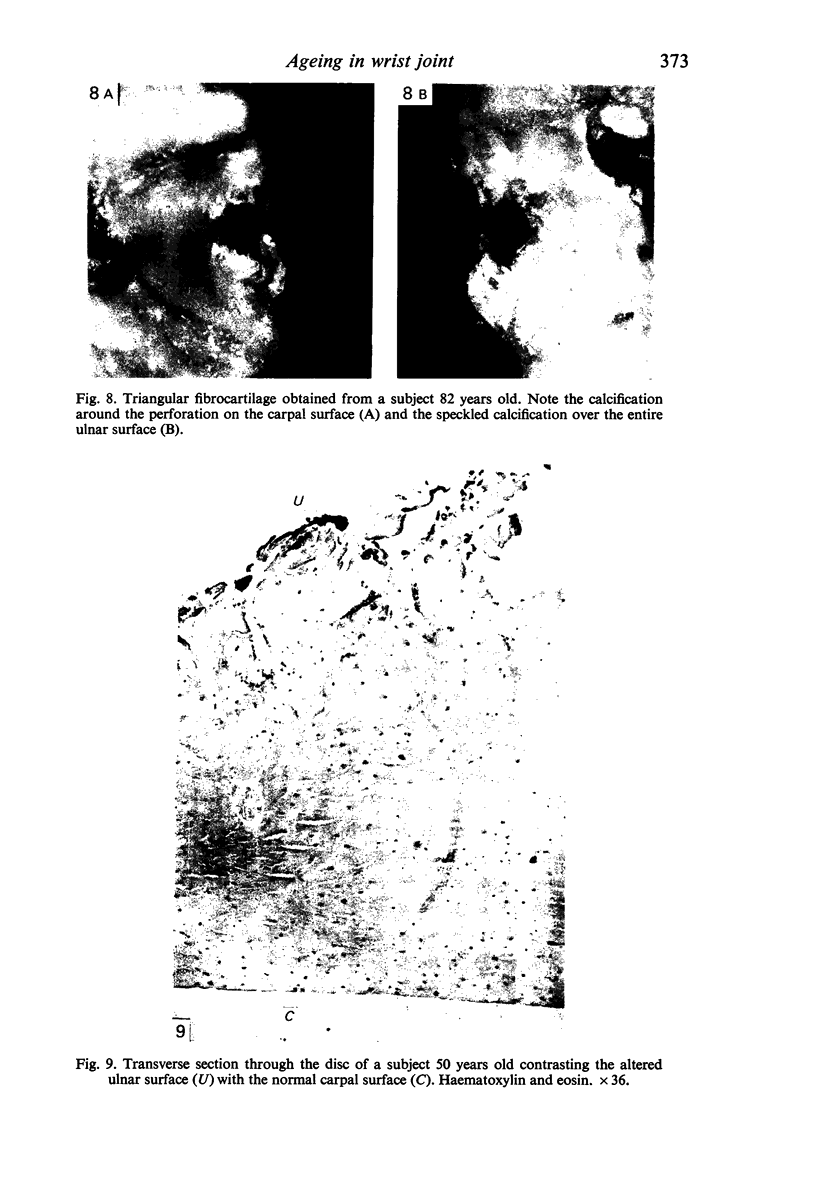
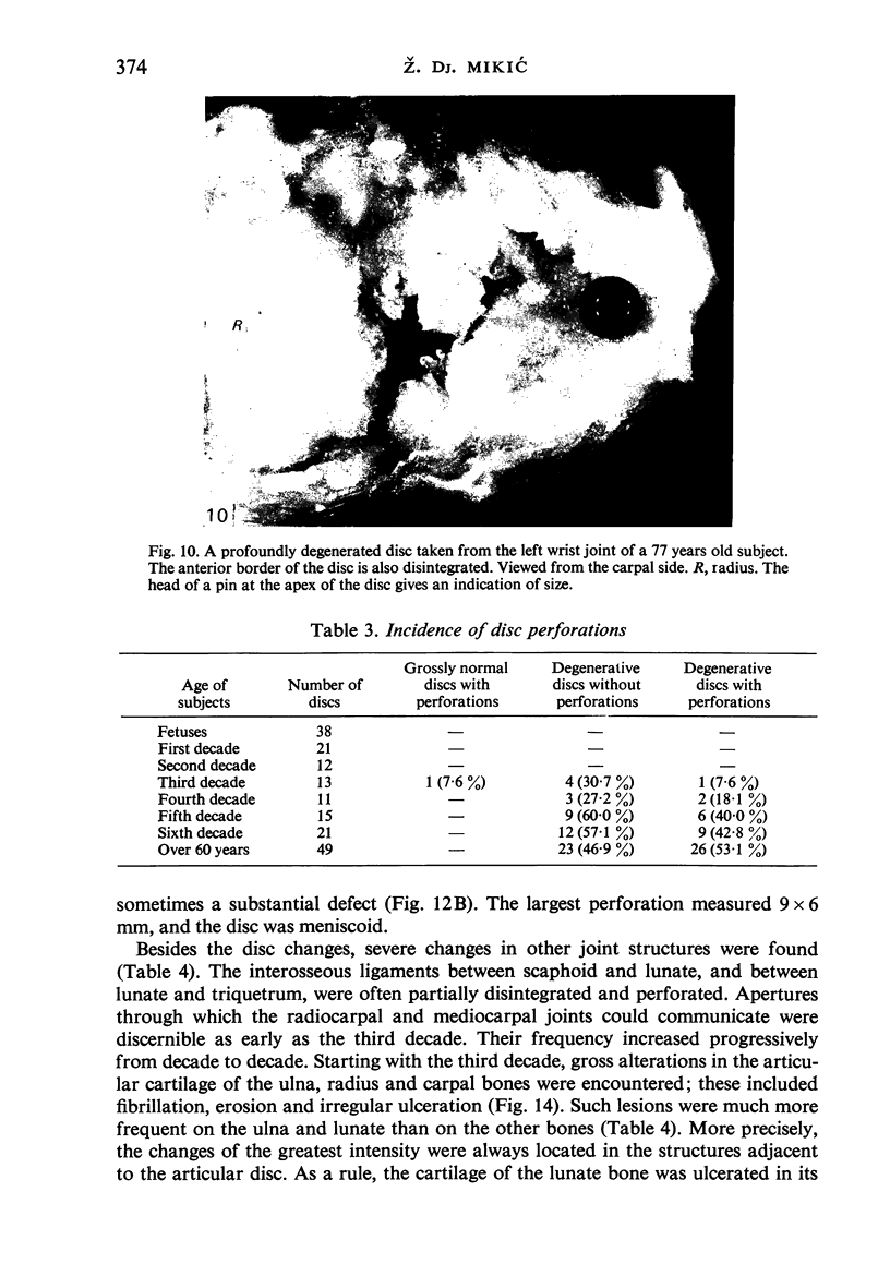

Images in this article
Selected References
These references are in PubMed. This may not be the complete list of references from this article.
- HOEGEN K., RESKE W. Veränderungen an der dreieckigen Bandscheibe des distalen Radio-Ulnar-Gelenkes; ein Beitrag zur Klinik und Pathologie. Z Orthop Ihre Grenzgeb. 1956;87(4):525–532. [PubMed] [Google Scholar]
- KESSLER I., SILBERMAN Z. An experimental study of the radiocarpal joint by arthrography. Surg Gynecol Obstet. 1961 Jan;112:33–40. [PubMed] [Google Scholar]
- LIEBOLT F. L. A new method for repair of the distal radioulnar ligaments. N Y State J Med. 1950 Dec 1;50(23):2817–2819. [PubMed] [Google Scholar]
- Lewis O. J., Hamshere R. J., Bucknill T. M. The anatomy of the wrist joint. J Anat. 1970 May;106(Pt 3):539–552. [PMC free article] [PubMed] [Google Scholar]
- Noble J., Hamblen D. L. The pathology of the degenerate meniscus lesion. J Bone Joint Surg Br. 1975 May;57(2):180–186. [PubMed] [Google Scholar]
- Pfeil E. Meniskusläsion und Alter. Z Orthop Ihre Grenzgeb. 1966 Dec;102(2):308–309. [PubMed] [Google Scholar]
- ROSE-INNES A. P. Anterior dislocation of the ulna at the inferior radio-ulnar joint. Case report, with a discussion of the anatomy of rotation of the forearm. J Bone Joint Surg Br. 1960 Aug;42-B:515–521. doi: 10.1302/0301-620X.42B3.515. [DOI] [PubMed] [Google Scholar]
- Ranawat C. S., Freiberger R. H., Jordan L. R., Straub L. R. Arthrography in the rheumatoid wrist joint. A preliminary report. J Bone Joint Surg Am. 1969 Oct;51(7):1269–1281. doi: 10.2106/00004623-196951070-00003. [DOI] [PubMed] [Google Scholar]
- Refior H. J., Fischer H. Velgleichende mikrostrukturelle Untersuchungen zur Degeneration der Kniegelenksmenisken. Z Orthop Ihre Grenzgeb. 1974 Feb;112(1):128–133. [PubMed] [Google Scholar]
- ZIPPEL H. MENISCUSSCHAEDEN UND MENISCUSVERLETZUNGEN. EINE UNTERSUCHUNG AN 1360 MENISCUSOPERATIONEN. Arch Orthop Unfallchir. 1964 May 27;56:236–247. doi: 10.1007/BF00415180. [DOI] [PubMed] [Google Scholar]


