Abstract
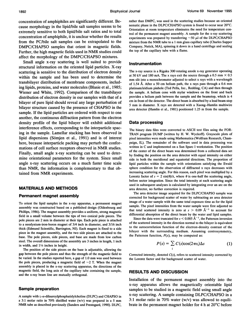
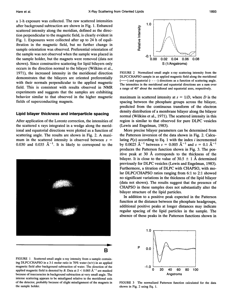
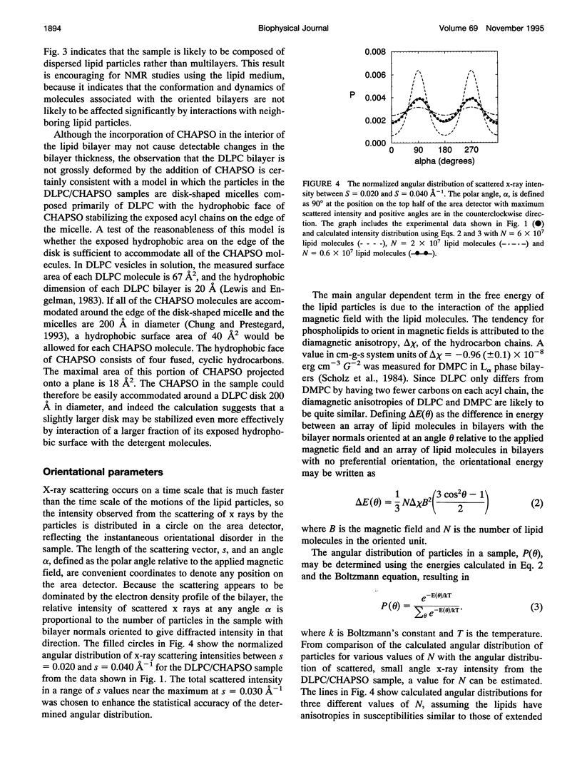
Magnetically oriented lipid/detergent bilayers are potentially useful for studies of membrane-associated molecules and complexes using x-ray scattering and nuclear magnetic resonance (NMR). To establish whether the system is a reasonable model of a phospholipid bilayer, we have studied the system using x-ray solution scattering to determine the bilayer thickness, interparticle spacing, and orientational parameters for magnetically oriented lipid bilayers. The magnetically orientable samples contain the phospholipid L-alpha-dilauroylphosphatidylcholine (DLPC) and the bile salt analog 3-[(3-cholamidopropyl)dimethylammonio]-2-hydroxy-1-propanesulfonate (CHAPSO) in a 3:1 molar ratio in 70% water (w/v) and are similar to magnetically orientable samples used as NMR media for structural studies of membrane-associated molecules. A bilayer thickness of 30 A was determined for the DLPC/CHAPSO particles, which is the same as the bilayer thickness of pure DLPC vesicles, suggesting that the CHAPSO is not greatly perturbing the lipid bilayer. These data, as well as NMR data on molecules incorporated in the oriented lipid particles, are consistent with the sample consisting of reasonably homogeneous and well dispersed lipid particles. Finally, the orientational energy of the sample suggests that the size of the cooperatively orienting unit in the samples is 2 x 10(7) phospholipid molecules.
Full text
PDF

Images in this article
Selected References
These references are in PubMed. This may not be the complete list of references from this article.
- Blasie J. K., Herbette L., Pachence J. Biological membrane structure as "seen" by X-ray and neutron diffraction techniques. J Membr Biol. 1985;86(1):1–7. doi: 10.1007/BF01871604. [DOI] [PubMed] [Google Scholar]
- Chabre M. X-ray diffraction studies of retinal rods. I. Structure of the disc membrane, effect of illumination. Biochim Biophys Acta. 1975 Mar 25;382(3):322–335. doi: 10.1016/0005-2736(75)90274-6. [DOI] [PubMed] [Google Scholar]
- Lewis B. A., Engelman D. M. Lipid bilayer thickness varies linearly with acyl chain length in fluid phosphatidylcholine vesicles. J Mol Biol. 1983 May 15;166(2):211–217. doi: 10.1016/s0022-2836(83)80007-2. [DOI] [PubMed] [Google Scholar]
- Mazer N. A., Benedek G. B., Carey M. C. Quasielastic light-scattering studies of aqueous biliary lipid systems. Mixed micelle formation in bile salt-lecithin solutions. Biochemistry. 1980 Feb 19;19(4):601–615. doi: 10.1021/bi00545a001. [DOI] [PubMed] [Google Scholar]
- Müller K. Structural dimorphism of bile salt/lecithin mixed micelles. A possible regulatory mechanism for cholesterol solubility in bile? X-ray structure analysis. Biochemistry. 1981 Jan 20;20(2):404–414. doi: 10.1021/bi00505a028. [DOI] [PubMed] [Google Scholar]
- Sanders C. R., 2nd, Prestegard J. H. Magnetically orientable phospholipid bilayers containing small amounts of a bile salt analogue, CHAPSO. Biophys J. 1990 Aug;58(2):447–460. doi: 10.1016/S0006-3495(90)82390-0. [DOI] [PMC free article] [PubMed] [Google Scholar]
- Scholz F., Boroske E., Helfrich W. Magnetic anisotropy of lecithin membranes. A new anisotropy susceptometer. Biophys J. 1984 Mar;45(3):589–592. doi: 10.1016/S0006-3495(84)84196-X. [DOI] [PMC free article] [PubMed] [Google Scholar]
- Speyer J. B., Sripada P. K., Das Gupta S. K., Shipley G. G., Griffin R. G. Magnetic orientation of sphingomyelin-lecithin bilayers. Biophys J. 1987 Apr;51(4):687–691. doi: 10.1016/S0006-3495(87)83394-5. [DOI] [PMC free article] [PubMed] [Google Scholar]
- Sömjen G. J., Coleman R., Koch M. H., Wachtel E., Billington D., Towns-Andrews E., Gilat T. The induction of lamellar stacking by cholesterol in lecithin-bile salt model systems and human bile studied by synchrotron X-radiation. FEBS Lett. 1991 Sep 9;289(2):163–166. doi: 10.1016/0014-5793(91)81060-l. [DOI] [PubMed] [Google Scholar]
- Torbet J. Solution behavior of DNA studied with magnetically induced birefringence. Methods Enzymol. 1992;211:518–532. doi: 10.1016/0076-6879(92)11029-i. [DOI] [PubMed] [Google Scholar]
- Walter A., Vinson P. K., Kaplun A., Talmon Y. Intermediate structures in the cholate-phosphatidylcholine vesicle-micelle transition. Biophys J. 1991 Dec;60(6):1315–1325. doi: 10.1016/S0006-3495(91)82169-5. [DOI] [PMC free article] [PubMed] [Google Scholar]
- Wiener M. C., White S. H. Structure of a fluid dioleoylphosphatidylcholine bilayer determined by joint refinement of x-ray and neutron diffraction data. III. Complete structure. Biophys J. 1992 Feb;61(2):434–447. doi: 10.1016/S0006-3495(92)81849-0. [DOI] [PMC free article] [PubMed] [Google Scholar]
- Wilkins M. H., Blaurock A. E., Engelman D. M. Bilayer structure in membranes. Nat New Biol. 1971 Mar 17;230(11):72–76. doi: 10.1038/newbio230072a0. [DOI] [PubMed] [Google Scholar]


