Abstract
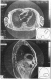
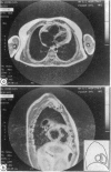
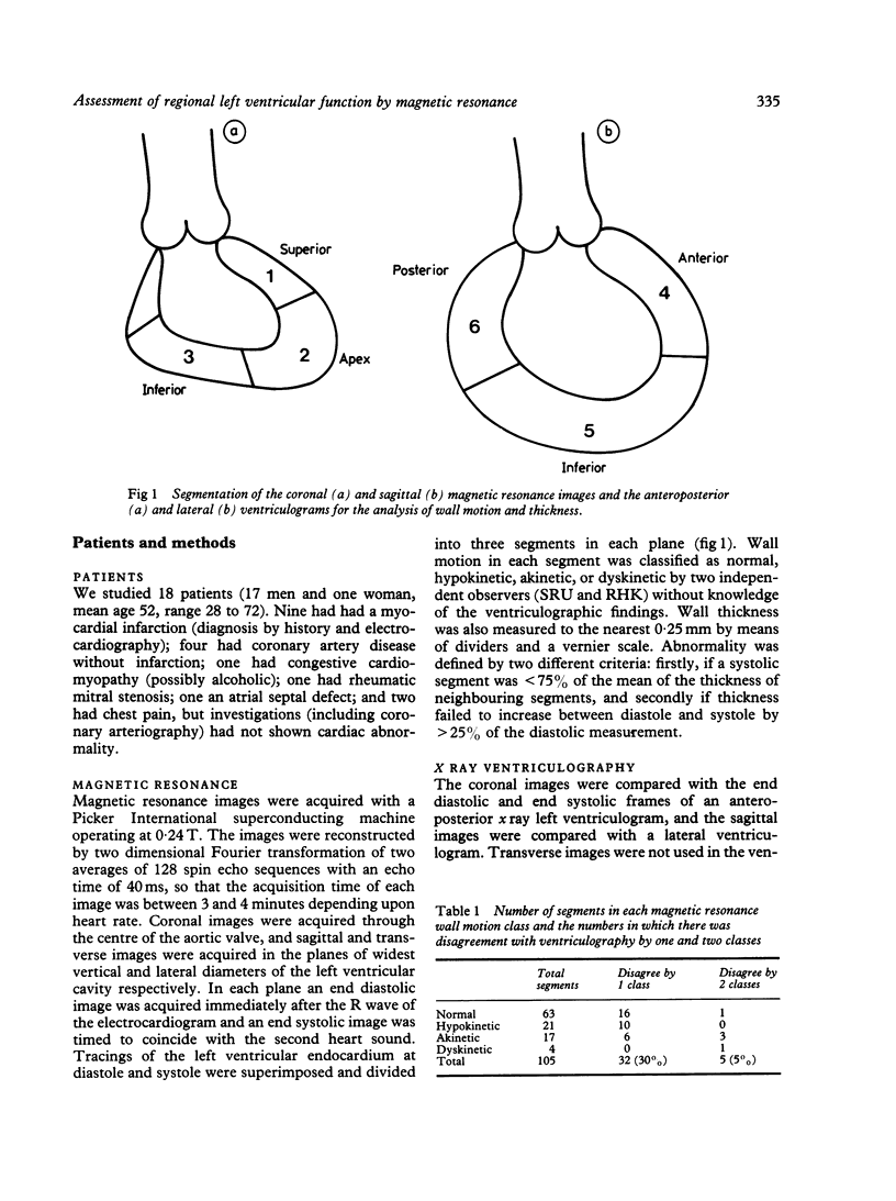
The ability of magnetic resonance to determine regional left ventricular function was investigated in 18 patients--13 with coronary artery disease (nine with previous infarction), one with congestive cardiomyopathy, one with mitral stenosis, one with an atrial septal defect, and two without detectable cardiac abnormality. Coronal magnetic resonance images were acquired through the aortic valve and sagittal images were acquired in the plane of widest diameter of the left ventricle seen in the coronal image, both at end diastole and end systole. Regional wall motion assessed by magnetic resonance was compared with the results of anteroposterior and left lateral x ray ventriculograms by two independent observers. The left ventricular wall was divided into three segments in each plane and the motion of the segments was classified as normal, hypokinetic, akinetic, or dyskinetic. Muscle thickness was measured in each segment of the magnetic resonance images and was considered to be abnormal if in the systolic images it was less than 75% of that in neighbouring segments or if it failed to increase by at least 25% between diastole and systole. Wall motion assessments by the two methods agreed in 68 of 105 segments analysed, but differed by one class in 32 segments and by two classes in five segments. The differences can be explained by the conditions under which the investigations were performed and by the disparity between a tomographic section and an x ray projection. Magnetic resonance showed 25 segments to have abnormal wall thickness. Only one patient with infarction did not have an area of wall thinning and no patient without infarction had an area of thinning. It is concluded that magnetic resonance allows an accurate non-invasive assessment of left ventricular wall motion and thickness.
Full text
PDF
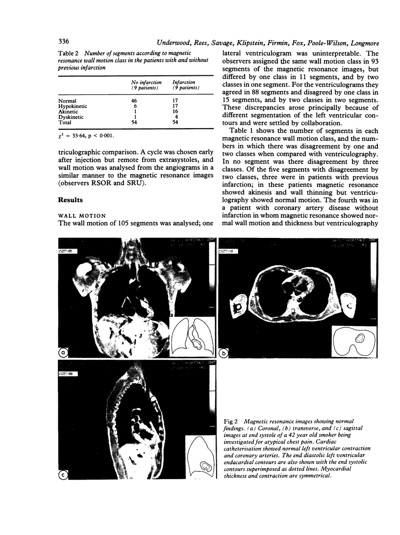
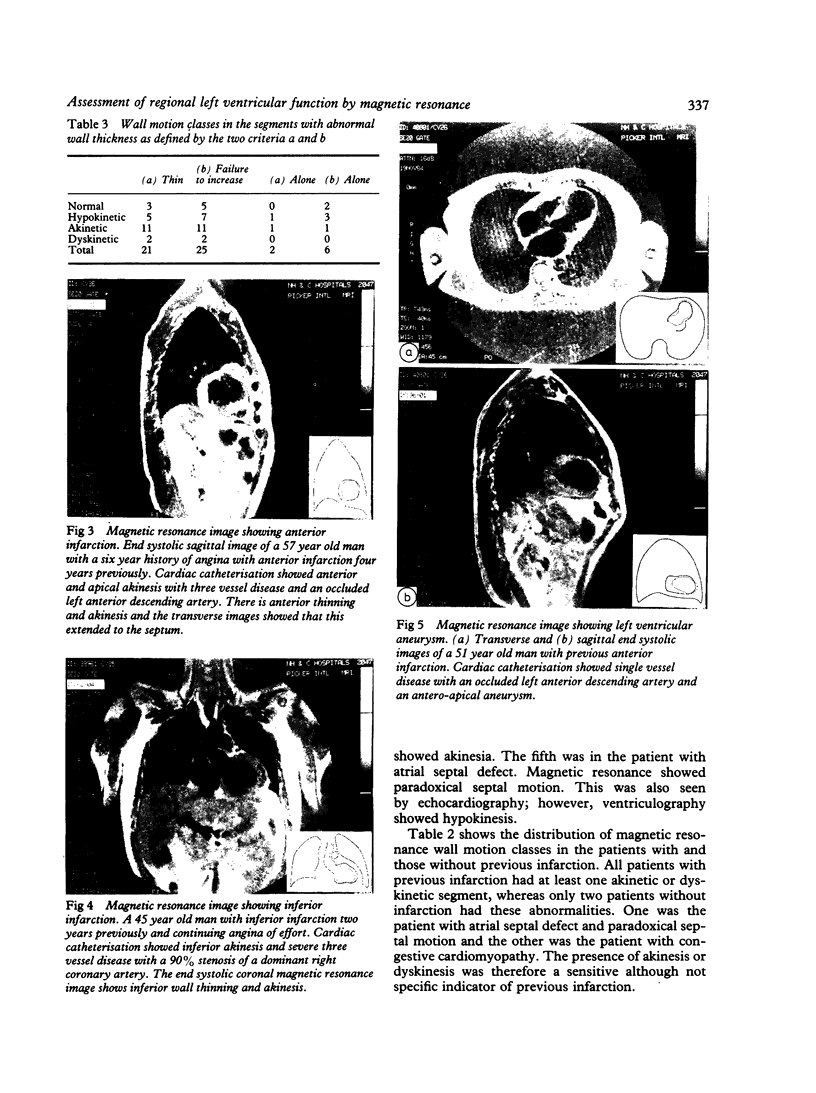
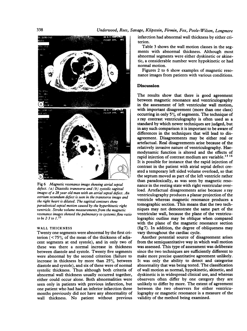
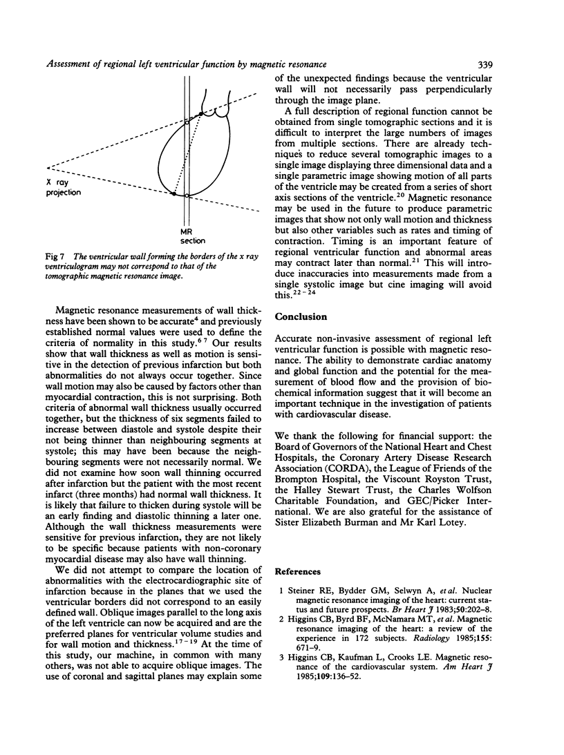
Images in this article
Selected References
These references are in PubMed. This may not be the complete list of references from this article.
- Akins E. W., Hill J. A., Fitzsimmons J. R., Pepine C. J., Williams C. M. Importance of imaging plane for magnetic resonance imaging of the normal left ventricle. Am J Cardiol. 1985 Aug 1;56(4):366–372. doi: 10.1016/0002-9149(85)90866-5. [DOI] [PubMed] [Google Scholar]
- Bradley W. G., Jr, Waluch V., Lai K. S., Fernandez E. J., Spalter C. The appearance of rapidly flowing blood on magnetic resonance images. AJR Am J Roentgenol. 1984 Dec;143(6):1167–1174. doi: 10.2214/ajr.143.6.1167. [DOI] [PubMed] [Google Scholar]
- Brody W. R., Macovski A., Lehmann L., DiBianca F. A., Volz D., Edelheit L. S. Intravenous angiography using scanned projection radiography: preliminary investigation of a new method. Invest Radiol. 1980 May-Jun;15(3):220–223. doi: 10.1097/00004424-198005000-00008. [DOI] [PubMed] [Google Scholar]
- Bryant D. J., Payne J. A., Firmin D. N., Longmore D. B. Measurement of flow with NMR imaging using a gradient pulse and phase difference technique. J Comput Assist Tomogr. 1984 Aug;8(4):588–593. doi: 10.1097/00004728-198408000-00002. [DOI] [PubMed] [Google Scholar]
- Byrd B. F., 3rd, Schiller N. B., Botvinick E. H., Higgins C. B. Normal cardiac dimensions by magnetic resonance imaging. Am J Cardiol. 1985 May 1;55(11):1440–1442. doi: 10.1016/0002-9149(85)90529-6. [DOI] [PubMed] [Google Scholar]
- Feiglin D. H., George C. R., MacIntyre W. J., O'Donnell J. K., Go R. T., Pavlicek W., Meaney T. F. Gated cardiac magnetic resonance structural imaging: optimization by electronic axial rotation. Radiology. 1985 Jan;154(1):129–132. doi: 10.1148/radiology.154.1.3155478. [DOI] [PubMed] [Google Scholar]
- Fisher M. R., von Schulthess G. K., Higgins C. B. Multiphasic cardiac magnetic resonance imaging: normal regional left ventricular wall thickening. AJR Am J Roentgenol. 1985 Jul;145(1):27–30. doi: 10.2214/ajr.145.1.27. [DOI] [PubMed] [Google Scholar]
- Friedman B. J., Waters J., Kwan O. L., DeMaria A. N. Comparison of magnetic resonance imaging and echocardiography in determination of cardiac dimensions in normal subjects. J Am Coll Cardiol. 1985 Jun;5(6):1369–1376. doi: 10.1016/s0735-1097(85)80350-8. [DOI] [PubMed] [Google Scholar]
- Higgins C. B., Byrd B. F., 2nd, McNamara M. T., Lanzer P., Lipton M. J., Botvinick E., Schiller N. B., Crooks L. E., Kaufman L. Magnetic resonance imaging of the heart: a review of the experience in 172 subjects. Radiology. 1985 Jun;155(3):671–679. doi: 10.1148/radiology.155.3.3159039. [DOI] [PubMed] [Google Scholar]
- Higgins C. B., Kaufman L., Crooks L. E. Magnetic resonance imaging of the cardiovascular system. Am Heart J. 1985 Jan;109(1):136–152. doi: 10.1016/0002-8703(85)90426-0. [DOI] [PubMed] [Google Scholar]
- Higgins C. B., Lanzer P., Stark D., Botvinick E., Schiller N. B., Crooks L., Kaufman L., Lipton M. J. Imaging by nuclear magnetic resonance in patients with chronic ischemic heart disease. Circulation. 1984 Mar;69(3):523–531. doi: 10.1161/01.cir.69.3.523. [DOI] [PubMed] [Google Scholar]
- Longmore D. B., Klipstein R. H., Underwood S. R., Firmin D. N., Hounsfield G. N., Watanabe M., Bland C., Fox K., Poole-Wilson P. A., Rees R. S. Dimensional accuracy of magnetic resonance in studies of the heart. Lancet. 1985 Jun 15;1(8442):1360–1362. doi: 10.1016/s0140-6736(85)91786-6. [DOI] [PubMed] [Google Scholar]
- McNamara M. T., Higgins C. B., Schechtmann N., Botvinick E., Lipton M. J., Chatterjee K., Amparo E. G. Detection and characterization of acute myocardial infarction in man with use of gated magnetic resonance. Circulation. 1985 Apr;71(4):717–724. doi: 10.1161/01.cir.71.4.717. [DOI] [PubMed] [Google Scholar]
- Schwimmer M., Heiken J. P., McClennan B. L., Friedrich E. R. Postoperative hysterosalpingogram: radiographic-surgical correlation. Radiology. 1985 Nov;157(2):313–317. doi: 10.1148/radiology.157.2.4048437. [DOI] [PubMed] [Google Scholar]
- Sheehan F. H., Stewart D. K., Dodge H. T., Mitten S., Bolson E. L., Brown B. G. Variability in the measurement of regional left ventricular wall motion from contrast angiograms. Circulation. 1983 Sep;68(3):550–559. doi: 10.1161/01.cir.68.3.550. [DOI] [PubMed] [Google Scholar]
- Steiner R. E., Bydder G. M., Selwyn A., Deanfield J., Longmore D. B., Klipsten R. H., Firmin D. Nuclear magnetic resonance imaging of the heart. Current status and future prospects. Br Heart J. 1983 Sep;50(3):202–208. doi: 10.1136/hrt.50.3.202. [DOI] [PMC free article] [PubMed] [Google Scholar]
- Underwood S. R., Walton S., Laming P. J., Jarritt P. H., Ell P. J., Emanuel R. W., Swanton R. H. Left ventricular volume and ejection fraction determined by gated blood pool emission tomography. Br Heart J. 1985 Feb;53(2):216–222. doi: 10.1136/hrt.53.2.216. [DOI] [PMC free article] [PubMed] [Google Scholar]
- Walton S., Yiannikas J., Jarritt P. H., Brown N. J., Swanton R. H., Ell P. J. Phasic abnormalities of left ventricular emptying in coronary artery disease. Br Heart J. 1981 Sep;46(3):245–253. doi: 10.1136/hrt.46.3.245. [DOI] [PMC free article] [PubMed] [Google Scholar]
- Waterton J. C., Jenkins J. P., Zhu X. P., Love H. G., Isherwood I., Rowlands D. J. Magnetic resonance (MR) cine imaging of the human heart. Br J Radiol. 1985 Aug;58(692):711–716. doi: 10.1259/0007-1285-58-692-711. [DOI] [PubMed] [Google Scholar]


