

Abstract
Background
Tracheobronchial injuries (TBI) are rare yet potentially fatal complications of blunt chest trauma, often underdiagnosed due to nonspecific clinical manifestations.
Case presentation
We report the case of an 11-year-old Arab girl who developed progressive dyspnea two months after a motor vehicle accident initially managed conservatively. Imaging revealed complete atelectasis of the right lung and obstruction of the right main bronchus by granulation tissue. Bronchoscopy confirmed complete bronchial occlusion, and surgical intervention revealed a delayed bronchial transection. Successful end-to-end anastomosis restored full lung expansion and respiratory function.
Clinical discussion
This case highlights the diagnostic challenge of TBIs in children, particularly when symptoms present late. Anatomical vulnerability of the right bronchus, delayed symptom onset, and nonspecific radiologic signs may obscure early recognition. Granulation-induced bronchial obstruction, a known complication of undiagnosed TBIs, was confirmed intraoperatively.
Conclusion
Delayed presentation of tracheobronchial injury may lead to progressive airway obstruction due to granulation tissue. High suspicion, timely bronchoscopy, and early surgical intervention are essential to prevent irreversible lung damage and restore pulmonary function.
Keywords: Tracheobronchial injury, Bronchial transection, Granulation tissue, Pediatric trauma, Bronchoscopy, Surgical repair
Highlights
-
•
Tracheobronchial injuries are rare but potentially fatal, especially when diagnosis is delayed.
-
•
We report a case of a complete right main bronchus transection in an 11-year-old Arab girl following blunt chest trauma.
-
•
The injury remained undiagnosed for nearly two months, eventually causing airway obstruction due to granuloma formation.
-
•
Bronchoscopy revealed complete bronchial occlusion, and definitive management was achieved through surgical end-to-end anastomosis.
-
•
This case highlights the importance of early suspicion and bronchoscopy in post-trauma respiratory deterioration.
1. Introduction
Tracheobronchial injuries (TBI), though rare, can be life-threatening and often involve the airway segment extending from the cricoid cartilage down to the bifurcation of the right and left mainstem bronchi [1]. TBI is a potentially fatal condition that is often overlooked in polytrauma patients. Its incidence is approximately 0.4 % in adults, while it is even rarer in children, representing only 0.05 % of cases in the pediatric population [2,3]. However, the true incidence is likely underestimated, as many patients succumb to their injuries before reaching medical care [4]. Even among those who reach the hospital alive, the overall mortality rate remains high, approaching 30 % [5]. Physical examination may reveal subcutaneous emphysema, pneumothorax, hemoptysis, and in some cases, active air leakage through a penetrating wound [6]. Diagnosis is confirmed by bronchoscopy, which remains the gold standard and should be performed in all cases of suspected airway injury to facilitate early detection and improve survival. Delayed diagnosis may allow for the development of granulation tissue and airway structures within the first 1 to 4 weeks, increasing the risk of complications such as pneumonia, bronchiectasis, atelectasis, and abscess formation. Even with successful airway repair, the distal lung tissue may become nonfunctional due to prolonged obstruction and irreversible damage [7]. We present the case of an 11-year-old girl from Syria who sustained a delayed bronchial fracture following a motor vehicle accident, leading to the formation of a granuloma and subsequent airway obstruction. The work has been reported in line with the SCARE criteria [8].
2. Case presentation
An 11-year-old female from Syria, with no significant past medical, surgical, or allergic history, was admitted to the emergency room due to progressive dyspnea. Approximately two months prior to admission, she was involved in a motor vehicle collision (MVC) that resulted in pulmonary contusion and right-sided pneumothorax, as confirmed by chest computed tomography (CT) performed at the initial hospital in her home region. She was hospitalized in the intensive care unit (ICU) for over a week and managed conservatively. After clinical improvement, the patient was discharged.
Six weeks following discharge, she developed gradually worsening shortness of breath—initially on exertion, later progressing to dyspnea at rest and orthopnea. These symptoms prompted referral for further evaluation. On presentation, her vital signs were: blood pressure 110/60 mmHg, heart rate 110 bpm, temperature 36.5 °C, respiratory rate 25 breaths/min, and oxygen saturation 92 % on room air. Physical examination revealed markedly diminished breath sounds over the right hemithorax, with preserved breath sounds on the left.
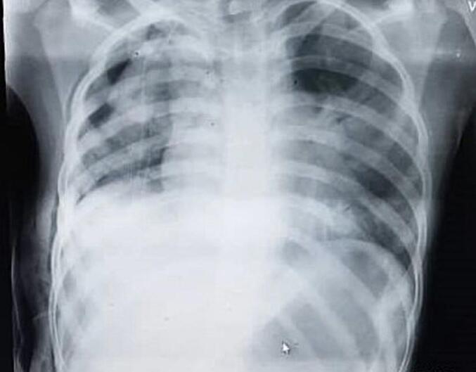
A chest X-ray showed near-complete hypoventilation of the right lung, small air pockets consistent with pulmonary contusion, and a right-sided pleural effusion [Fig. 1]. A contrast-enhanced chest CT scan revealed a mediastinal shift to the right, absence of aeration in the right lung, associated pleural effusion, consolidation consistent with atelectasis, and a mass-like lesion occluding the right main bronchus [Fig. 2]. Preoperative flexible bronchoscopy demonstrated complete obstruction of the right main bronchus by granulomatous tissue, located approximately 0.5 cm distal to the carinal spur. The lesion corresponded to the site of bronchial transection and was the cause of the patient's progressive dyspnea. A transbronchial biopsy revealed nonspecific inflammatory changes, with no evidence of malignancy.
Fig. 1.

Frontal chest X-ray showing near-complete opacification of the right hemithorax due to hypoventilation and collapse. Small air lucencies are visible within the opacity, suggestive of pulmonary contusion. A moderate right-sided pleural effusion and mild mediastinal shift to the right are also noted.
Fig. 2.

Axial contrast-enhanced chest CT scan showing complete absence of aeration in the right lung, right-sided pleural effusion, and mediastinal shift toward the right side. A soft tissue density is noted in the right main bronchus, suggestive of an obstructing lesion, consistent with granulation tissue formation following traumatic bronchial transection.
Surgical repair was undertaken approximately one week after admission, via a right posterolateral thoracotomy through the 5th intercostal space. Extensive pleural adhesions were encountered and carefully dissected. The transection was identified approximately 0.5 cm distal to the carinal spur at the origin of the right main bronchus. No bronchial resection was required; only debridement of obstructing granulation tissue at the proximal stump was performed. End-to-end anastomosis was then achieved using 3-0 Vicryl sutures placed in an interrupted fashion, with all knots tied externally on the bronchial wall [Fig. 3]. A 20 French chest tube was inserted for drainage. Intraoperative findings confirmed a complete traumatic transection of the right main bronchus with granulomatous obstruction. Following completion of the anastomosis, the right lung fully re-expanded intraoperatively, demonstrating restoration of airway continuity [Fig. 4].
Fig. 3.

Intraoperative view showing complete transection of the right main bronchus.
Fig. 4.

Intraoperative view following completion of end-to-end anastomosis of the right main bronchus. The right lung is seen fully re-expanded, indicating restoration of airway continuity and adequate ventilation.
Postoperative imaging showed significant improvement in right lung expansion and resolution of the pleural effusion [Fig. 5]. The patient's respiratory status improved markedly, with normalized oxygen saturation and complete resolution of dyspnea. Given the excellent radiological re-expansion of the lung and absence of any respiratory symptoms, postoperative bronchoscopy was deemed unnecessary. Follow-up imaging performed approximately two months after surgery confirmed sustained aeration of the right lung and no evidence of recurrence or new complications [Fig. 6].
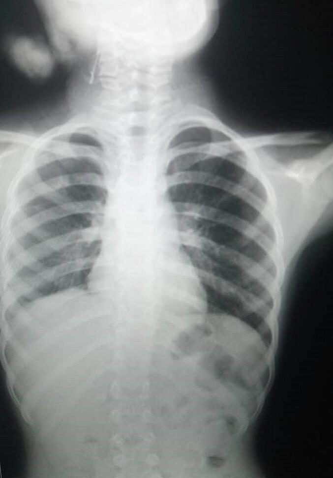
Fig. 5.
Postoperative chest X-ray demonstrating significant re-expansion of the right lung with improved aeration and resolution of the previously noted pleural effusion. The mediastinum is centrally positioned, and lung fields appear well inflated bilaterally.
Fig. 6.
Follow-up chest X-ray obtained two months postoperatively showing sustained re-expansion and aeration of the right lung. No evidence of pleural effusion, atelectasis, or other complications is observed. The mediastinum remains centrally aligned, and lung fields appear clear bilaterally.
The sequence of events is summarized in [Table 1].
Table 1.
Timeline of clinical events.
| Date/Period | Event | Details |
|---|---|---|
| Day 0 | Motor vehicle accident | Patient sustained blunt chest trauma, right-sided pneumothorax, and pulmonary contusion; managed conservatively with chest tube drainage at local facility. |
| Week 1 | Discharge | Patient discharged after initial improvement and removal of chest tube. |
| Week 6 | Symptom progression | Progressive dyspnea and exertional intolerance developed, later worsening to dyspnea at rest and orthopnea. |
| Week 8 | Referral & evaluation | Admitted to specialized center; chest X-ray showed right lung collapse, CT revealed obstructing lesion; bronchoscopy confirmed complete right main bronchus obstruction 0.5 cm from carina. |
| Week 9 | Surgery | Right posterolateral thoracotomy; debridement of granulation tissue and end-to-end bronchial anastomosis performed. |
| Immediate postoperative | Recovery | Full right lung re-expansion noted intraoperatively; patient's respiratory function improved markedly. |
| 2 months postoperative | Follow-up | Chest X-ray confirmed sustained aeration of the right lung with no recurrence or complications; patient asymptomatic. |
3. Patient perspective
“We were very worried when our daughter's breathing kept getting worse weeks after the accident. At first, we thought the problem was over after the chest tube was removed. When doctors at the specialized center explained that her main bronchus was completely blocked and needed surgery, it was frightening, but we were relieved to finally have answers. After the operation, her breathing improved dramatically, and we are grateful that she has returned to normal life.”
4. Discussion
Despite the substantial rise in trauma cases in recent years, tracheobronchial injuries remain rare and are often challenging to diagnose in the early stages [9]. They often occur in the context of severe polytrauma but may remain undiagnosed and therefore untreated, as the clinical signs and symptoms frequently fail to reflect the true severity of the injury [10]. Tracheobronchial injuries most commonly occur within 2.5 cm of the carina, accounting for approximately 40–80 % of reported cases [11]. According to Symbas et al., the right main bronchus is affected more frequently than the left, with reported rates of 26 % and 17.5 %, respectively [12]. This has been attributed to the anatomical position of the aorta, which offers a degree of protection to the left main bronchus [13]. The distal trachea and right main bronchus lie over the vertebral bodies, which act as an anvil during impact, thereby increasing the likelihood of rupture in these airway segments [14]. This anatomical and epidemiological pattern is reflected in our case, where the patient sustained a complete transection of the right main bronchus within close proximity to the carina following blunt chest trauma.
There are three main theories explaining the pathophysiology of blunt tracheobronchial injuries. One suggests that sudden anteroposterior chest compression causes transverse expansion, pulling the lungs away from the carina. Another proposes that a rapid rise in intrathoracic pressure against a closed glottis leads to rupture of the membranous portion of the airway [15]. The third hypothesis attributes tracheobronchial injury to rapid deceleration—commonly seen in motor vehicle collisions—which creates shearing forces between the fixed carina and the more mobile lung tissue [16].
The clinical presentation of tracheobronchial injuries is often subtle and non-specific, necessitating a high index of suspicion based on trauma mechanisms [10,17]. Common findings include dyspnea, tachypnea, and respiratory distress (59–100 % of cases) [17]. Subcutaneous emphysema is the most specific sign of tracheal disruption, while persistent air leak is pathognomonic of near-total or total tracheal avulsion, reported in about 60 % of patients [12]. In such cases, the ipsilateral lung may collapse peripherally rather than centrally, producing the “falling lung sign of Kumpe,” a hallmark radiologic finding of bronchial avulsion [18]. Nearly 50 % of injuries are missed in the first 24–48 h due to nonspecific presentation and potential airway obstruction [19]. Delayed diagnosis can lead to granulation tissue formation within 1–4 weeks, resulting in complications such as pneumonia, bronchiectasis, atelectasis, or abscess [7]. This was evident in our patient, who initially presented with nonspecific symptoms—progressive dyspnea and exertional intolerance—despite apparent early recovery. The diagnosis was missed at first because she was managed in a rural primary care setting with limited resources. Initial chest radiography revealed a pneumothorax, which was treated with tube thoracostomy. The drain was later removed once the pneumothorax regressed, creating a false impression of complete resolution. In reality, progressive intraluminal granulation tissue gradually occluded the right main bronchus, and the underlying transection remained undetected until respiratory deterioration occurred six weeks later. This scenario underscores the diagnostic challenges in low-resource and post-conflict healthcare systems, where advanced imaging and early bronchoscopy are often unavailable. At initial presentation, several differential diagnoses could have been considered, including mucus plugging, post-contusional collapse, and airway edema. Each of these can produce radiological atelectasis or consolidation, potentially mimicking the effects of bronchial transection and further contributing to diagnostic delay.
Prompt diagnosis and initial management of airway injuries are crucial for patient stabilization [1]. On initial chest radiography, subcutaneous emphysema may be evident, while CT scans can reveal signs such as mediastinal air, airway discontinuity, or tracheal deviation. However, CT is not ideal for hemodynamically unstable or severely dyspneic patients, and a negative result does not exclude injury or eliminate the need for further testing [20]. Flexible fiberoptic bronchoscopy remains the gold standard for diagnosing tracheobronchial injuries, as it accurately identifies the location, type, and severity of damage [7]. While some authors suggest bronchoscopy may not always be necessary when clinical and radiological signs are clear [21], it is essential that such procedures be performed by experienced bronchoscopes to avoid missed injuries [22].
Optimal management of tracheobronchial injuries necessitates a multidisciplinary approach involving trauma surgeons, intensivists, anesthesiologists, and cardiothoracic surgeons within specialized centers equipped for complex airway care [23]. The overarching goal is to establish a secure and patent airway, which remains the central challenge in these cases [24]. Management strategies may be either operative or non-operative, depending on the nature and extent of the injury [7]. However, due to the intricate anatomy of the airway and the urgency in stabilizing patients with severe associated trauma, surgical intervention remains the cornerstone of treatment [11]. In the acute setting, prompt surgical repair is considered standard practice [16]. Postoperative complications are primarily related to anastomotic integrity, with 5–6 % of patients experiencing issues such as dehiscence or restenosis following tracheal reconstruction [25].
5. Conclusion
Tracheobronchial injuries in pediatric patients may present with delayed and subtle symptoms, complicating timely diagnosis. This case emphasizes the importance of maintaining clinical suspicion even after apparent recovery from blunt chest trauma. Early bronchoscopy is critical for diagnosis, and definitive surgical repair remains the cornerstone of management in complete transections. Timely intervention can prevent complications such as irreversible airway obstruction and ensure full functional recovery of the affected lung.
Author contribution
First Author (Mohammad Alaa Aldakak): Conceived the report, planned and contributed to manuscript writing, drafted the initial manuscript, and led the revision process.
Second Author (Nawwar Fallouh): Contributed to manuscript writing, assisted in data collection and figure preparation, and approved the final version.
Third Author (Bassel Ibrahim): Participated in drafting and editing the manuscript, contributed to the interpretation of imaging and intraoperative findings, and approved the final version.
Fourth Author (Raneem Ahmad): Reviewed all manuscript versions critically for important intellectual content.
Fifth Author (Youssef Abbas): Participated in writing the report, prepared the figures, and approved the final version.
The supervisor (Kamal Al kateb): Ensured the accuracy of clinical data, and approved the final version.
Parental consent for minors
Written informed consent was obtained from the patient's parents/legal guardian for publication and any accompanying images. A copy of the written consent is available for review by the Editor-in-Chief of this journal on request.
Ethical approval
Ethical approval for this study (Ethical Committee MD-0910011235-123) was provided by the Biomedical research Ethics Committee BMREC of Damascus University on 1 June 2025.
Guarantor
The First Author.
Research registration number
Not applicable.
Declaration of Generative AI and AI-assisted technologies in the writing process
Artificial intelligence tools (ChatGPT, OpenAI) were used to assist in language editing and improving the clarity of the manuscript. All AI-generated content was reviewed and verified by the authors to ensure accuracy and integrity, and no AI tools were used to generate or fabricate clinical data.
Funding
This research did not receive any specific grant from funding agencies in the public, commercial, or not-for-profit sectors.
Conflict of interest statement
The authors declared no potential conflicts of interest concerning the research, authorship, and/or publication of this article.
Contributor Information
Mohammad Alaa Aldakak, Email: alaa.aldakak@damascusuniversity.edu.sy.
Nawwar Fallouh, Email: nawwarfallouh@gmail.com.
Bassel Ibrahim, Email: drbasselmibrahim@gmail.com.
Raneem Ahmad, Email: raneemahmad095@gmail.com.
Youssef Abbas, Email: youssefabbas370@gmail.com.
Kamal Al Kateb, Email: alkatebk@gmail.com.
References
- 1.Altinok T., Can A. Management of tracheobronchial injuries. Eurasian J. Med. 2014;46(3):209–215. doi: 10.5152/eajm.2014.42. [DOI] [PMC free article] [PubMed] [Google Scholar]
- 2.Dominguez E., De La Torre C., Sánchez A.V., Hernandez F., Ortiz R., Moreno A.M., et al. Severe tracheobronchial injuries: our experience. Eur. J. Pediatr. Surg. 2015;25(1):71–76. doi: 10.1055/s-0034-1386642. [DOI] [PubMed] [Google Scholar]
- 3.Prokakis C., Koletsis E.N., Dedeilias P., Fligou F., Filos K., Dougenis D. Airway trauma: a review on epidemiology, mechanisms of injury, diagnosis and treatment. J. Cardiothorac. Surg. 2014;9:117. doi: 10.1186/1749-8090-9-117. [DOI] [PMC free article] [PubMed] [Google Scholar]
- 4.Iwasaki M., Kaga K., Ogawa J., Inoue H., Shohtsu A. Bronchoscopy findings and early treatment of patients with blunt tracheo-bronchial trauma. J. Cardiovasc. Surg. 1994;35(3):269–271. [PubMed] [Google Scholar]
- 5.Chesterman J.T., Satsangi P.N. Rupture of the trachea and bronchi by closed injury. Thorax. 1966;21(1):21–27. doi: 10.1136/thx.21.1.21. [DOI] [PMC free article] [PubMed] [Google Scholar]
- 6.Carratola M., Hart C.K. Pediatric tracheal trauma. Semin. Pediatr. Surg. 2021;30(3) doi: 10.1016/j.sempedsurg.2021.151057. [DOI] [PubMed] [Google Scholar]
- 7.Aljehani Y., Aldossary I., AlQatari A.A., Alreshaid F., Alsadery H.A. Blunt traumatic tracheobronchial injury: a clinical pathway. Mediev. Archaeol. 2022;76(6):430–437. doi: 10.5455/medarh.2022.76.430-437. [DOI] [PMC free article] [PubMed] [Google Scholar]
- 8.Kerwan A., Al-Jabir A., Mathew G., Sohrabi C., Rashid R., Franchi T., Nicola M., Agha M., Agha R.A. Revised Surgical CAse REport (SCARE) guideline: an update for the age of Artificial Intelligence. Premier J. Sci. 2025;10 [Google Scholar]
- 9.Saad R., Jr., Gonçalves R., Dorgan V., Neto J.A.G.P., Rivaben J.H., Botter M., et al. Tracheobronchial injuries in chest trauma: a 17-year experience. Rev. Col. Bras. Cir. 2017;44(2):194–201. doi: 10.1590/0100-69912017002014. [DOI] [PubMed] [Google Scholar]
- 10.Moonsamy P., Sachdeva U.M., Morse C.R. Management of laryngotracheal trauma. Ann. Cardiothorac. Surg. 2018;7(2):210–216. doi: 10.21037/acs.2018.03.03. [DOI] [PMC free article] [PubMed] [Google Scholar]
- 11.Chu C.P., Chen P.P. Tracheobronchial injury secondary to blunt chest trauma: diagnosis and management. Anaesth. Intensive Care. 2002;30(2):145–152. doi: 10.1177/0310057X0203000204. [DOI] [PubMed] [Google Scholar]
- 12.Symbas P.N., Justicz A.G., Ricketts R.R. Rupture of the airways from blunt trauma: treatment of complex injuries. Ann. Thorac. Surg. 1992;54(1):177–183. doi: 10.1016/0003-4975(92)91177-b. [DOI] [PubMed] [Google Scholar]
- 13.Corsten G., Berkowitz R.G. Membranous tracheal rupture in children following minor blunt cervical trauma. Ann. Otol. Rhinol. Laryngol. 2002;111(3 Pt 1):197–199. doi: 10.1177/000348940211100301. [DOI] [PubMed] [Google Scholar]
- 14.Pratt L.W., Smith R.J., Guite L.A., Jr., Tryzelaar J.F. Blunt chest trauma with tracheobronchial rupture. Ann. Otol. Rhinol. Laryngol. 1984;93(4 Pt 1):357–363. doi: 10.1177/000348948409300415. [DOI] [PubMed] [Google Scholar]
- 15.Alassal M.A., Ibrahim B.M., Elsadeck N. Traumatic intrathoracic tracheobronchial injuries: a study of 78 cases. Asian Cardiovasc. Thorac. Ann. 2014;22(7):816–823. doi: 10.1177/0218492313516777. [DOI] [PubMed] [Google Scholar]
- 16.Kiser A.C., O’Brien S.M., Detterbeck F.C. Blunt tracheobronchial injuries: treatment and outcomes. Ann. Thorac. Surg. 2001;71(6):2059–2065. doi: 10.1016/s0003-4975(00)02453-x. [DOI] [PubMed] [Google Scholar]
- 17.Heldenberg E., Vishne T.H., Pley M., Simansky D., Refaeli Y., Binun A., Saute M., Yellin A. Major bronchial trauma in the pediatric age group. World J. Surg. 2005;29(2):149–154. doi: 10.1007/s00268-004-7381-9. [DOI] [PubMed] [Google Scholar]
- 18.Unger J.M., Schuchmann G.G., Grossman J.E., Pellett J.R. Tears of the trachea and main bronchi caused by blunt trauma: radiologic findings. AJR Am. J. Roentgenol. 1989;153(6):1175–1180. doi: 10.2214/ajr.153.6.1175. [DOI] [PubMed] [Google Scholar]
- 19.Rezende-Neto J.B., Hoffmann J., Al Mahroos M., Tien H., Hsee L.C., Spencer Netto F., Speers V., Rizoli S.B. Occult pneumomediastinum in blunt chest trauma: clinical significance. Injury. 2010;41(1):40–43. doi: 10.1016/j.injury.2009.06.161. [DOI] [PubMed] [Google Scholar]
- 20.Jones C.M., Athanasiou T. Is virtual bronchoscopy an efficient diagnostic tool for the thoracic surgeon? Ann. Thorac. Surg. 2005;79(1):365–374. doi: 10.1016/j.athoracsur.2004.03.013. [DOI] [PubMed] [Google Scholar]
- 21.Poli-Merol M.L., Belouadah M., Parvy F., Chauvet P., Egreteau L., Daoud S. Tracheobronchial injury by blunt trauma in children: is emergency tracheobronchoscopy always necessary? Eur. J. Pediatr. Surg. 2003;13(6):398–402. doi: 10.1055/s-2003-44730. [DOI] [PubMed] [Google Scholar]
- 22.Baumgartner F., Sheppard B., de Virgilio C., Esrig B., Harrier D., Nelson R.J., Robertson J.M. Tracheal and main bronchial disruptions after blunt chest trauma: presentation and management. Ann. Thorac. Surg. 1990;50(4):569–574. doi: 10.1016/0003-4975(90)90191-8. [DOI] [PubMed] [Google Scholar]
- 23.Aprile V., Korasidis S., Ambrogi M.C., Lucchi M. Extracorporeal membrane oxygenation in traumatic tracheal injuries: a bold life-saving option. J. Thorac. Dis. 2019;11(7):2660–2663. doi: 10.21037/jtd.2019.05.61. [DOI] [PMC free article] [PubMed] [Google Scholar]
- 24.Kuhne C.A., Kaiser G.M., Flohe S., Beiderlinden M., Kuehl H., Stavrou G.A., Waydhas C., Lendemanns S., Paffrath T., Nast-Kolb D. Nonoperative management of tracheobronchial injuries in severely injured patients. Surg. Today. 2005;35(7):518–523. doi: 10.1007/s00595-005-3001-z. [DOI] [PubMed] [Google Scholar]
- 25.Grillo H.C., Zannini P., Michelassi F. Complications of tracheal reconstruction. Incidence, treatment, and prevention. J. Thorac. Cardiovasc. Surg. 1986;91(3):322–328. [PubMed] [Google Scholar]