

Abstract
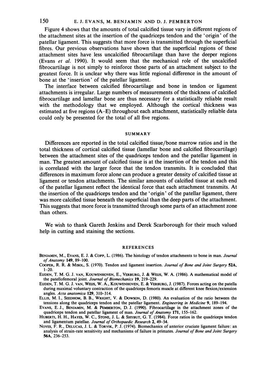
Differences are reported in the total calcified tissue/bone marrow ratios and in the total thickness of cortical calcified tissue (lamellar bone and calcified fibrocartilage) between the attachment sites of the quadriceps tendon and the patellar ligament in man. The greatest amount of calcified tissue is at the insertion of the tendon and this is correlated with the larger force that the tendon transmits. It is concluded that differences in maximum force alone can produce a greater density of calcified tissue at ligament or tendon attachments. The similar amounts of calcified tissue at each end of the patellar ligament reflect the identical force that each attachment transmits. At the insertion of the quadriceps tendon and the 'origin' of the patellar ligament, there was more calcified tissue beneath the superficial than the deep parts of the attachment. This suggest that more force is transmitted through some parts of an attachment zone than others.
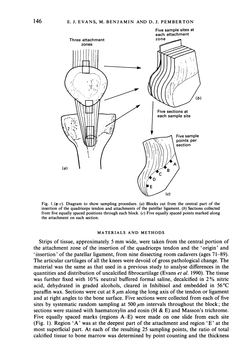
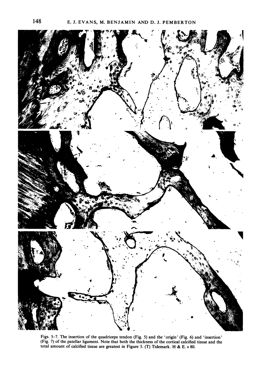
Full text
PDF


Images in this article
Selected References
These references are in PubMed. This may not be the complete list of references from this article.
- Benjamin M., Evans E. J., Copp L. The histology of tendon attachments to bone in man. J Anat. 1986 Dec;149:89–100. [PMC free article] [PubMed] [Google Scholar]
- Cooper R. R., Misol S. Tendon and ligament insertion. A light and electron microscopic study. J Bone Joint Surg Am. 1970 Jan;52(1):1–20. [PubMed] [Google Scholar]
- Evans E. J., Benjamin M., Pemberton D. J. Fibrocartilage in the attachment zones of the quadriceps tendon and patellar ligament of man. J Anat. 1990 Aug;171:155–162. [PMC free article] [PubMed] [Google Scholar]
- Huberti H. H., Hayes W. C., Stone J. L., Shybut G. T. Force ratios in the quadriceps tendon and ligamentum patellae. J Orthop Res. 1984;2(1):49–54. doi: 10.1002/jor.1100020108. [DOI] [PubMed] [Google Scholar]
- Noyes F. R., DeLucas J. L., Torvik P. J. Biomechanics of anterior cruciate ligament failure: an analysis of strain-rate sensitivity and mechanisms of failure in primates. J Bone Joint Surg Am. 1974 Mar;56(2):236–253. [PubMed] [Google Scholar]
- Noyes F. R., Grood E. S. The strength of the anterior cruciate ligament in humans and Rhesus monkeys. J Bone Joint Surg Am. 1976 Dec;58(8):1074–1082. [PubMed] [Google Scholar]
- Pedley R. B., Meachim G. Topographical variation in patellar subarticular calcified tissue density. J Anat. 1979 Jun;128(Pt 4):737–745. [PMC free article] [PubMed] [Google Scholar]
- van Eijden T. M., Kouwenhoven E., Verburg J., Weijs W. A. A mathematical model of the patellofemoral joint. J Biomech. 1986;19(3):219–229. doi: 10.1016/0021-9290(86)90154-5. [DOI] [PubMed] [Google Scholar]
- van Eijden T. M., Weijs W. A., Kouwenhoven E., Verburg J. Forces acting on the patella during maximal voluntary contraction of the quadriceps femoris muscle at different knee flexion/extension angles. Acta Anat (Basel) 1987;129(4):310–314. doi: 10.1159/000146421. [DOI] [PubMed] [Google Scholar]