Abstract
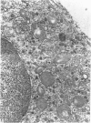
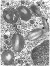
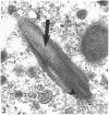
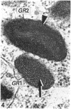
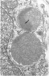
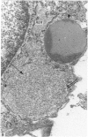
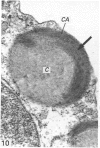
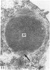
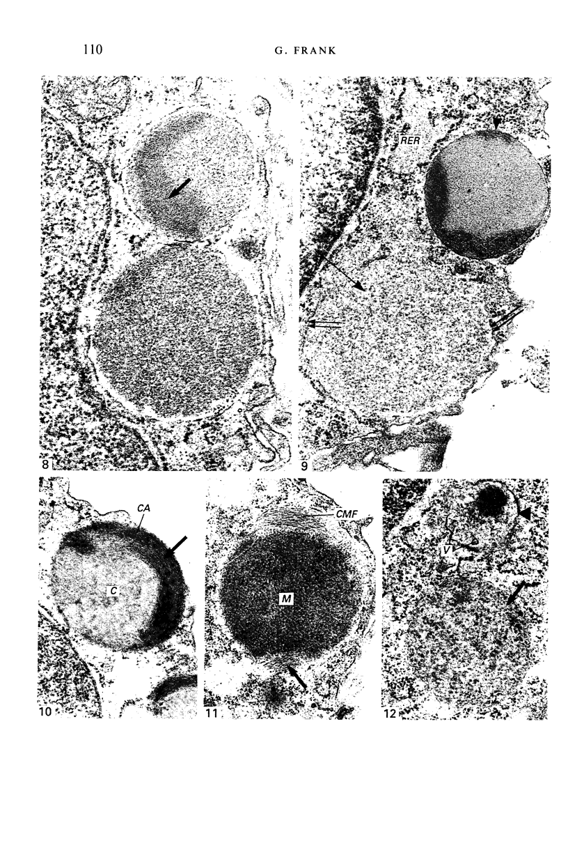
The morphology and development of heterophil and basophil granules from the trunk kidneys of Rana esculenta tadpoles were studied at the electron microscopic level. Cells of the heterophils series contain granules displaying either spheroid profiles with homogeneous content (Type A), or elongate profiles with a crystalloid interior (Type B). Type A granules apparently originate directly from Golgi-derived vesicles, which, gaining slightly in size and density, transform into the mature granules. Type B granules could be traced back to vacuolate structures showing an irregular content. Their development could be traced through increasingly elongated forms with the appearance of a characteristic crystalloid core. Fully developed basophil granules are considerably larger in size than heterophil granules and contain heterogeneous interna showing a central-cortical organisation: a finely stippled medullary zone of varying density is surrounded by a sickle-shaped and lamellate cortex (Type L), or a moderately dense and uniformly stippled medulla is enclosed by two diametrically opposed, cap-shaped, filamentous regions (Type F). The heterophil and basophil granules are compared to those in other vertebrates and possible phylogenetic aspects are discussed.
Full text
PDF
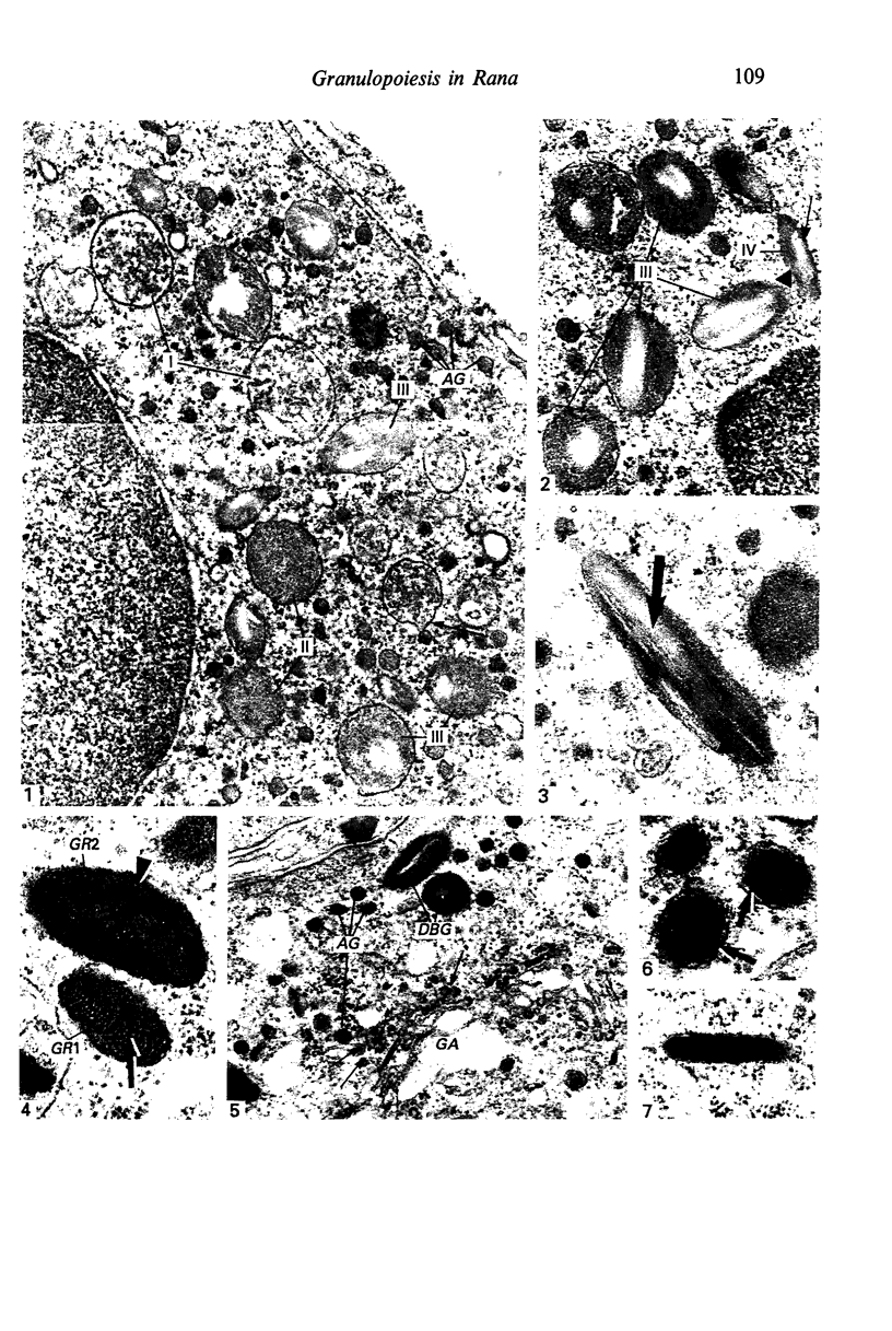
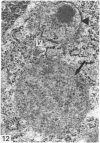
Images in this article
Selected References
These references are in PubMed. This may not be the complete list of references from this article.
- Bainton D. F. Differentiation of human neutrophilic granulocytes: normal and abnormal. Prog Clin Biol Res. 1977;13:1–27. [PubMed] [Google Scholar]
- Bainton D. F., Farquhar M. G. Segregation and packaging of granule enzymes in eosinophilic leukocytes. J Cell Biol. 1970 Apr;45(1):54–73. doi: 10.1083/jcb.45.1.54. [DOI] [PMC free article] [PubMed] [Google Scholar]
- Bainton D. F., Ullyot J. L., Farquhar M. G. The development of neutrophilic polymorphonuclear leukocytes in human bone marrow. J Exp Med. 1971 Oct 1;134(4):907–934. doi: 10.1084/jem.134.4.907. [DOI] [PMC free article] [PubMed] [Google Scholar]
- Bentfeld M. E., Nichols B. A., Bainton D. F. Ultrastructural localization of peroxidase in leukocytes of rat bone marrow and blood. Anat Rec. 1977 Feb;187(2):219–240. doi: 10.1002/ar.1091870208. [DOI] [PubMed] [Google Scholar]
- Bielek E. Developmental stages and localization of peroxidatic activity in the leucocytes of three teleost species (Cyprinus carpio L.; Tinca tinca L.; Salmo gairdneri Richardson). Cell Tissue Res. 1981;220(1):163–180. doi: 10.1007/BF00209975. [DOI] [PubMed] [Google Scholar]
- Brederoo P., Daems W. T. The ultrastructure of guinea pig heterophil granulocytes and the heterogeneity of the granules. Cell Tissue Res. 1978 Nov 20;194(2):183–205. doi: 10.1007/BF00220388. [DOI] [PubMed] [Google Scholar]
- Breton-Gorius J., Reyes F. Ultrastructure of human bone marrow cell maturation. Int Rev Cytol. 1976;46:251–321. doi: 10.1016/s0074-7696(08)60993-6. [DOI] [PubMed] [Google Scholar]
- Campbell F. R. Electron microscopic studies on granulocytopoiesis in the slender salamander. Anat Rec. 1969 Mar;163(3):427–441. doi: 10.1002/ar.1091630306. [DOI] [PubMed] [Google Scholar]
- Campbell F. R. Ultrastructure of the bone marrow of the frog. Am J Anat. 1970 Nov;129(3):329–355. doi: 10.1002/aja.1001290306. [DOI] [PubMed] [Google Scholar]
- Campbell F. Fine structure of the bone marrow of the chicken and pigeon. J Morphol. 1967 Dec;123(4):405–439. doi: 10.1002/jmor.1051230407. [DOI] [PubMed] [Google Scholar]
- Daems W. T. On the fine structure of human neutrophilic leukocyte granules. J Ultrastruct Res. 1968 Aug;24(3):343–348. doi: 10.1016/s0022-5320(68)90070-1. [DOI] [PubMed] [Google Scholar]
- Desser S. S., Weller I. Ultrastructural observations on the granular leucocytes of the tuatara Sphenodon punctatus (Gray). Tissue Cell. 1979;11(4):703–715. doi: 10.1016/0040-8166(79)90025-9. [DOI] [PubMed] [Google Scholar]
- Frank G. Granulopoiesis in tadpoles of Rana esculenta. Survey of the organs involved. J Anat. 1988 Oct;160:59–66. [PMC free article] [PubMed] [Google Scholar]
- Frank G. Granulopoiesis in tadpoles of Rana esculenta. Ultrastructural observations on the developing granulocytes and on the development of eosinophil granules. J Anat. 1989 Apr;163:97–105. [PMC free article] [PubMed] [Google Scholar]
- Hightower J. A., Haar J. L. A light and electron microscopic study of myelopoietic cells in the perihepatic subcapsular region of the liver in the adult aquatic newt, Notophthalmus viridescens. Cell Tissue Res. 1975 May 27;159(1):63–71. doi: 10.1007/BF00231995. [DOI] [PubMed] [Google Scholar]
- Ishizeki K., Nawa T., Tachibana T., Sakakura Y., Iida S. Hemopoietic sites and development of eosinophil granulocytes in the loach, Misgurnus anguillicaudatus. Cell Tissue Res. 1984;235(2):419–426. doi: 10.1007/BF00217868. [DOI] [PubMed] [Google Scholar]
- Kelényi G., Németh A. Comparative histochemistry and electron microscopy of the eosinophil leucocytes of vertebrates. I. A study of avian, reptile, amphibian and fish leucocytes. Acta Biol Acad Sci Hung. 1969;20(4):405–422. [PubMed] [Google Scholar]
- Kelényi G. Phylogenesis of the azurophil leucocyte granules in vertebrates. Experientia. 1972 Sep 15;28(9):1094–1096. doi: 10.1007/BF01918695. [DOI] [PubMed] [Google Scholar]
- MITSUI T. LIGHT MICROSCOPE AND ELECTRON MICROSCOPE STUDY OF THE PEROXIDASE REACTION OF THE EOSINOPHIL LEUKOCYTES IN COLD-BLOODED ANIMALS. THE 30TH REPORT OF HISTOCHEMICAL STUDY OF PEROXIDASE. Okajimas Folia Anat Jpn. 1965 Jan;40:893–909. doi: 10.2535/ofaj1936.40.4-6_893. [DOI] [PubMed] [Google Scholar]
- Meseguer J., Lozano M. T., Agulleiro B. Ultrastructure of the renal granulopoietic tissue of the Rana ridibunda tadpole. J Submicrosc Cytol. 1985 Jul;17(3):391–401. [PubMed] [Google Scholar]
- Murata F., Spicer S. S. Morphologic and cytochemical studies of rabbit heterophilic leukocytes. Evidence for tertiary granules. Lab Invest. 1973 Jul;29(1):65–72. [PubMed] [Google Scholar]
- Scott R. E., Horn R. G. Fine structural features of eosinophile granulocyte development in human bone marrow. Evidence for granule secretion. J Ultrastruct Res. 1970 Oct;33(1):16–28. doi: 10.1016/s0022-5320(70)90116-4. [DOI] [PubMed] [Google Scholar]
- Setoguti T., Fujii H., Isono H. An electron microscopic study on neutrophil leukocytes of the toad, Bufo vulgaris japonicus. Arch Histol Jpn. 1970 Jun;32(1):87–94. doi: 10.1679/aohc1950.32.87. [DOI] [PubMed] [Google Scholar]
- Temmink J. H., Bayne C. J. Ultrastructural characterization of leucocytes in the pronephros of carp (Cyprinus carpio, L.). Dev Comp Immunol. 1987 Winter;11(1):125–137. doi: 10.1016/0145-305x(87)90014-0. [DOI] [PubMed] [Google Scholar]
- Terry R. W., Bainton D. F., Farquhar M. G. Formation and structure of specific granules in basophilic leukocytes of the guinea pig. Lab Invest. 1969 Jul;21(1):65–76. [PubMed] [Google Scholar]
- WEINREB E. L. STUDIES ON THE FINE STRUCTURE OF TELEOST BLOOD CELLS. I. PERIPHERALBLOOD. Anat Rec. 1963 Oct;147:219–238. doi: 10.1002/ar.1091470206. [DOI] [PubMed] [Google Scholar]
- WINQVIST G. The ultrastructure of the granules of the basophil granulocyte. Z Zellforsch Mikrosk Anat. 1960;52:475–481. doi: 10.1007/BF00339760. [DOI] [PubMed] [Google Scholar]
- Wetzel B. K., Horn R. G., Spicer S. S. Fine structural studies on the development of heterophil, eosinophil, and basophil granulocytes in rabbits. Lab Invest. 1967 Mar;16(3):349–382. [PubMed] [Google Scholar]


