Abstract
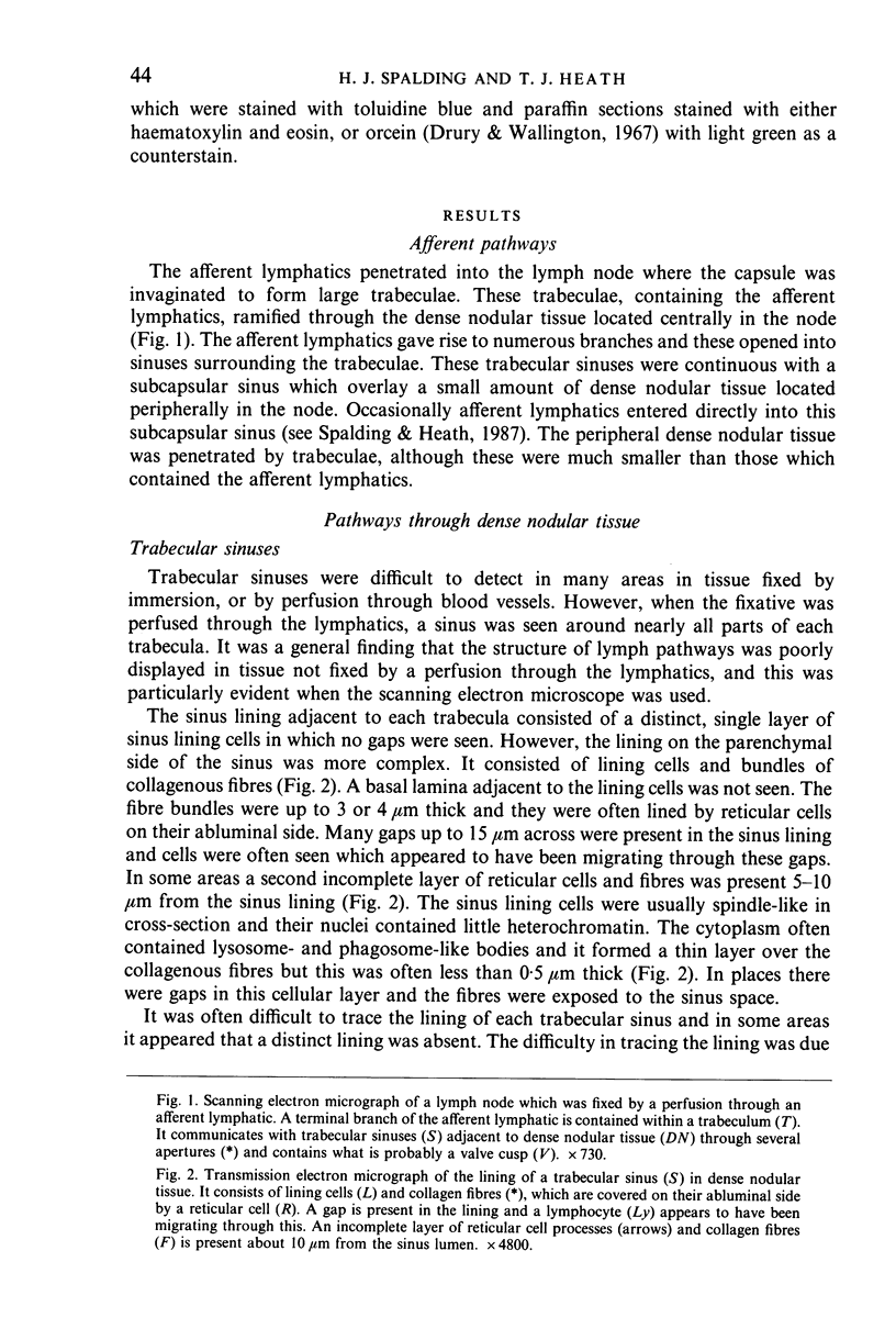
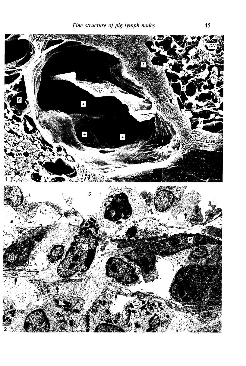
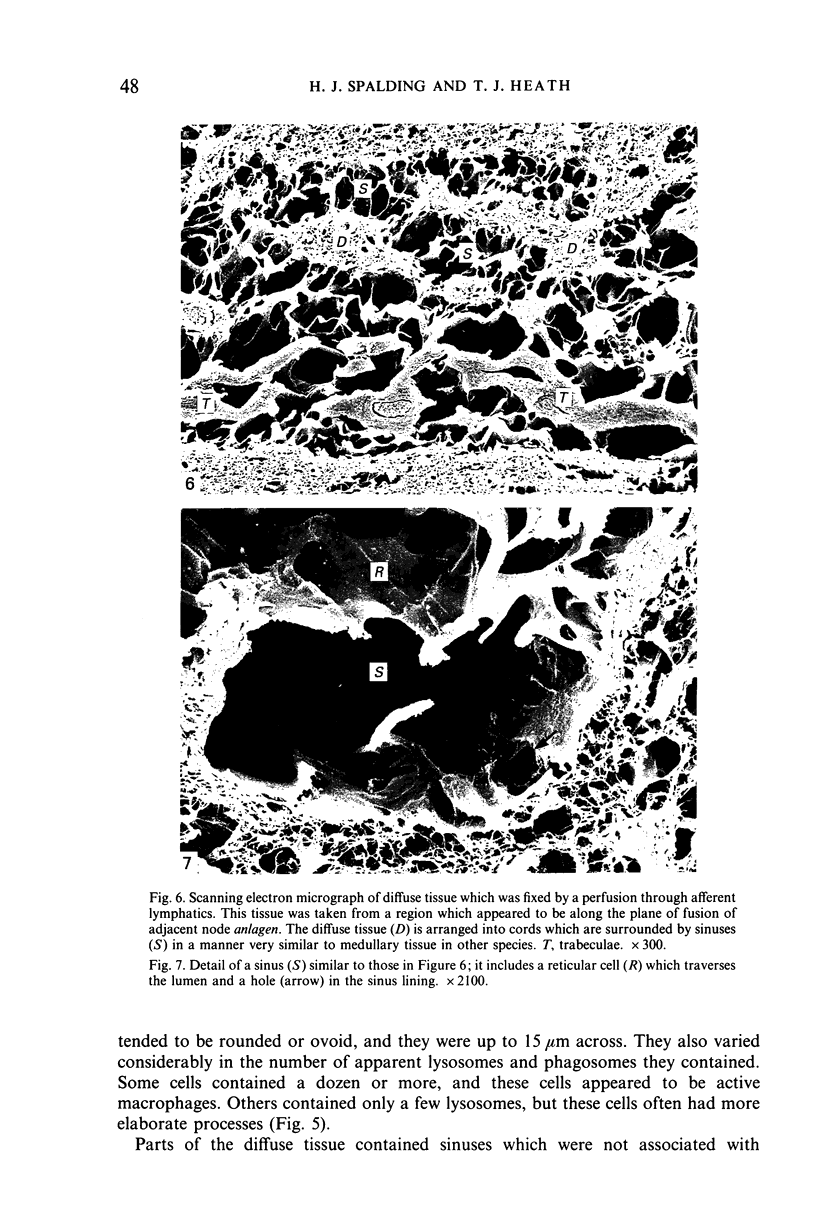
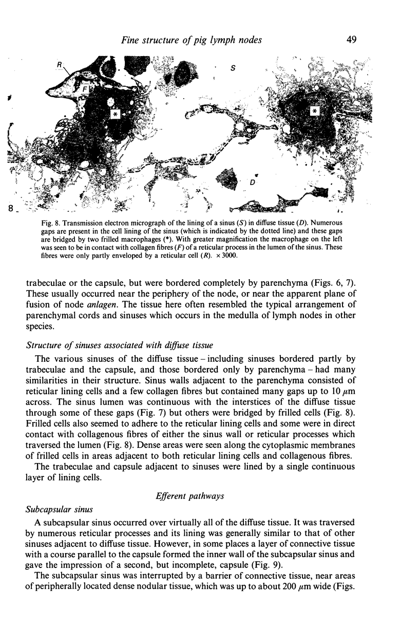
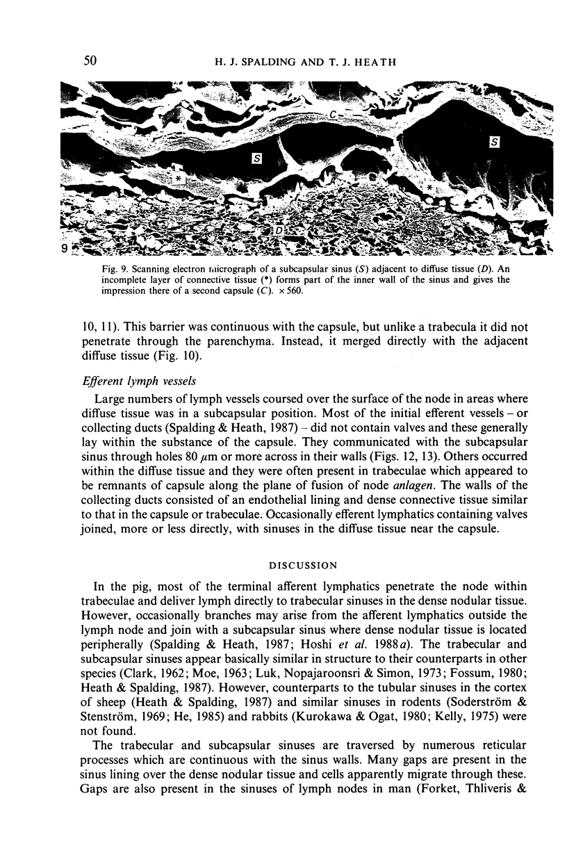
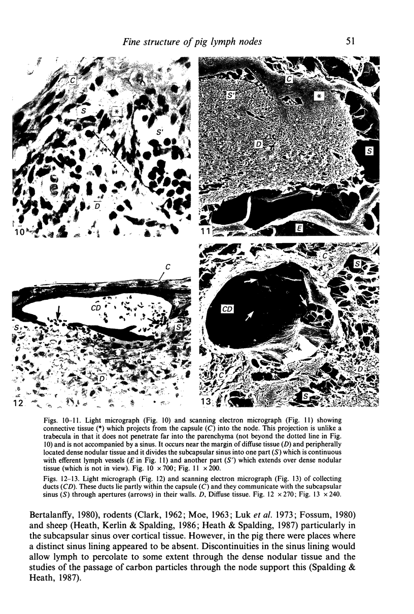
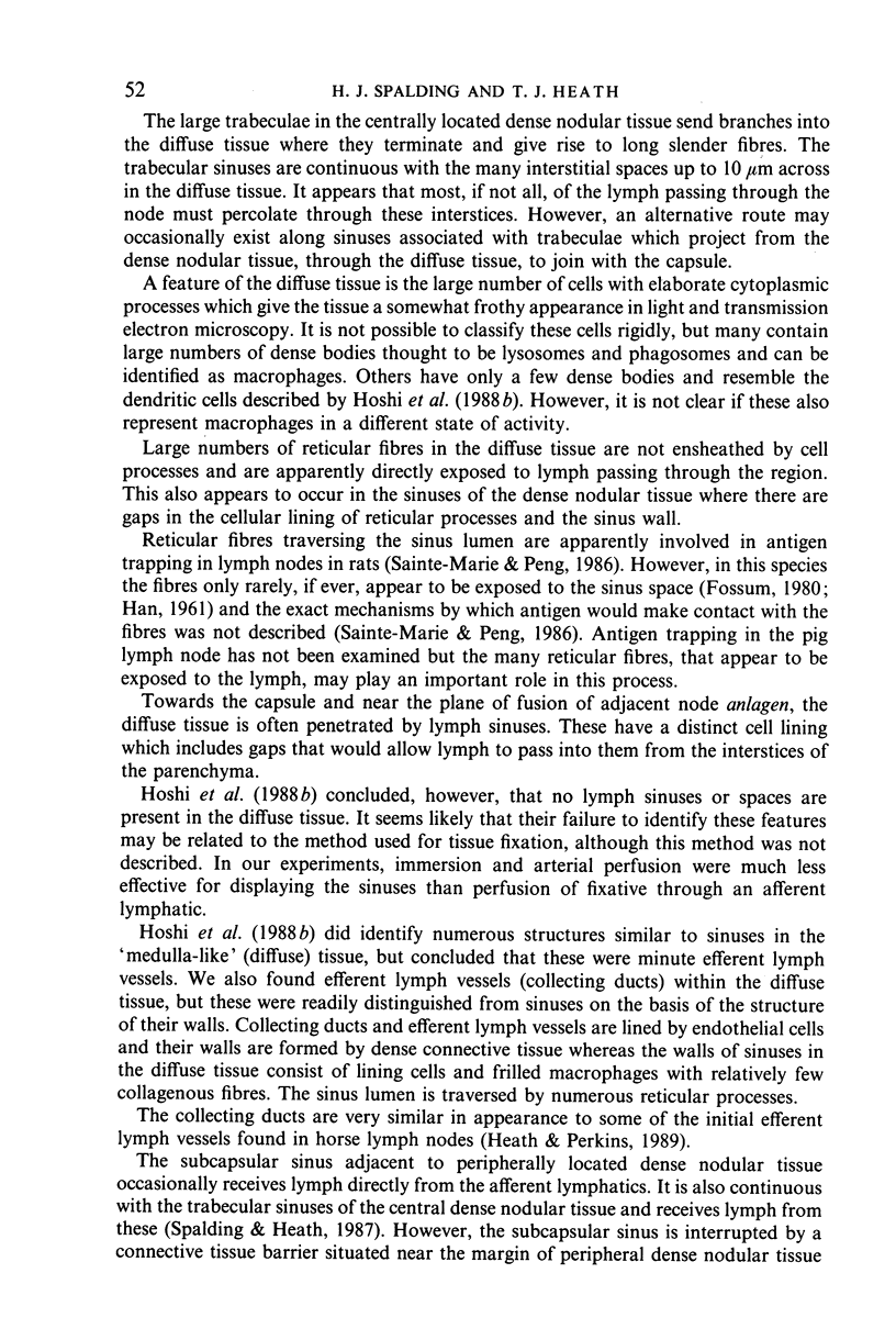
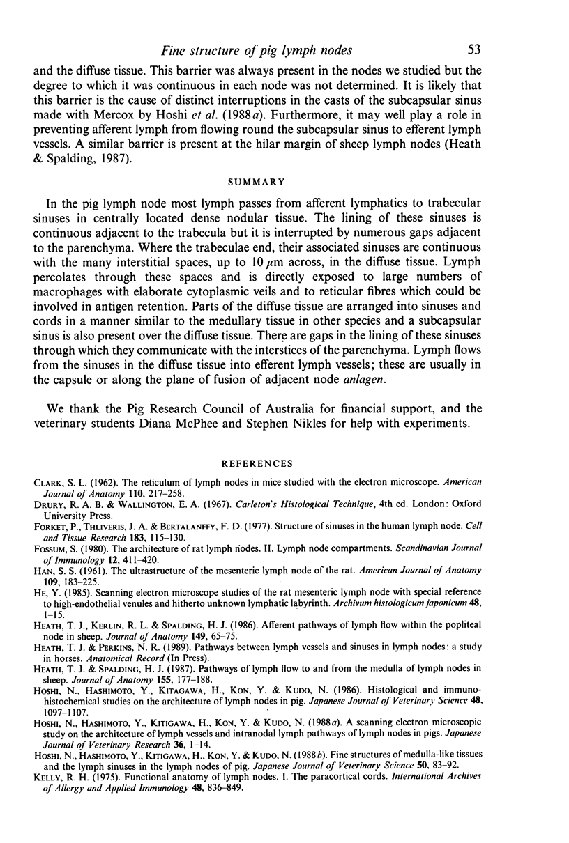
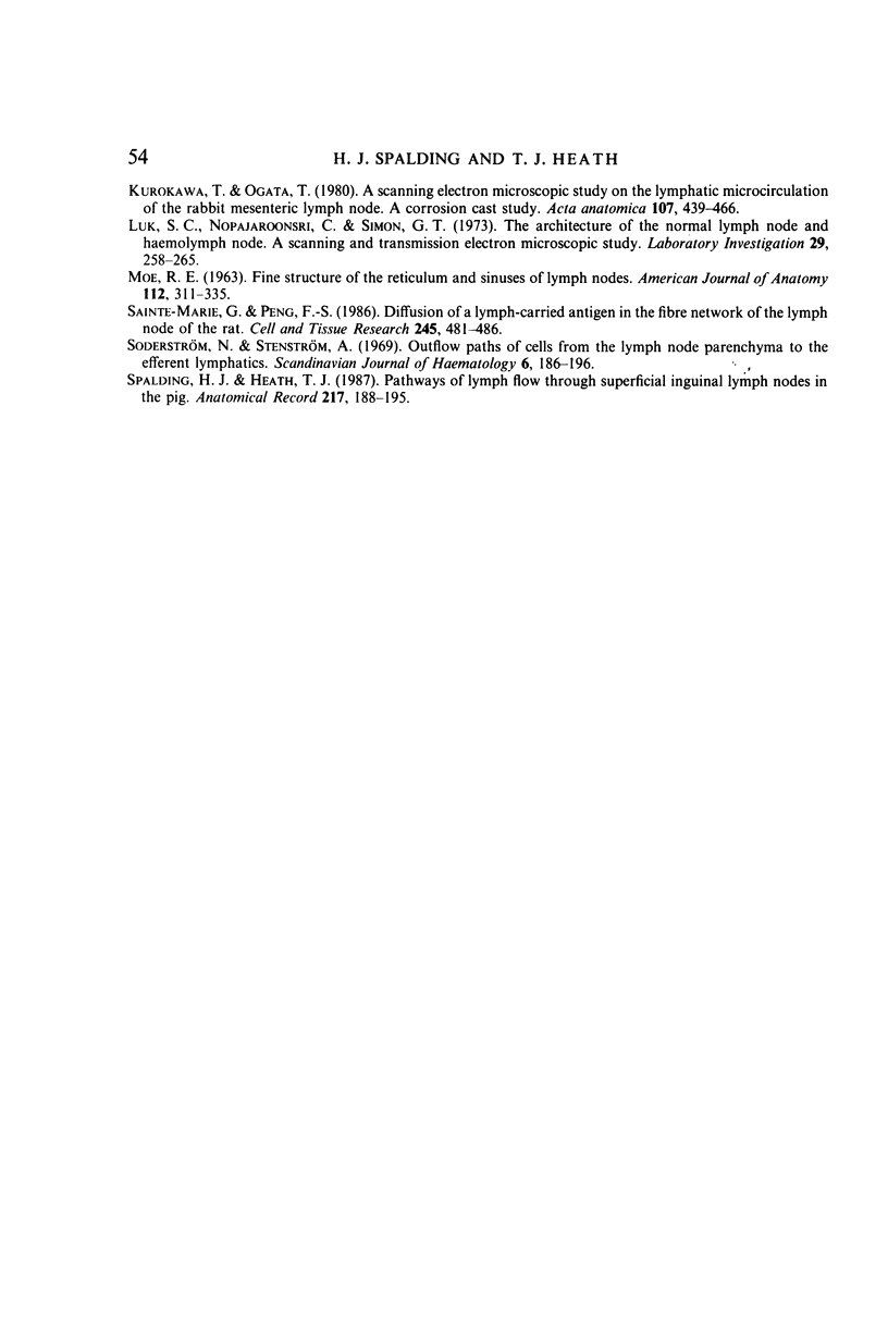
In the pig lymph node most lymph passes from afferent lymphatics to trabecular sinuses in centrally located dense nodular tissue. The lining of these sinuses is continuous adjacent to the trabecula but it is interrupted by numerous gaps adjacent to the parenchyma. Where the trabeculae end, their associated sinuses are continuous with the many interstitial spaces, up to 10 microns across, in the diffuse tissue. Lymph percolates through these spaces and is directly exposed to large numbers of macrophages with elaborate cytoplasmic veils and to reticular fibres which could be involved in antigen retention. Parts of the diffuse tissue are arranged into sinuses and cords in a manner similar to the medullary tissue in other species and a subcapsular sinus is also present over the diffuse tissue. There are gaps in the lining of these sinuses through which they communicate with the interstices of the parenchyma. Lymph flows from the sinuses in the diffuse tissue into efferent lymph vessels; these are usually in the capsule or along the plane of fusion of adjacent node anlagen.
Full text
PDF

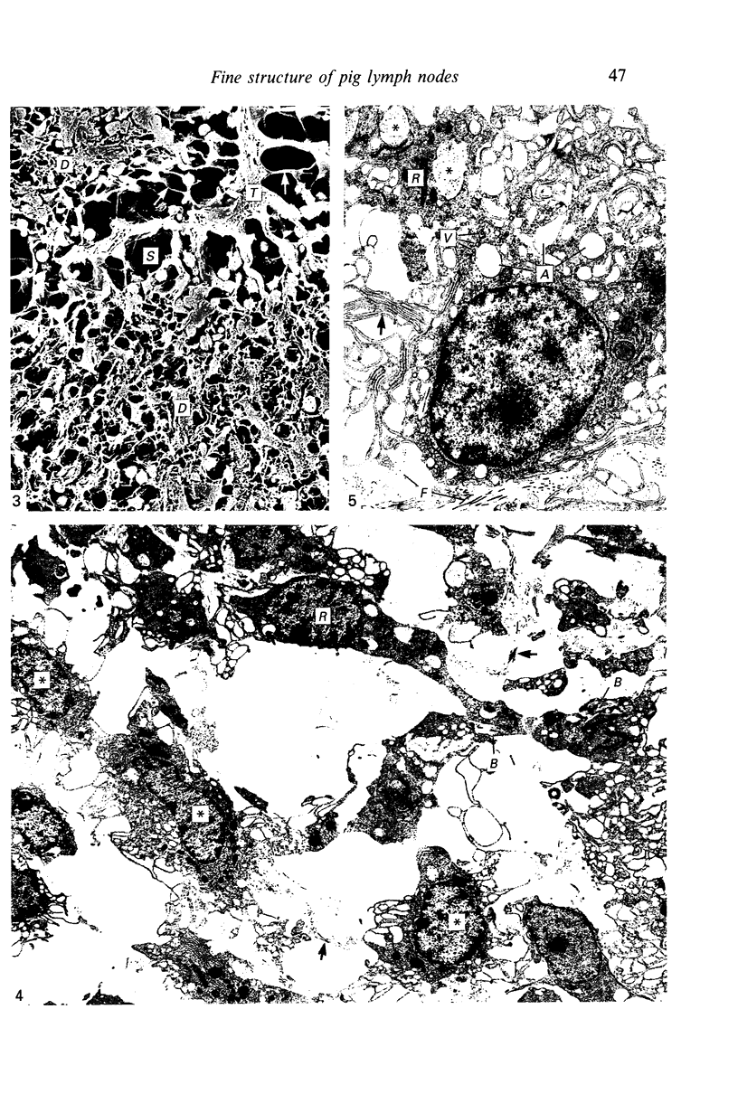
Images in this article
Selected References
These references are in PubMed. This may not be the complete list of references from this article.
- CLARK S. L., Jr The reticulum of lymph nodes in mice studied with the electron microscope. Am J Anat. 1962 May;110:217–257. doi: 10.1002/aja.1001100303. [DOI] [PubMed] [Google Scholar]
- Forkert P. G., Thliveris J. A., Bertalanffy F. D. Structure of sinuses in the human lymph node. Cell Tissue Res. 1977 Sep 14;183(1):115–130. doi: 10.1007/BF00219996. [DOI] [PubMed] [Google Scholar]
- HAN S. S. The ultrastructure of the mesenteric lymph node of the rat. Am J Anat. 1961 Sep;109:183–225. doi: 10.1002/aja.1001090208. [DOI] [PubMed] [Google Scholar]
- Heath T. J., Kerlin R. L., Spalding H. J. Afferent pathways of lymph flow within the popliteal node in sheep. J Anat. 1986 Dec;149:65–75. [PMC free article] [PubMed] [Google Scholar]
- Heath T. J., Spalding H. J. Pathways of lymph flow to and from the medulla of lymph nodes in sheep. J Anat. 1987 Dec;155:177–188. [PMC free article] [PubMed] [Google Scholar]
- Hoshi N., Hashimoto Y., Kitagawa H., Kon Y., Kudo N. A scanning electron microscopic study on the architecture of lymph vessels and intranodal lymph pathways of lymph nodes in pigs. Jpn J Vet Res. 1988 Jan;36(1):1–14. [PubMed] [Google Scholar]
- Hoshi N., Hashimoto Y., Kitagawa H., Kon Y., Kudo N. Fine structures of the medulla-like tissues and the lymph sinuses in the lymph nodes of pig. Nihon Juigaku Zasshi. 1988 Feb;50(1):83–92. doi: 10.1292/jvms1939.50.83. [DOI] [PubMed] [Google Scholar]
- Hoshi N., Hashimoto Y., Kitagawa H., Kon Y., Kudo N. Histological and immunohistochemical studies on the architecture of lymph nodes in pig. Nihon Juigaku Zasshi. 1986 Dec;48(6):1097–1107. doi: 10.1292/jvms1939.48.1097. [DOI] [PubMed] [Google Scholar]
- Kelly R. H. Functional anatomy of lymph nodes. I. The paracortical cords. Int Arch Allergy Appl Immunol. 1975;48(6):836–849. doi: 10.1159/000231371. [DOI] [PubMed] [Google Scholar]
- Kurokawa T., Ogata T. A scanning electron microscopic study on the lymphatic microcirculation of the rabbit mesenteric lymph node. A corrosion cast study. Acta Anat (Basel) 1980;107(4):439–466. doi: 10.1159/000145272. [DOI] [PubMed] [Google Scholar]
- Luk S. C., Nopajaroonsri C., Simon G. T. The architecture of the normal lymph node and hemolymph node. A scanning and transmission electron microscopic study. Lab Invest. 1973 Aug;29(2):258–265. [PubMed] [Google Scholar]
- Sainte-Marie G., Peng F. S. Diffusion of a lymph-carried antigen in the fiber network of the lymph node of the rat. Cell Tissue Res. 1986;245(3):481–486. doi: 10.1007/BF00218547. [DOI] [PubMed] [Google Scholar]
- Spalding H., Heath T. Pathways of lymph flow through superficial inguinal lymph nodes in the pig. Anat Rec. 1987 Feb;217(2):188–195. doi: 10.1002/ar.1092170211. [DOI] [PubMed] [Google Scholar]


