Abstract
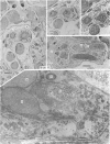
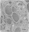
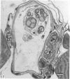
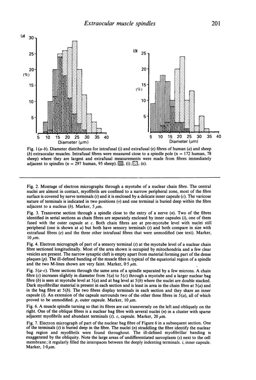
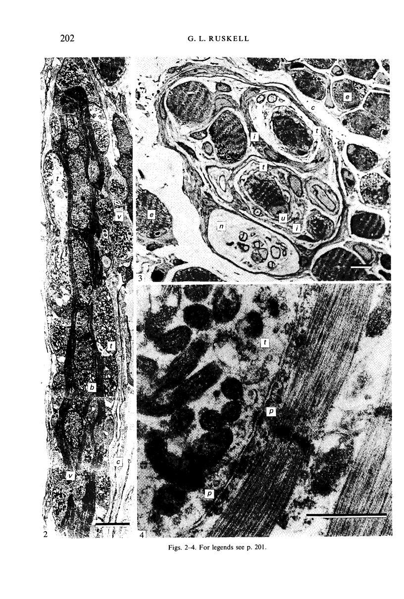
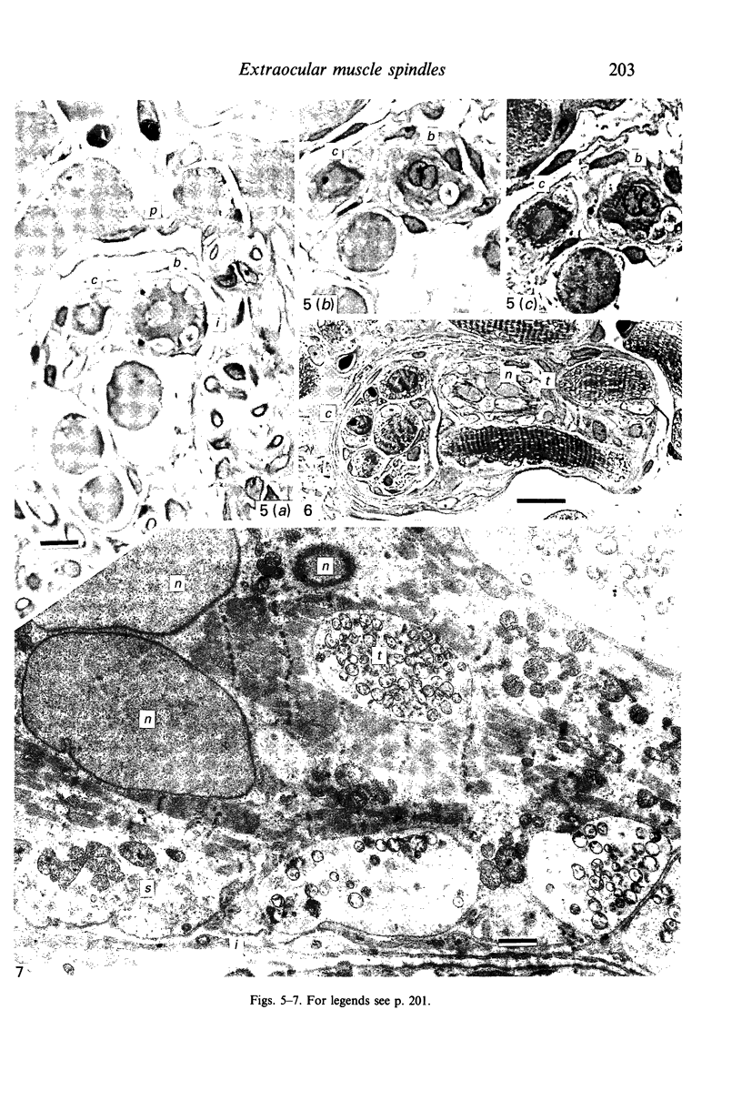
Twenty seven muscle spindles from six extraocular muscles removed following ocular enucleation from patients aged 58, 76 and 74 years were examined throughout all or most of their length by means of light and electron microscopy using serial transverse sections. Five others were prepared in longitudinal section. Twelve spindles of the superior rectus muscle from three sheep orbits were studied in a similar manner to provide a comparison. The human spindles contained a total of 90 (42%) nuclear chain and 5 (2%) nuclear bag fibres with the usual complement of sensory endings, and 120 (56%) fibres were anomalous with continuous, unattenuated myofibrils throughout their length, a constant width and peripherally placed nuclei. Eight anomalous fibres received sensory terminals similar in form to those of chain and bag fibres. Most (26) spindles contained at least one chain and one anomalous fibre. The periaxial space was limited or absent and the inner capsule was often segmented and in contact with the outer capsule. Abrupt termination of some chain fibres including several with one pole missing, together with evidence of fibre fragmentation and other structural anomalies, were indicative of degeneration. Eight further encapsulated fibre groups were identified as false spindles containing only anomalous fibres; associated nerves failed to terminate in the encapsulations. Sheep spindle content was of regular form, all spindles containing several chain and at least one bag fibre enclosed by an inner capsule and surrounded by a substantial periaxial space equatorially. The human extraocular muscle spindles have lost, either by aging or phylogenetically, the privilege of contractile chambers isolated by a fluid periaxial space from extrafusal fibre activity and sensory terminals are subject to the direct mechanical influences of anomalous intrafusal fibres. These, and the other departures from normal structure described, must jeopardize monitoring of muscle activity in the manner normally attributed to spindles and their capacity to provide useful proprioceptive information is questionable.
Full text
PDF
Images in this article
Selected References
These references are in PubMed. This may not be the complete list of references from this article.
- Banker B. Q., Girvin J. P. The ultrastructural features of the mammalian muscle spindle. J Neuropathol Exp Neurol. 1971 Apr;30(2):155–195. doi: 10.1097/00005072-197104000-00001. [DOI] [PubMed] [Google Scholar]
- COOPER S., DANIEL P. M., WHITTERIDGE D. Muscle spindles and other sensory endings in the extrinsic eye muscles; the physiology and anatomy of these receptors and of their connexions with the brain-stem. Brain. 1955;78(4):564–583. doi: 10.1093/brain/78.4.564. [DOI] [PubMed] [Google Scholar]
- Cazzato G., Walton J. N. The pathology of the muscle spindle. A study of biopsy material in various muscular and neuromuscular diseases. J Neurol Sci. 1968 Jul-Aug;7(1):15–70. doi: 10.1016/0022-510x(68)90003-8. [DOI] [PubMed] [Google Scholar]
- Daniel P. Spiral nerve endings in the extrinsic eye muscles of man. J Anat. 1946 Oct;80(Pt 4):189–193. [PMC free article] [PubMed] [Google Scholar]
- MERRILLEES N. C. R., SUNDERLAND S., HAYHOW W. Neuromuscular spindles in the extraocular muscles in man. Anat Rec. 1950 Sep;108(1):23–30. doi: 10.1002/ar.1091080103. [DOI] [PubMed] [Google Scholar]
- Maier A., DeSantis M., Eldred E. The occurrence of muscle spindles in extraocular muscles of various vertebrates. J Morphol. 1974 Aug;143(4):397–408. doi: 10.1002/jmor.1051430404. [DOI] [PubMed] [Google Scholar]
- Mukuno K., Nomura T. [Fine structure of the muscle spindles in the human extraocular muscles. (Preliminary report)]. Nippon Ganka Gakkai Zasshi. 1969 Oct;73(10):2119–2127. [PubMed] [Google Scholar]
- Richmond F. J., Johnston W. S., Baker R. S., Steinbach M. J. Palisade endings in human extraocular muscles. Invest Ophthalmol Vis Sci. 1984 Apr;25(4):471–476. [PubMed] [Google Scholar]
- Ruskell G. L. Sheathing of muscle fibres at neuromuscular junctions and at extra-junctional loci in human extra-ocular muscles. J Anat. 1984 Jan;138(Pt 1):33–44. [PMC free article] [PubMed] [Google Scholar]
- Ruskell G. L. Spiral nerve endings in human extraocular muscles terminate in motor end plates. J Anat. 1984 Aug;139(Pt 1):33–43. [PMC free article] [PubMed] [Google Scholar]
- Ruskell G. L. The incidence and variety of Golgi tendon organs in extraocular muscles of the rhesus monkey. J Neurocytol. 1979 Oct;8(5):639–653. doi: 10.1007/BF01208514. [DOI] [PubMed] [Google Scholar]
- Sodi A., Corsi M., Faussone Pellegrini M. S., Salvi G. Fine structure of the receptors at the myotendinous junction of human extraocular muscles. Histol Histopathol. 1988 Apr;3(2):103–113. [PubMed] [Google Scholar]
- Swash M., Fox K. P. The effect of age on human skeletal muscle. Studies of the morphology and innervation of muscle spindles. J Neurol Sci. 1972 Aug;16(4):417–432. doi: 10.1016/0022-510x(72)90048-2. [DOI] [PubMed] [Google Scholar]


