Abstract
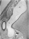
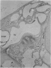
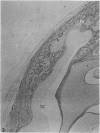
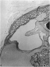
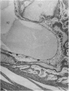
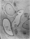
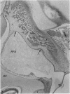
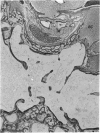
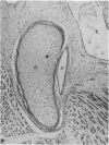
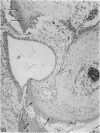
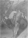
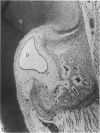
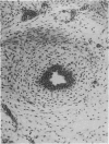
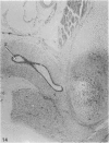
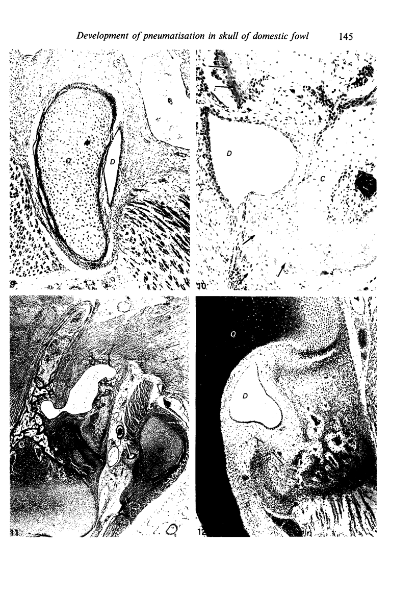
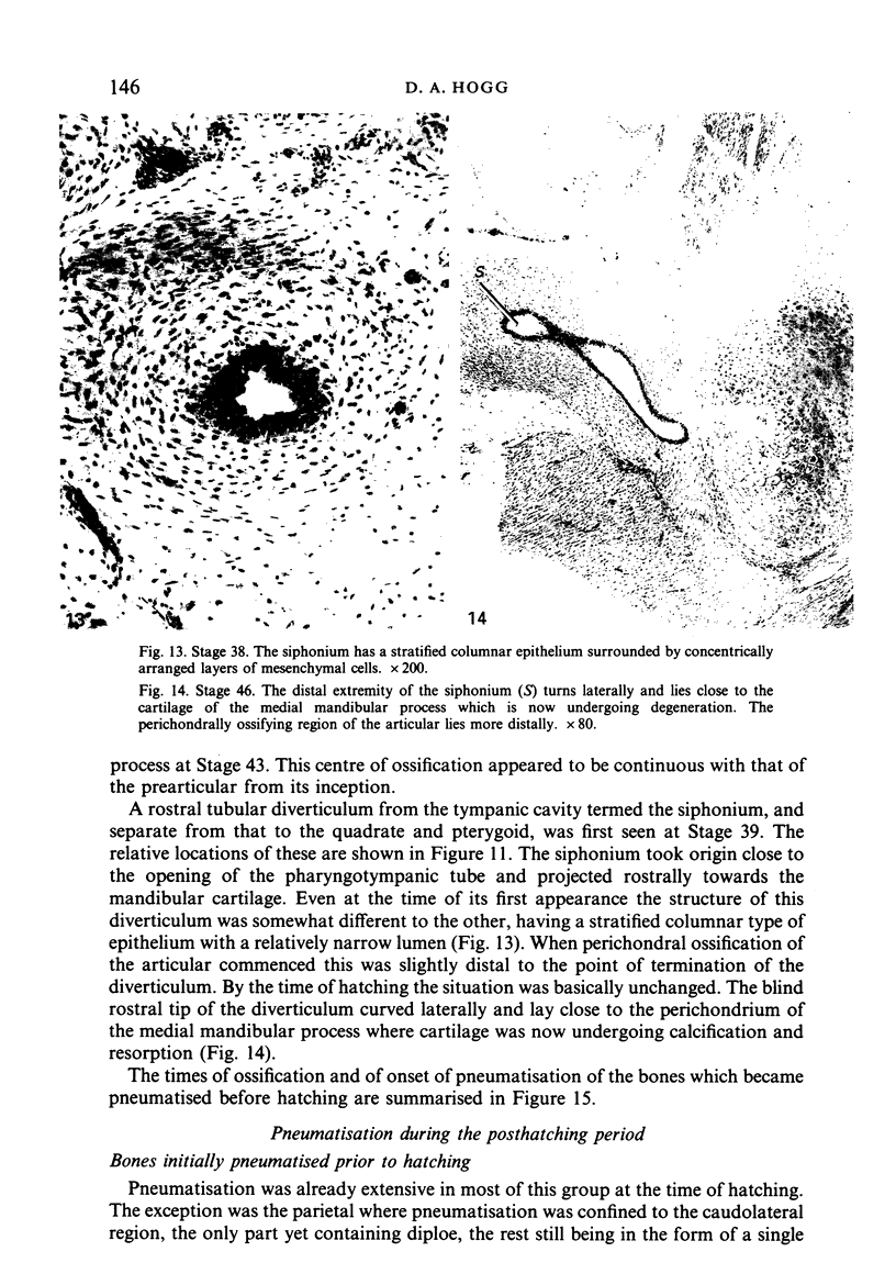
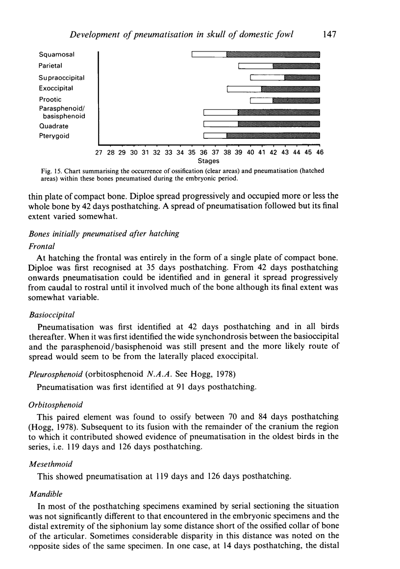
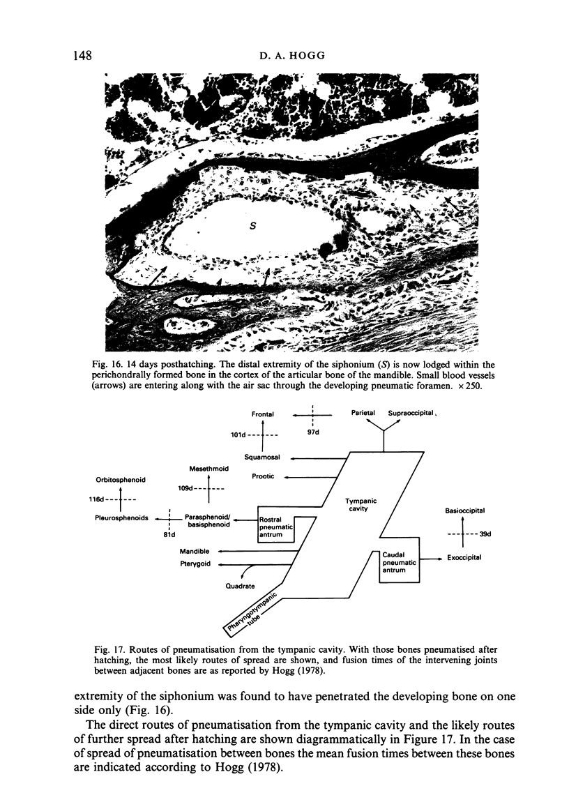
The development of pneumatisation in the skull of the domestic fowl has been studied in a series of chick embryos from 7-20 days incubation (Hamburger & Hamilton Stages 29-46) and in birds from hatching to 126 days posthatching. During the embryonic period primary pneumatisation developed by 3 routes. (i) The tympanic cavity directly invaded surrounding bones-squamosal, parietal, supraoccipital and prootic. (ii) Extensions of the tympanic cavity invaded the bones in which these occurred-the caudal pneumatic antrum in the exoccipital and the rostral pneumatic antrum in the parasphenoid/basisphenoid. (iii) A tubular diverticulum from the tympanic cavity grew rostrally and invaded the quadrate and pterygoid. A similar diverticulum grew rostrally towards the cartilaginous mandible but was only found to invade it in one case after the time of hatching. In most instances onset of pneumatisation occurred three stages subsequent to the onset of ossification. In bones in which ossification is intramembranous bone tissue often formed around small air sac outgrowths, resulting in multiple sites of invasion while, in bones ossifying perichondrally, cartilage resorption was a necessary prerequisite and air sac invasion frequently occurred in common with a vascular bud resulting in a single pneumatic foramen. After hatching secondary pneumatisation spread from the already pneumatised bones to involve the whole cranium. Spread throughout the parietal and frontal was preceded by the establishment of dipole within these bones and the final extent of pneumatisation was variable. Spread to the most distal parts of the cranium was only accomplished after the intervening sychondroses had fused.
Full text
PDF
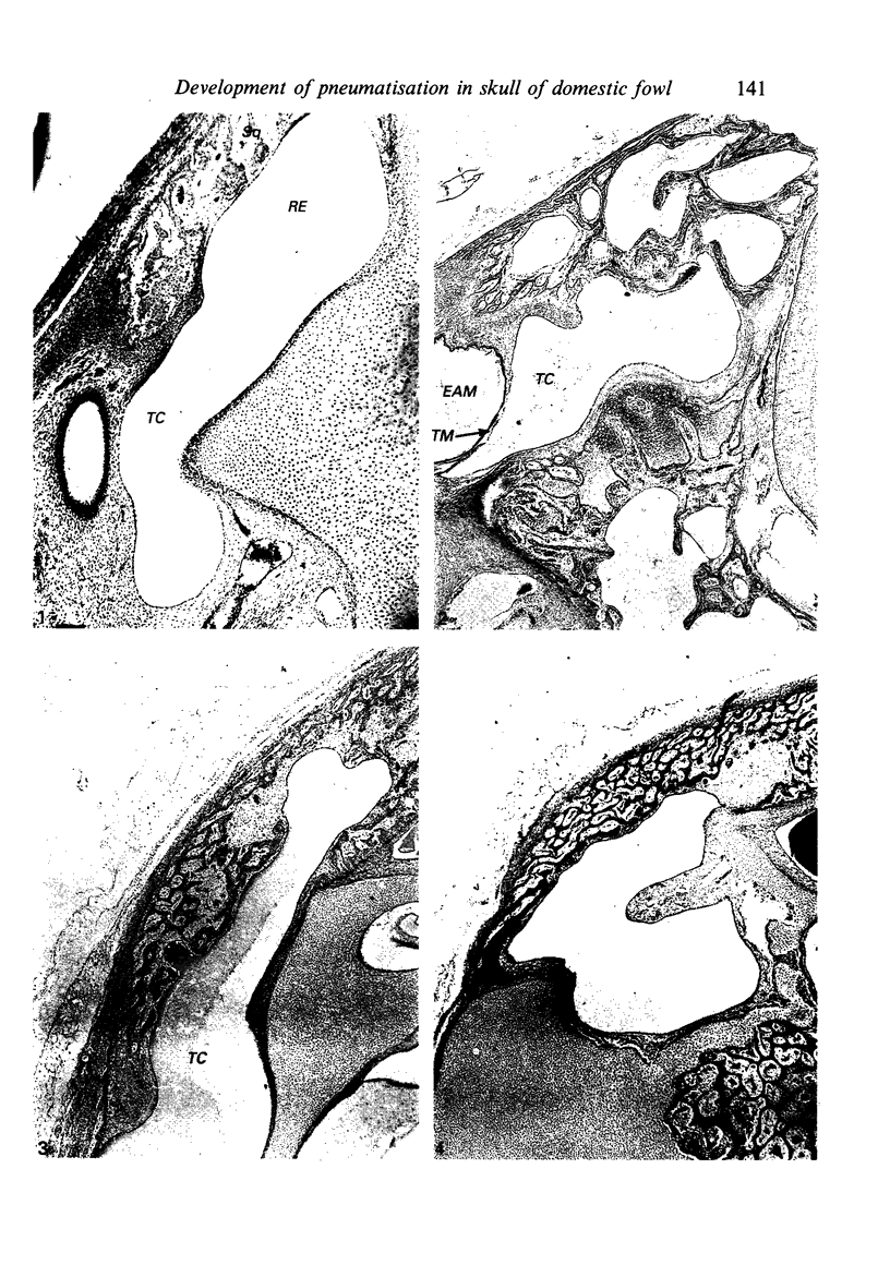
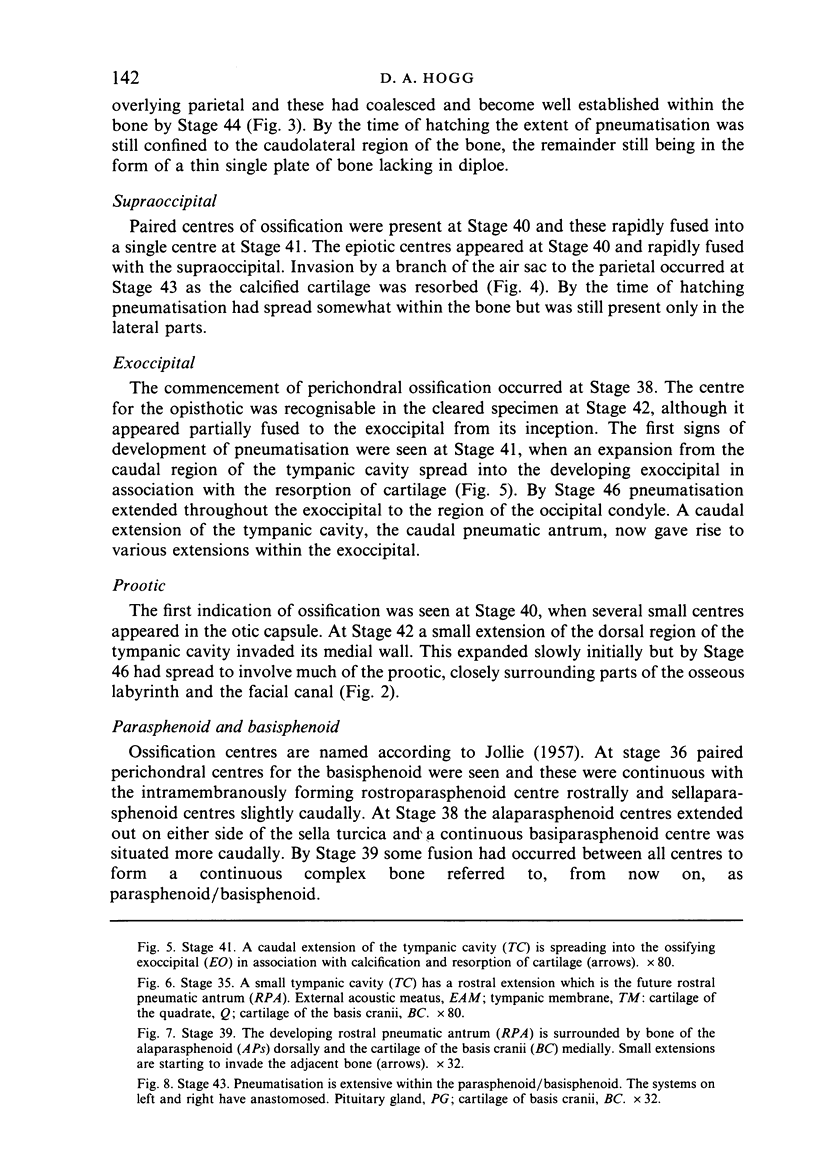
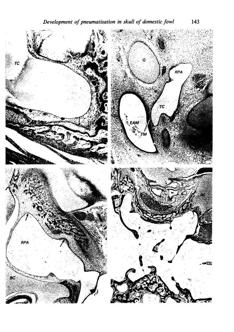
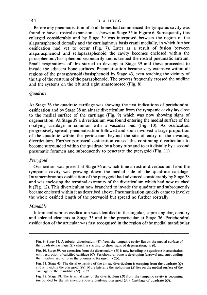
Images in this article
Selected References
These references are in PubMed. This may not be the complete list of references from this article.
- Dingerkus G., Uhler L. D. Enzyme clearing of alcian blue stained whole small vertebrates for demonstration of cartilage. Stain Technol. 1977 Jul;52(4):229–232. doi: 10.3109/10520297709116780. [DOI] [PubMed] [Google Scholar]
- Hogg D. A. The articulations of the neurocranium in the postnatal skeleton of the domestic fowl (Gallus gallus domesticus). J Anat. 1978 Sep;127(Pt 1):53–63. [PMC free article] [PubMed] [Google Scholar]
- Hogg D. A. The development of pneumatisation in the postcranial skeleton of the domestic fowl. J Anat. 1984 Aug;139(Pt 1):105–113. [PMC free article] [PubMed] [Google Scholar]
- Hogg D. A. The distribution of pneumatisation in the skeleton of the adult domestic fowl. J Anat. 1984 Jun;138(Pt 4):617–629. [PMC free article] [PubMed] [Google Scholar]


