

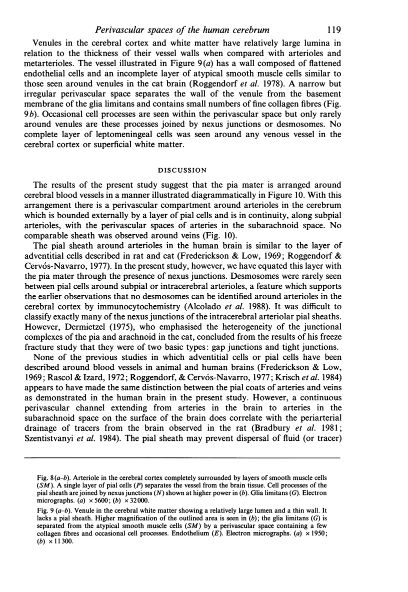
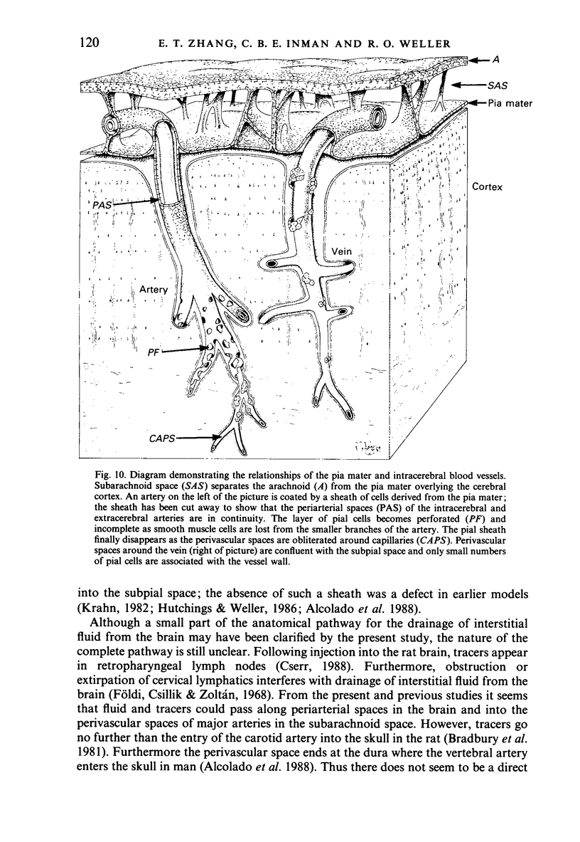
Abstract
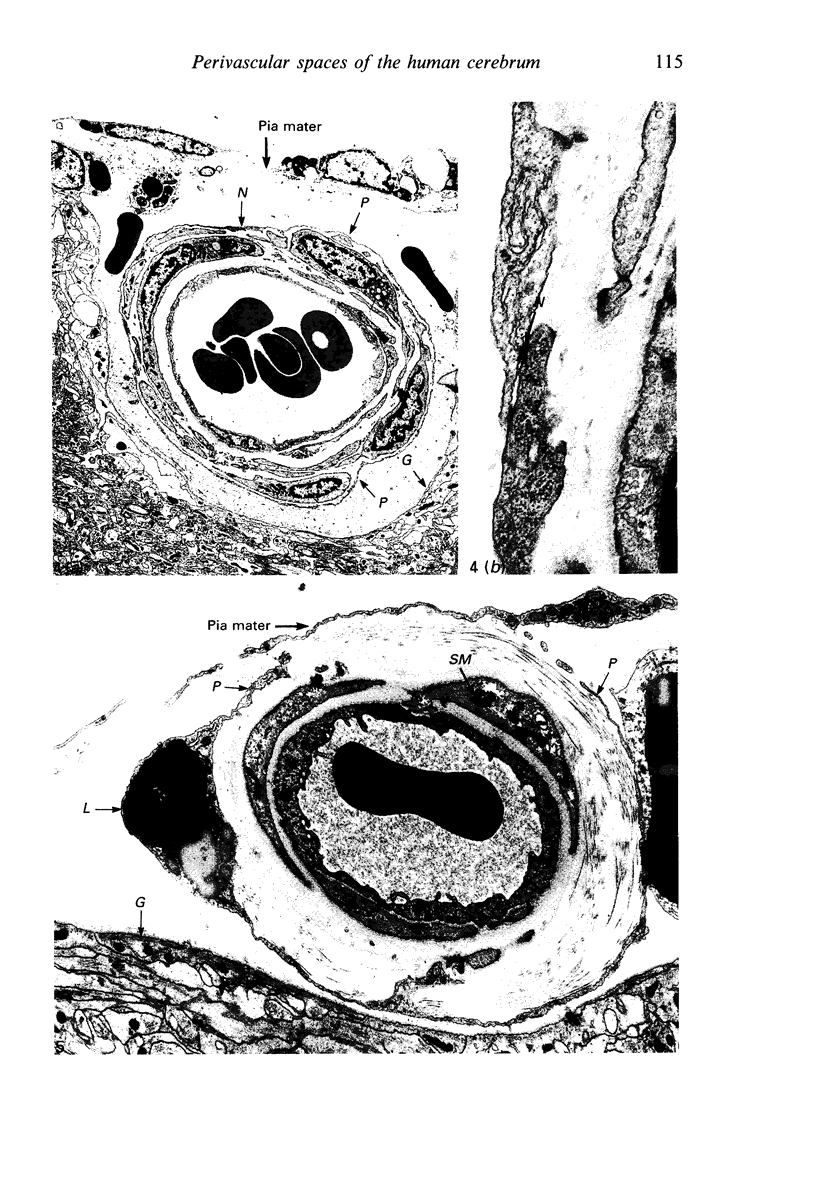
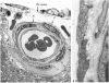
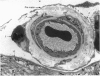
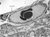
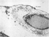
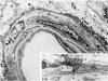
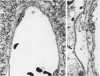
Biopsies of histologically normal adult human cerebral cortex, underlying white matter and overlying leptomeninges were taken from frontal and temporal lobectomy specimens excised during the removal of cerebral tumours. Multiple blocks from 6 patients (aged 18-53 years) were examined by light and transmission electron microscopy. A thin sheath of pia mater cells was found to surround completely arterioles and arteries in the brain, in the subpial space and in the subarachnoid space. Pia mater cells, forming the perivascular sheath, were identified by the presence of desmosomes or small nexus junctions and by continuity with the pia mater itself. The presence of the pial sheath suggests that the perivascular spaces around intracerebral arteries are in direct continuity with the perivascular spaces around subarachnoid arteries. No similar pial sheath was observed around intracerebral or subpial venules. The role of the periarterial spaces, enclosed by the pial sheath, is discussed in relation to the results of physiological experiments suggesting drainage of interstitial fluid from brain tissue into the perivascular pathways along major cerebral arteries in the subarachnoid space. As arterioles in the brain become smaller and lose their smooth muscle coats, the pial sheath becomes incomplete. The anatomical relationships between the pia mater and blood vessels in the human cerebrum is summarised diagrammatically, and a possible role for pial cells as an enzymic barrier protecting the brain from exogenous catecholamines is discussed.
Full text
PDF
Images in this article
Selected References
These references are in PubMed. This may not be the complete list of references from this article.
- Alcolado R., Weller R. O., Parrish E. P., Garrod D. The cranial arachnoid and pia mater in man: anatomical and ultrastructural observations. Neuropathol Appl Neurobiol. 1988 Jan-Feb;14(1):1–17. doi: 10.1111/j.1365-2990.1988.tb00862.x. [DOI] [PubMed] [Google Scholar]
- Andres K. H. Uber die Feinstruktur der Arachnoidea und Dura mater von Mammalia. Z Zellforsch Mikrosk Anat. 1967;79(2):272–295. [PubMed] [Google Scholar]
- Bradbury M. W., Cserr H. F., Westrop R. J. Drainage of cerebral interstitial fluid into deep cervical lymph of the rabbit. Am J Physiol. 1981 Apr;240(4):F329–F336. doi: 10.1152/ajprenal.1981.240.4.F329. [DOI] [PubMed] [Google Scholar]
- Cserr H. F. Role of secretion and bulk flow of brain interstitial fluid in brain volume regulation. Ann N Y Acad Sci. 1988;529:9–20. doi: 10.1111/j.1749-6632.1988.tb51415.x. [DOI] [PubMed] [Google Scholar]
- DAHL E., FLORA G., NELSON E. ELECTRON MICROSCOPIC OBSERVATIONS ON NORMAL HUMAN INTRACRANIAL ARTERIES. Neurology. 1965 Feb;15:132–140. doi: 10.1212/wnl.15.2.132. [DOI] [PubMed] [Google Scholar]
- Dahl E. The fine structure of intracerebral vessels. Z Zellforsch Mikrosk Anat. 1973 Dec 21;145(4):577–586. doi: 10.1007/BF00306725. [DOI] [PubMed] [Google Scholar]
- Dahl E. The ultrastructure of cerebral blood vessels in man. Cephalalgia. 1986;6 (Suppl 4):45–48. [PubMed] [Google Scholar]
- Dermietzel R. Junctions in the central nervous system of the cat. V. The junctional complex of the pia-arachnoid membrane. Cell Tissue Res. 1975 Dec 10;164(3):309–329. doi: 10.1007/BF00223012. [DOI] [PubMed] [Google Scholar]
- Edvinsson L. Innervation of the cerebral circulation. Ann N Y Acad Sci. 1987;519:334–348. doi: 10.1111/j.1749-6632.1987.tb36308.x. [DOI] [PubMed] [Google Scholar]
- Frederickson R. G., Low F. N. Blood vessels and tissue space associated with the brain of the rat. Am J Anat. 1969 Jun;125(2):123–145. doi: 10.1002/aja.1001250202. [DOI] [PubMed] [Google Scholar]
- Földi M., Csillik B., Zoltán O. T. Lymphatic drainage of the brain. Experientia. 1968 Dec 15;24(12):1283–1287. doi: 10.1007/BF02146675. [DOI] [PubMed] [Google Scholar]
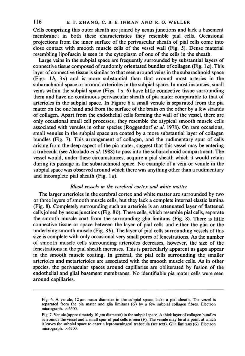
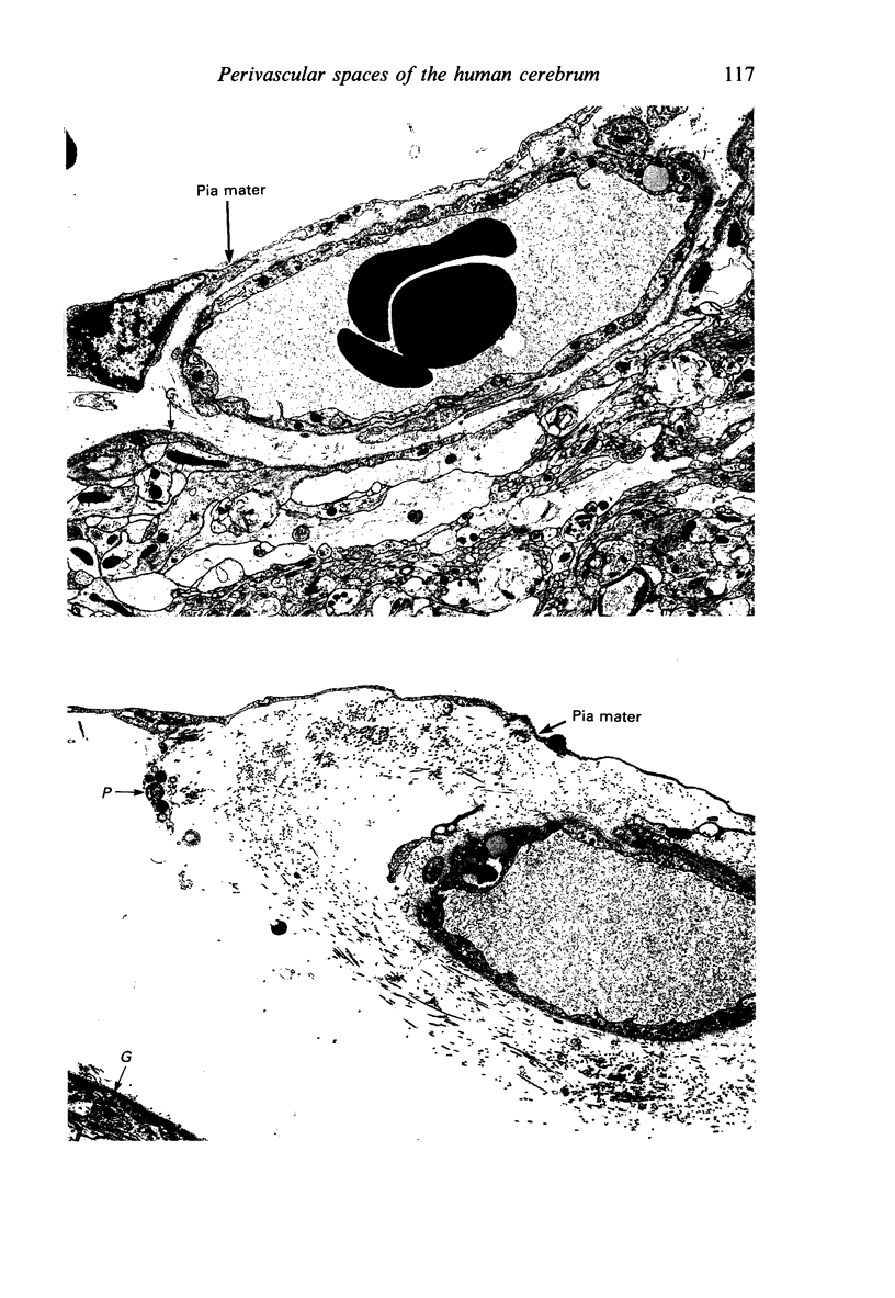
- Hutchings M., Weller R. O. Anatomical relationships of the pia mater to cerebral blood vessels in man. J Neurosurg. 1986 Sep;65(3):316–325. doi: 10.3171/jns.1986.65.3.0316. [DOI] [PubMed] [Google Scholar]
- Jones E. G. On the mode of entry of blood vessels into the cerebral cortex. J Anat. 1970 May;106(Pt 3):507–520. [PMC free article] [PubMed] [Google Scholar]
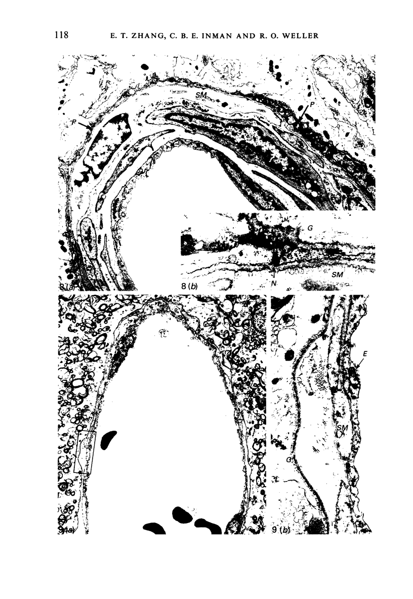
- Kaplan G. P., Hartman B. K., Creveling C. R. Localization of catechol-O-methyltransferase in the leptomeninges, choroid plexus and ciliary epithelium: implications for the separation of central and peripheral catechols. Brain Res. 1981 Jan 12;204(2):353–360. doi: 10.1016/0006-8993(81)90594-1. [DOI] [PubMed] [Google Scholar]
- Kartenbeck J., Schwechheimer K., Moll R., Franke W. W. Attachment of vimentin filaments to desmosomal plaques in human meningiomal cells and arachnoidal tissue. J Cell Biol. 1984 Mar;98(3):1072–1081. doi: 10.1083/jcb.98.3.1072. [DOI] [PMC free article] [PubMed] [Google Scholar]
- Krahn V. The pia mater at the site of the entry of blood vessels into the central nervous system. Anat Embryol (Berl) 1982;164(2):257–263. doi: 10.1007/BF00318509. [DOI] [PubMed] [Google Scholar]
- Krisch B., Leonhardt H., Oksche A. Compartments and perivascular arrangement of the meninges covering the cerebral cortex of the rat. Cell Tissue Res. 1984;238(3):459–474. doi: 10.1007/BF00219861. [DOI] [PubMed] [Google Scholar]
- Krisch B. Ultrastructure of the meninges at the site of penetration of veins through the dura mater, with particular reference to Pacchionian granulations. Investigations in the rat and two species of New-World monkeys (Cebus apella, Callitrix jacchus). Cell Tissue Res. 1988 Mar;251(3):621–631. doi: 10.1007/BF00214011. [DOI] [PubMed] [Google Scholar]
- NELSON E., BLINZINGER K., HAGER H. Electron microscopic observations on subarachnoid and perivascular spaces of the Syrian hamster brain. Neurology. 1961 Apr;11(4):285–295. doi: 10.1212/wnl.11.4.285. [DOI] [PubMed] [Google Scholar]
- Nabeshima S., Reese T. S., Landis D. M., Brightman M. W. Junctions in the meninges and marginal glia. J Comp Neurol. 1975 Nov 15;164(2):127–169. doi: 10.1002/cne.901640202. [DOI] [PubMed] [Google Scholar]
- Nicholas D. S., Weller R. O. The fine anatomy of the human spinal meninges. A light and scanning electron microscopy study. J Neurosurg. 1988 Aug;69(2):276–282. doi: 10.3171/jns.1988.69.2.0276. [DOI] [PubMed] [Google Scholar]
- Parrish E. P., Garrod D. R., Mattey D. L., Hand L., Steart P. V., Weller R. O. Mouse antisera specific for desmosomal adhesion molecules of suprabasal skin cells, meninges, and meningioma. Proc Natl Acad Sci U S A. 1986 Apr;83(8):2657–2661. doi: 10.1073/pnas.83.8.2657. [DOI] [PMC free article] [PubMed] [Google Scholar]
- Ramsey H. J. Fine structure of the surface of the cerebral cortex of human brain. J Cell Biol. 1965 Aug;26(2):323–334. doi: 10.1083/jcb.26.2.323. [DOI] [PMC free article] [PubMed] [Google Scholar]
- Rascol M., Izard J. La jonction cortico-pie-mérienne et la pénétration des vaisseaux dans le cortex cérébral chez l'Homme. Structure et ultrastructure. Z Zellforsch Mikrosk Anat. 1972;123(3):337–355. [PubMed] [Google Scholar]
- Rennels M. L., Gregory T. F., Blaumanis O. R., Fujimoto K., Grady P. A. Evidence for a 'paravascular' fluid circulation in the mammalian central nervous system, provided by the rapid distribution of tracer protein throughout the brain from the subarachnoid space. Brain Res. 1985 Feb 4;326(1):47–63. doi: 10.1016/0006-8993(85)91383-6. [DOI] [PubMed] [Google Scholar]
- Roggendorf W., Cervós-Navarro J., Lazaro-Lacalle M. D. Ultrastructure of venules in the cat brain. Cell Tissue Res. 1978 Sep 26;192(3):461–474. doi: 10.1007/BF00212326. [DOI] [PubMed] [Google Scholar]
- Roggendorf W., Cervós-Navarro J. Ultrastructure of arterioles in the cat brain. Cell Tissue Res. 1977 Mar 24;178(4):495–515. doi: 10.1007/BF00219571. [DOI] [PubMed] [Google Scholar]
- Szentistványi I., Patlak C. S., Ellis R. A., Cserr H. F. Drainage of interstitial fluid from different regions of rat brain. Am J Physiol. 1984 Jun;246(6 Pt 2):F835–F844. doi: 10.1152/ajprenal.1984.246.6.F835. [DOI] [PubMed] [Google Scholar]
- WOOLLAM D. H., MILLEN J. W. The perivascular spaces of the mammalian central nervous system and their relation to the perineuronal and subarachnoid spaces. J Anat. 1955 Apr;89(2):193–200. [PMC free article] [PubMed] [Google Scholar]
- Zervas N. T., Liszczak T. M., Mayberg M. R., Black P. M. Cerebrospinal fluid may nourish cerebral vessels through pathways in the adventitia that may be analogous to systemic vasa vasorum. J Neurosurg. 1982 Apr;56(4):475–481. doi: 10.3171/jns.1982.56.4.0475. [DOI] [PubMed] [Google Scholar]