

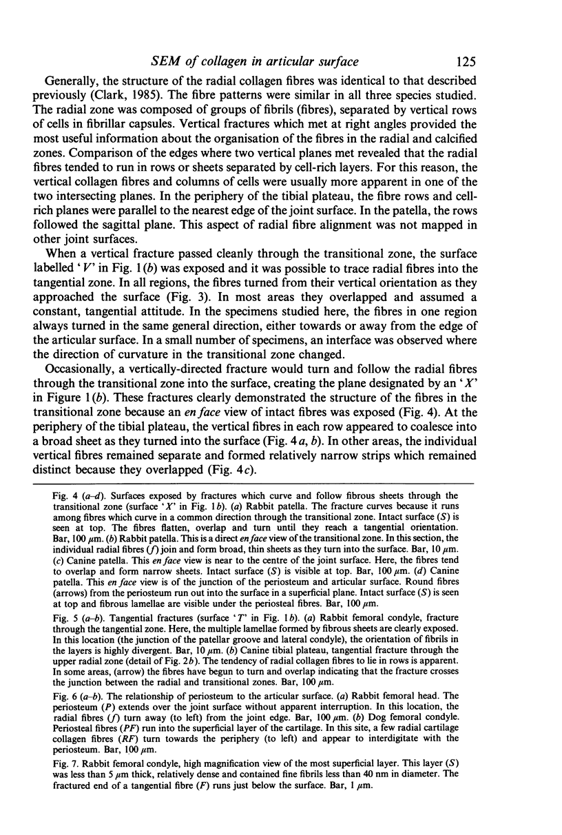
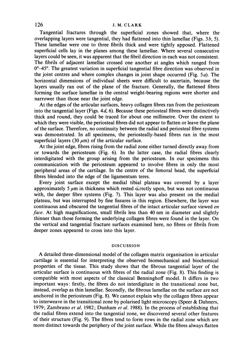
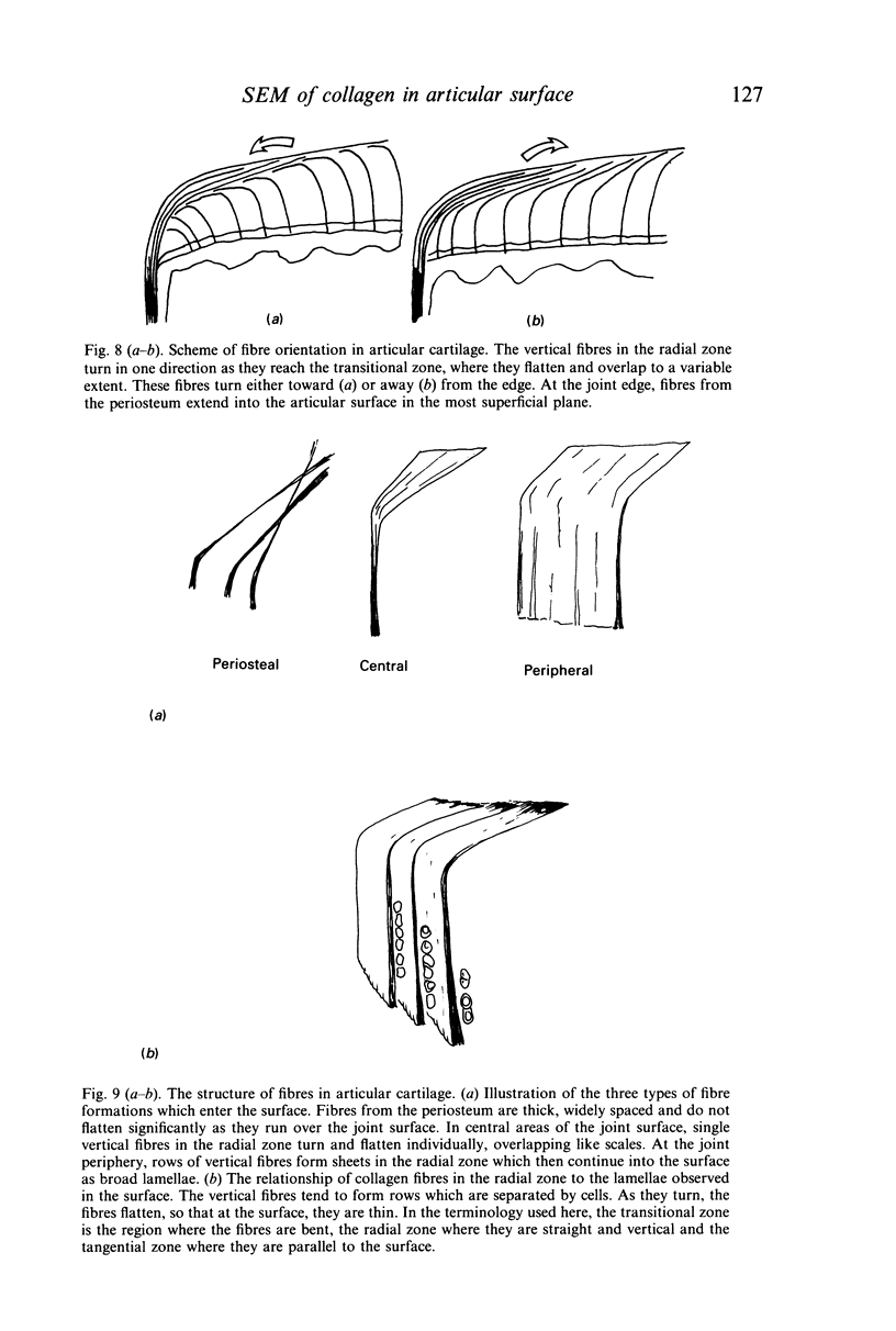
Abstract
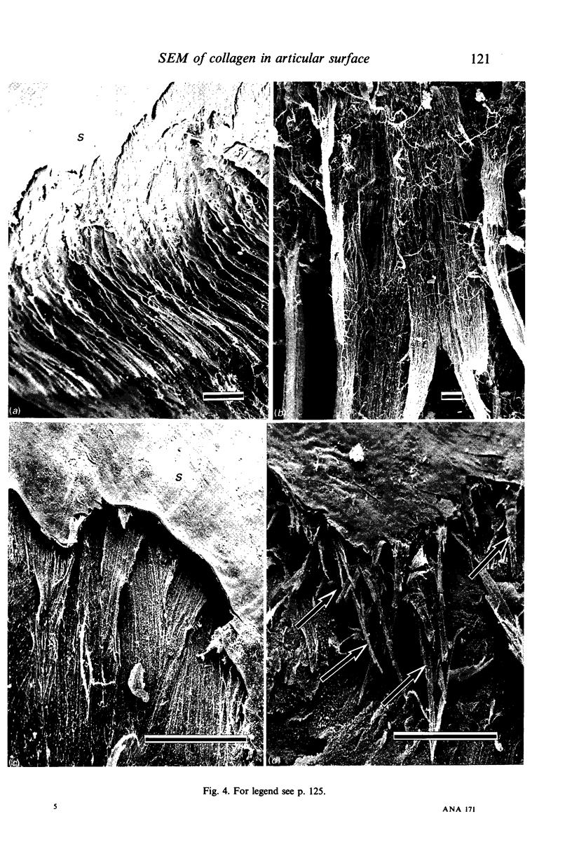
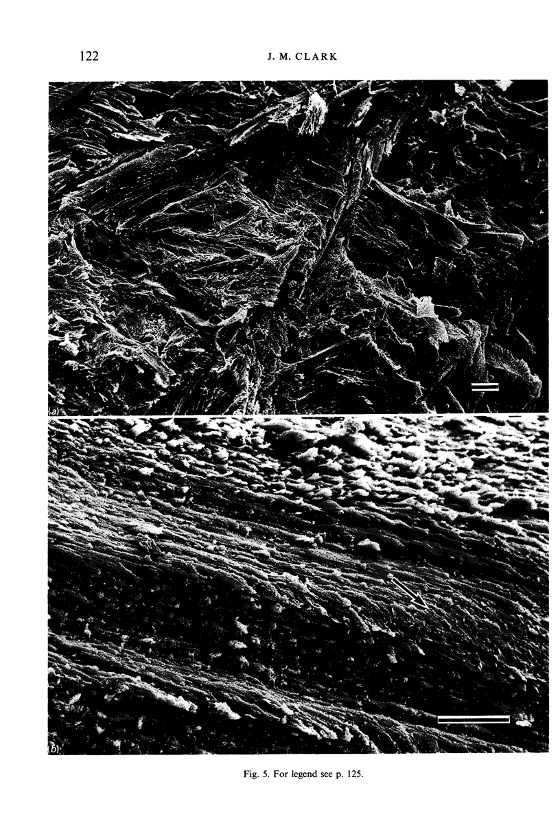
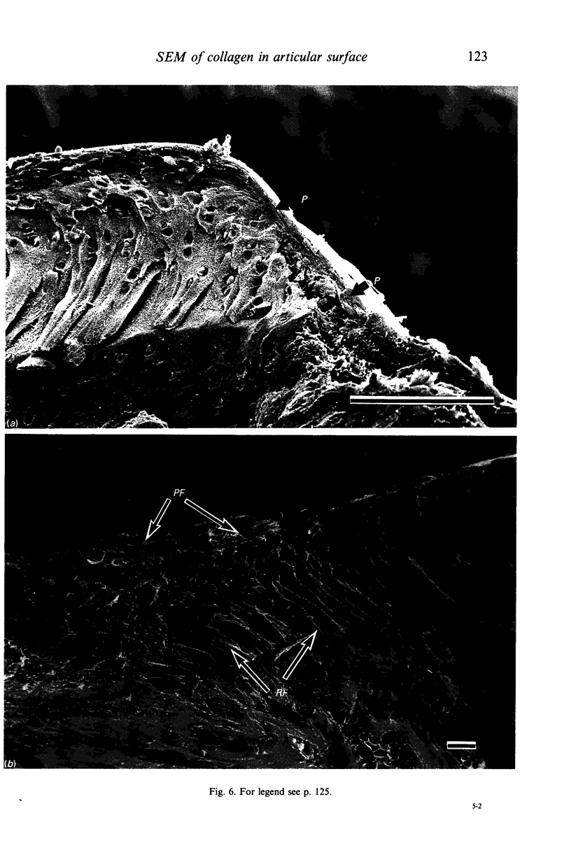
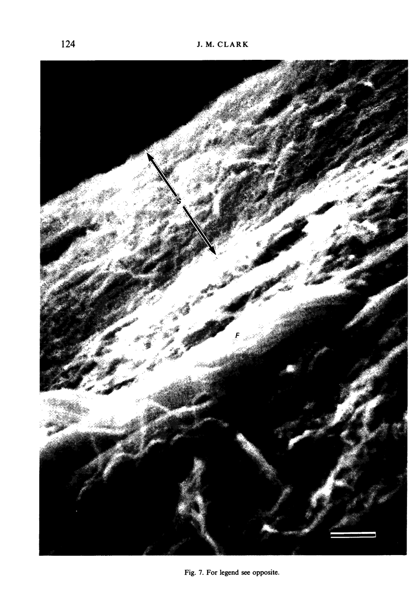
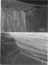
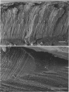
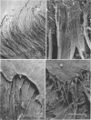
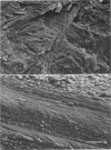
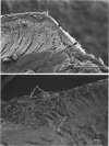
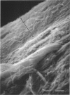
The origin and structure of collagen fibres in the surface of articular cartilage were studied using SEM. Cryofracture was used to create orthogonal fracture surfaces in three planes. Fibres which originated in the radial zone could be traced into the surface where they flattened and overlapped in a common direction. Thick fibres from the periosteum ran into the surface as well, but apparently ended there and did not enter the radial zone. The tangential fibres were covered by a dense, separate layer of small fibrils. The fundamental aspects of the model proposed by Benninghoff are supported by these findings.
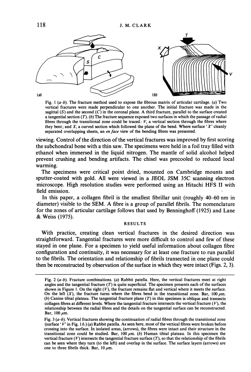
Full text
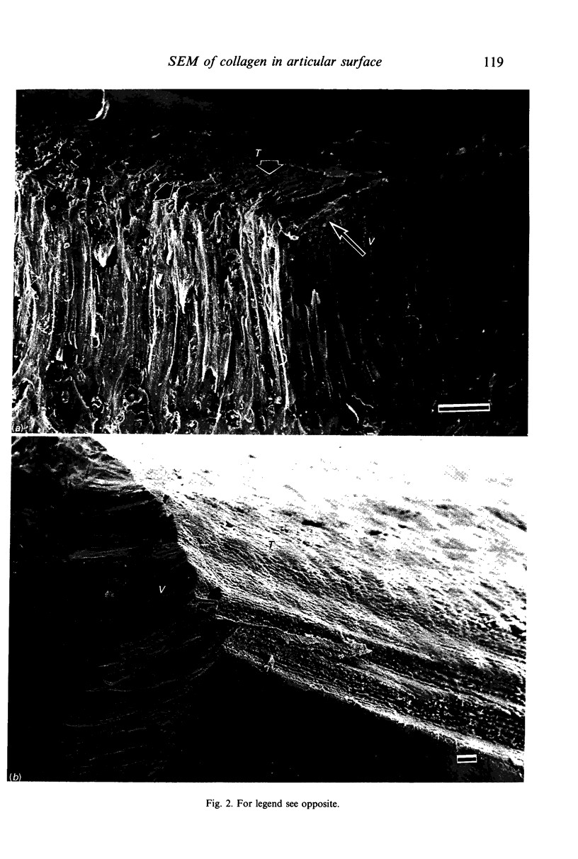
PDF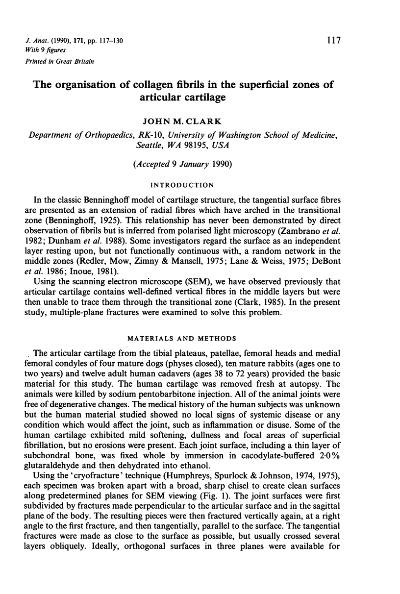
Images in this article
Selected References
These references are in PubMed. This may not be the complete list of references from this article.
- Broom N. D., Myers D. D. Fibrous waveforms or crimp in surface and subsurface layers of hyaline cartilage maintained in its wet functional condition. Connect Tissue Res. 1980;7(3):165–175. doi: 10.3109/03008208009152108. [DOI] [PubMed] [Google Scholar]
- Clark J. M. The organization of collagen in cryofractured rabbit articular cartilage: a scanning electron microscopic study. J Orthop Res. 1985;3(1):17–29. doi: 10.1002/jor.1100030102. [DOI] [PubMed] [Google Scholar]
- Dunham J., Shackleton D. R., Billingham M. E., Bitensky L., Chayen J., Muir I. H. A reappraisal of the structure of normal canine articular cartilage. J Anat. 1988 Apr;157:89–99. [PMC free article] [PubMed] [Google Scholar]
- Humphreys W. J., Spurlock B. O., Johnson J. S. Transmission electron microscopy of tissue prepared for scanning electron microscopy by ethanol-cryofracturing. Stain Technol. 1975 Mar;50(2):119–125. doi: 10.3109/10520297509117045. [DOI] [PubMed] [Google Scholar]
- Inoue H. Alterations in the collagen framework of osteoarthritic cartilage and subchondral bone. Int Orthop. 1981;5(1):47–52. doi: 10.1007/BF00286099. [DOI] [PubMed] [Google Scholar]
- Kempson G. E., Muir H., Pollard C., Tuke M. The tensile properties of the cartilage of human femoral condyles related to the content of collagen and glycosaminoglycans. Biochim Biophys Acta. 1973 Feb 28;297(2):456–472. doi: 10.1016/0304-4165(73)90093-7. [DOI] [PubMed] [Google Scholar]
- Lane J. M., Weiss C. Review of articular cartilage collagen research. Arthritis Rheum. 1975 Nov-Dec;18(6):553–562. doi: 10.1002/art.1780180605. [DOI] [PubMed] [Google Scholar]
- Meachim G., Denham D., Emery I. H., Wilkinson P. H. Collagen alignments and artificial splits at the surface of human articular cartilage. J Anat. 1974 Sep;118(Pt 1):101–118. [PMC free article] [PubMed] [Google Scholar]
- Meachim G., Sheffield S. R. Surface ultrastructure of mature adult human articular cartilage. J Bone Joint Surg Br. 1969 Aug;51(3):529–539. [PubMed] [Google Scholar]
- Minns R. J., Steven F. S. The collagen fibril organization in human articular cartilage. J Anat. 1977 Apr;123(Pt 2):437–457. [PMC free article] [PubMed] [Google Scholar]
- Muir H., Bullough P., Maroudas A. The distribution of collagen in human articular cartilage with some of its physiological implications. J Bone Joint Surg Br. 1970 Aug;52(3):554–563. [PubMed] [Google Scholar]
- O'Connor P., Bland C., Gardner D. L. Fine structure of artificial splits in femoral condylar cartilage of the rat: a scanning electron microscopic study. J Pathol. 1980 Oct;132(2):169–179. doi: 10.1002/path.1711320207. [DOI] [PubMed] [Google Scholar]
- Ortmann R. Use of polarized light for quantitative determination of the adjustment of the tangential fibres in articular cartilage. Anat Embryol (Berl) 1975 Dec 23;148(2):109–120. doi: 10.1007/BF00315264. [DOI] [PubMed] [Google Scholar]
- Redler I., Mow V. C., Zimny M. L., Mansell J. The ultrastructure and biomechanical significance of the tidemark of articular cartilage. Clin Orthop Relat Res. 1975 Oct;(112):357–362. [PubMed] [Google Scholar]
- Speer D. P., Dahners L. The collagenous architecture of articular cartilage. Correlation of scanning electron microscopy and polarized light microscopy observations. Clin Orthop Relat Res. 1979 Mar-Apr;(139):267–275. [PubMed] [Google Scholar]
- Weiss C., Rosenberg L., Helfet A. J. An ultrastructural study of normal young adult human articular cartilage. J Bone Joint Surg Am. 1968 Jun;50(4):663–674. doi: 10.2106/00004623-196850040-00002. [DOI] [PubMed] [Google Scholar]
- Zambrano N. Z., Montes G. S., Shigihara K. M., Sanchez E. M., Junqueira L. C. Collagen arrangement in cartilages. Acta Anat (Basel) 1982;113(1):26–38. doi: 10.1159/000145534. [DOI] [PubMed] [Google Scholar]
- de Bont L. G., Boering G., Havinga P., Liem R. S. Spatial arrangement of collagen fibrils in the articular cartilage of the mandibular condyle: a light microscopic and scanning electron microscopic study. J Oral Maxillofac Surg. 1984 May;42(5):306–313. doi: 10.1016/0278-2391(84)90110-1. [DOI] [PubMed] [Google Scholar]
- de Bont L. G., Liem R. S., Havinga P., Boering G., van der Korst J. Collagenous network in cartilage of human femoral condyles. A light microscopic and scanning electron microscopic study. Acta Anat (Basel) 1986;126(1):41–47. doi: 10.1159/000146184. [DOI] [PubMed] [Google Scholar]