Abstract
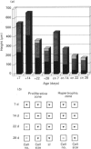
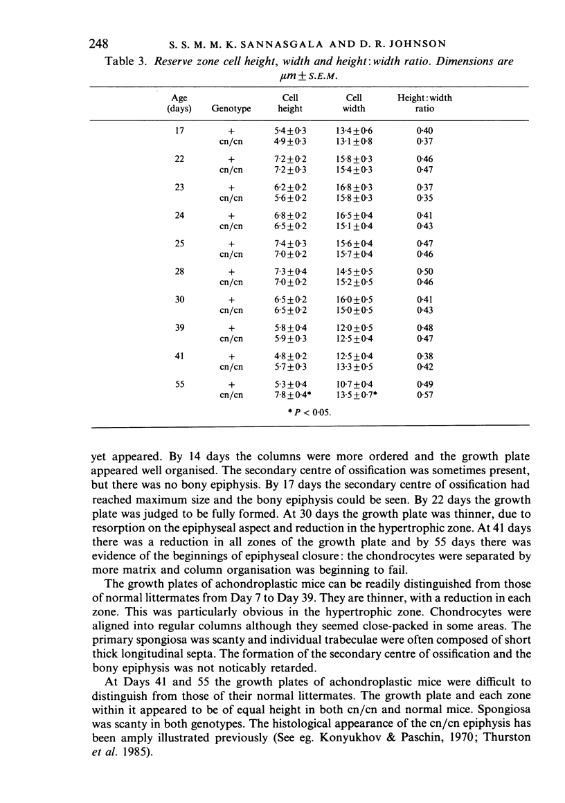
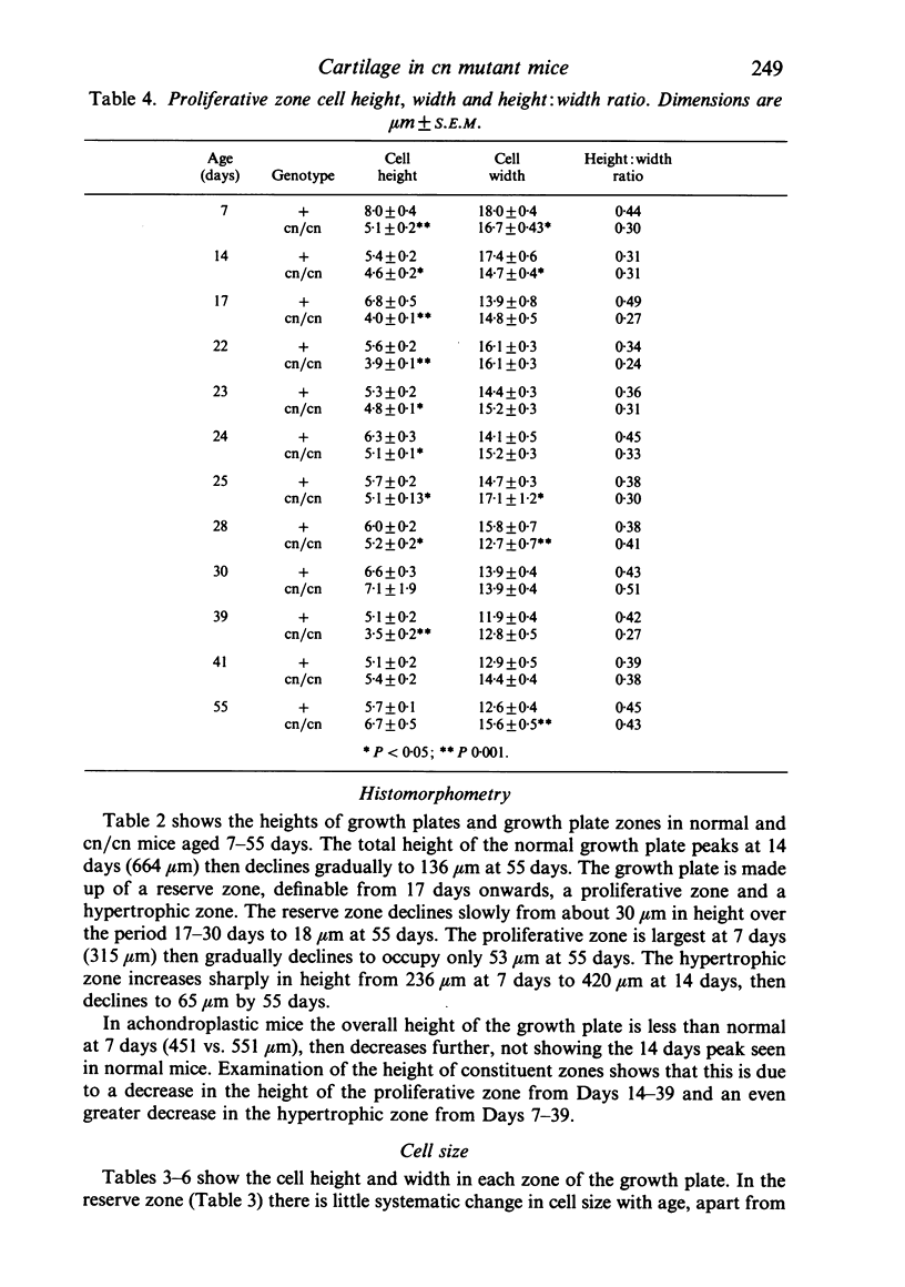
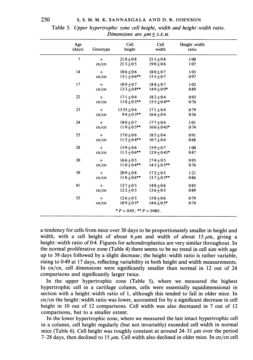
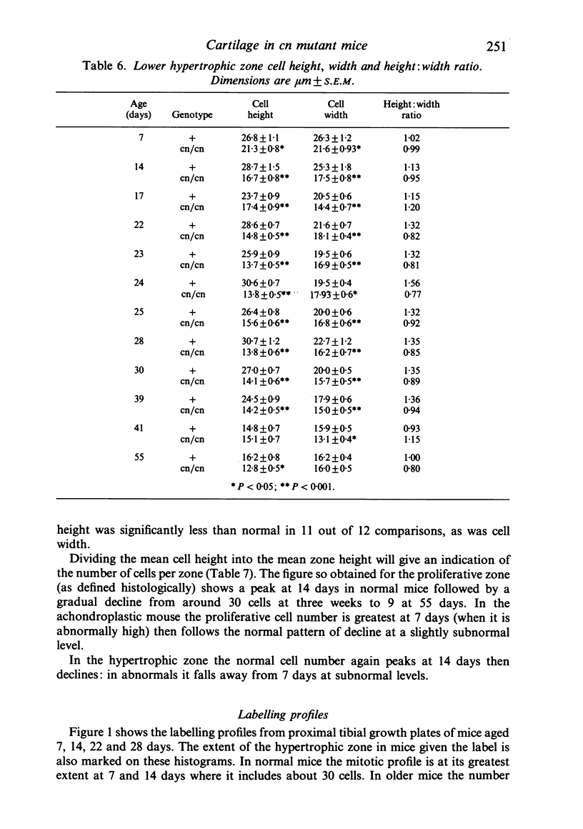
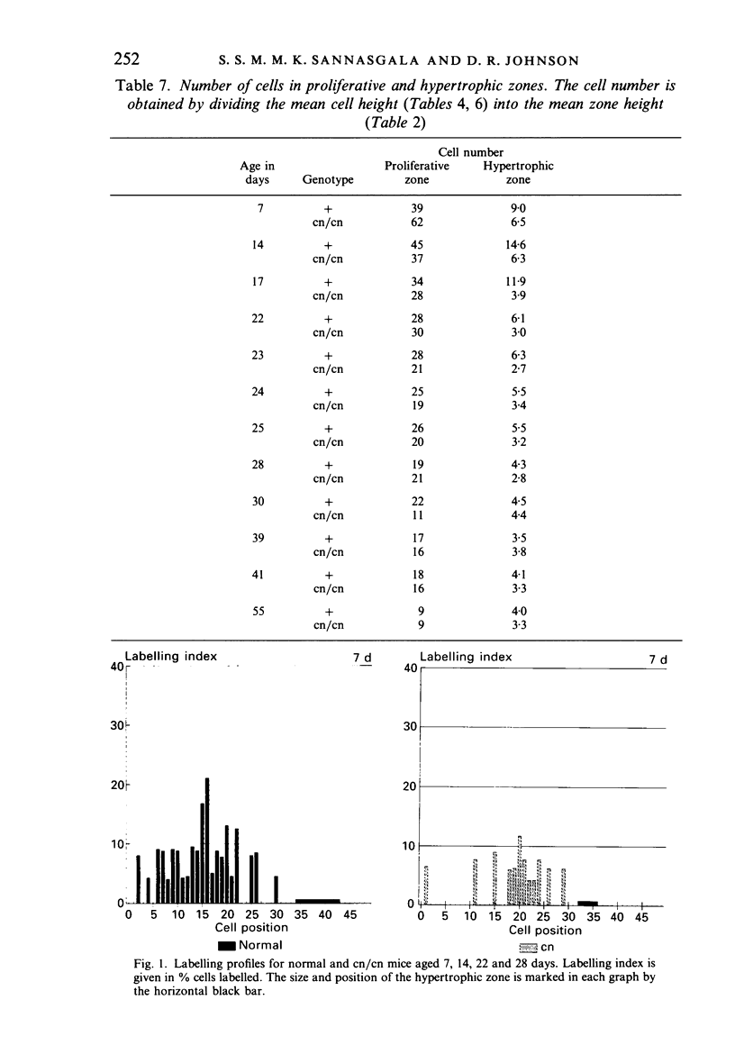
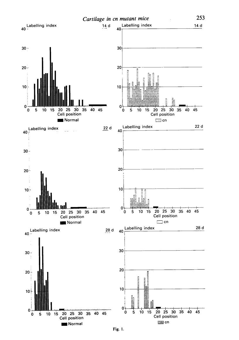
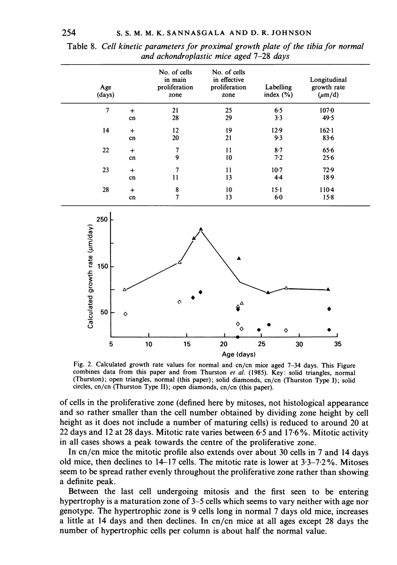
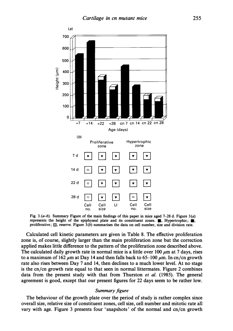
We have reinvestigated the overall size, zone size, mitotic rate and growth rate in the tibial epiphysis of normal and achondroplastic (cn/cn) mice aged 7-55 days. Our present sample did not contain the two phenotypes characteristic of some stocks carrying the gene: cn/cn mice showed smaller zone sizes within the growth plates, smaller cells, decreased mitotic indices and growth rates.
Full text
PDF
Images in this article
Selected References
These references are in PubMed. This may not be the complete list of references from this article.
- Bonucci E., Marco A. D., Nicoletti B., Petrinelli P., Pozzi L. Histological and histochemical investigations of achondroplastic mice: a possible model of human achondroplasia. Growth. 1976 Sep;40(3):241–251. [PubMed] [Google Scholar]
- Buckwalter J. A., Mower D., Schafer J., Ungar R., Ginsberg B., Moore K. Growth-plate-chondrocyte profiles and their orientation. J Bone Joint Surg Am. 1985 Jul;67(6):942–955. [PubMed] [Google Scholar]
- KEMBER N. F. Cell division in endochondral ossification. A study of cell proliferation in rat bones by the method of tritiated thymidine autoradiography. J Bone Joint Surg Br. 1960 Nov;42B:824–839. [PubMed] [Google Scholar]
- Kember N. F. Cell population kinetics of bone growth: the first ten years of autoradiographic studies with tritiated thymidine. Clin Orthop Relat Res. 1971 May;76:213–230. doi: 10.1097/00003086-197105000-00029. [DOI] [PubMed] [Google Scholar]
- Kember N. F. Comparative patterns of cell division in epiphyseal cartilage plates in the rat. J Anat. 1972 Jan;111(Pt 1):137–142. [PMC free article] [PubMed] [Google Scholar]
- Konyukhov B. V., Paschin Y. V. Abnormal growth of the body, internal organs and skeleton in the achondroplastic mice. Acta Biol Acad Sci Hung. 1970;21(4):347–354. [PubMed] [Google Scholar]
- Lane P. W., Dickie M. M. Three recessive mutations producing disproportionate dwarfing in mice: achondroplasia, brachymorphic, and stubby. J Hered. 1968 Sep-Oct;59(5):300–308. doi: 10.1093/oxfordjournals.jhered.a107725. [DOI] [PubMed] [Google Scholar]
- Miller W. A., Flynn-Miller K. L. A chondroplastic, brachymorphic and stubby chondrodystophies in mice. J Comp Pathol. 1976 Jul;86(3):349–363. doi: 10.1016/0021-9975(76)90002-5. [DOI] [PubMed] [Google Scholar]
- Silberberg R., Hasler M., Lesker P. Ultrastructure of articular cartilage of achondroplastic mice. Acta Anat (Basel) 1976;96(2):162–175. doi: 10.1159/000144670. [DOI] [PubMed] [Google Scholar]
- Silberberg R., Lesker P. Skeletal growth and development of achondroplastic mice. Growth. 1975 Mar;39(1):17–33. [PubMed] [Google Scholar]
- Thurston M. N., Johnson D. R., Kember N. F. Cell kinetics of growth cartilage of achondroplastic (cn) mice. J Anat. 1985 May;140(Pt 3):425–434. [PMC free article] [PubMed] [Google Scholar]


