

Abstract
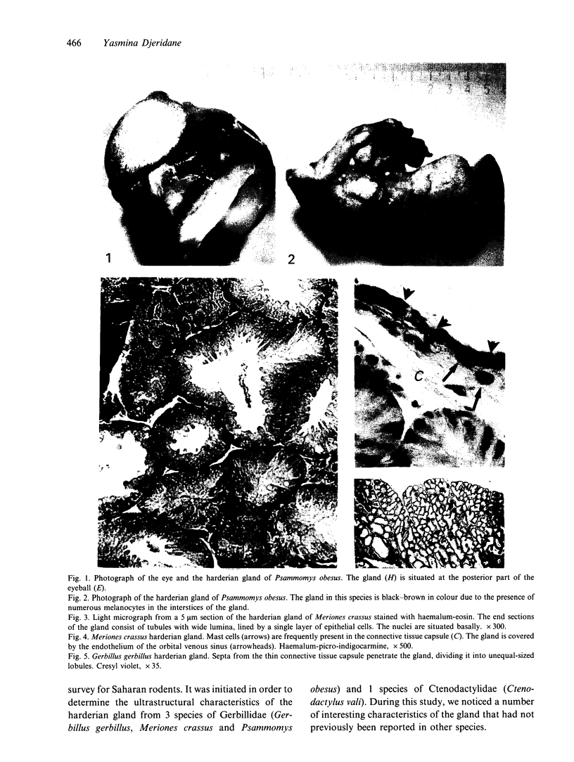
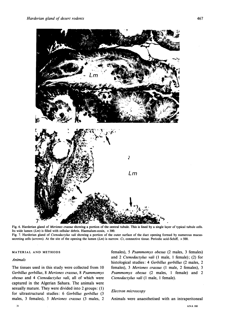
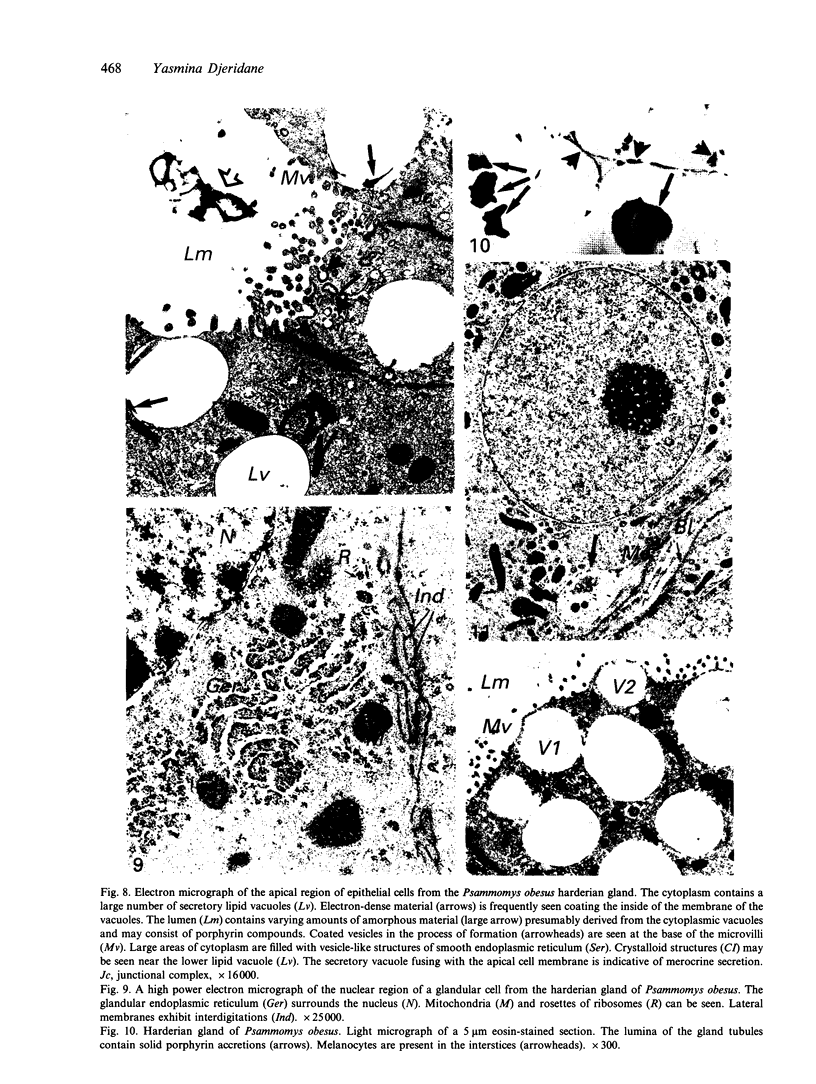
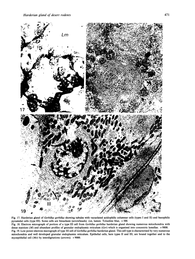
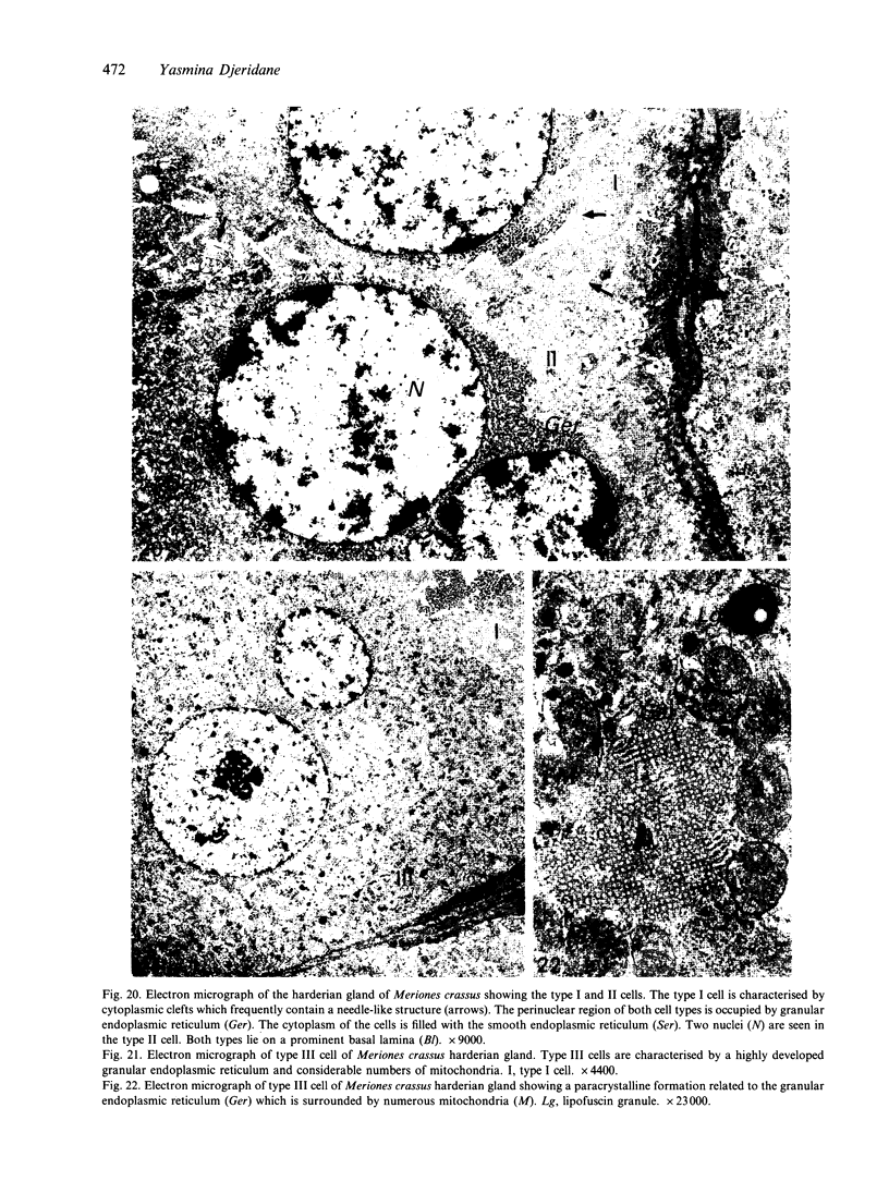
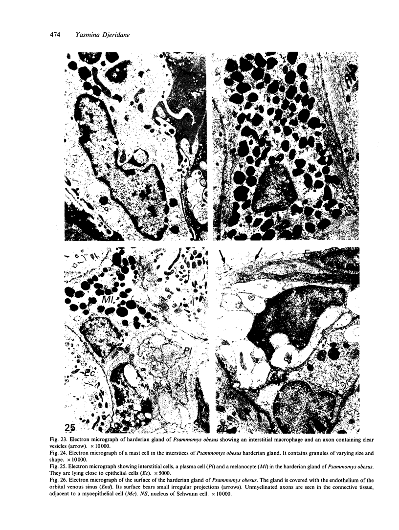
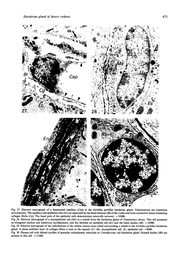
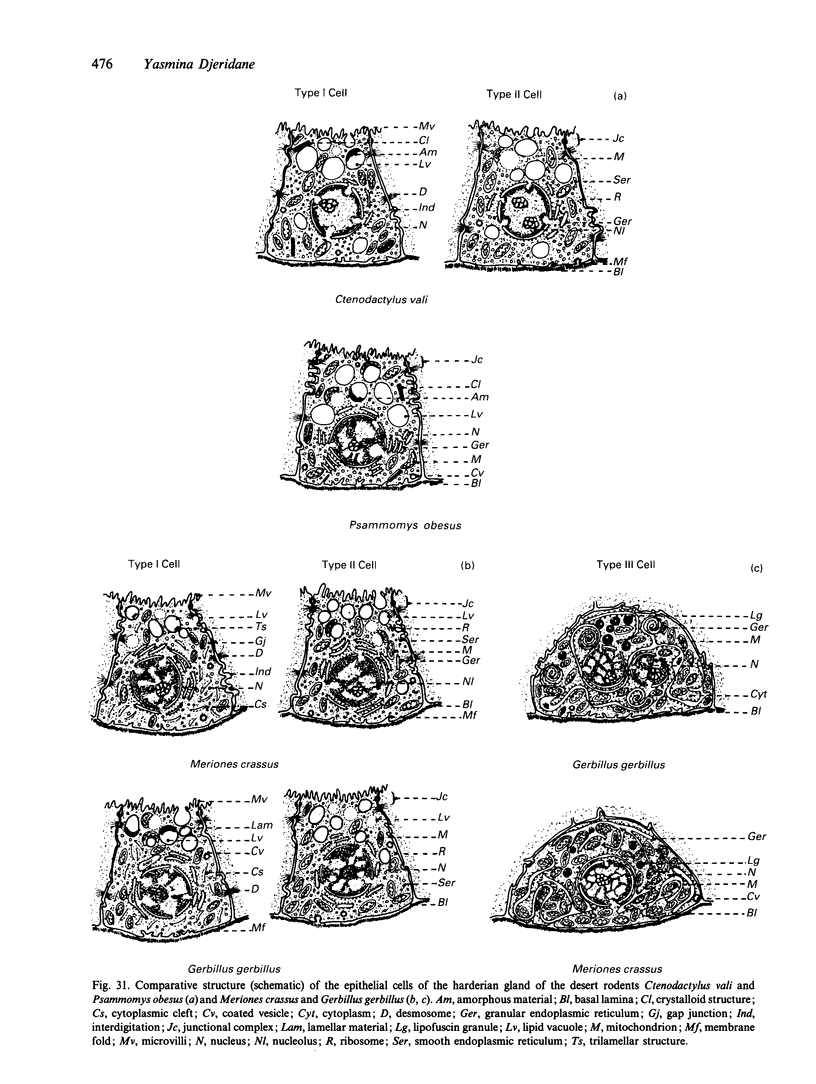
This study describes the structure of the harderian gland in desert rodents: 3 Gerbillidae species (Gerbillus gerbillus, Meriones crassus, Psammomys obesus) and 1 Ctenodactylidae species (Ctenodactylus vali). In all these species the gland consists of tubules lined by a single layer of epithelial cells and possesses myoepithelial cells within their basal laminae. The gland contains porphyrin which is stored as solid intraluminal deposits. The glandular epithelium presents a single cell type (type I) in Psammomys obesus, 2 cell types (I and II) in Ctenodactylus vali and 3 (I, II and III) in Gerbillus gerbillus and Meriones crassus. The type I and II cells are columnar. They are characterised by many lipid vacuoles and a well developed vesicle-like structure of smooth endoplasmic reticulum. In Gerbillus gerbillus and Meriones crassus the type I cells can be distinguished from type II cells by cytoplasmic clefts approximately 1 micron in length. In Ctenodactylus vali type I cells are characterised by cytoplasmic rod-shaped crystalloid structures approximately 0.5 microns in length which are frequently observed in the mitochondrial matrix. These structures are also present in the sole cell type of Psammomys obesus. Most of the secretory lipid vacuoles of the type I cell contain an electron-dense material, possibly porphyrin, which presents different appearances according to species: it is lamellar in Gerbillus gerbillus, trilamellar in Meriones crassus, and amorphous in Psammomys obesus and Ctenodactylus vali. Secretory lipid vacuoles are released primarily by exocytosis, but holocrine and apocrine secretion is also observed. The type III cells are pyramidal. This cell type is characterised by the presence of an extraordinarily well developed granular endoplasmic reticulum, organised in concentric lamellae in Gerbillus gerbillus, and very numerous mitochondria. Epithelial cells are frequently binucleate. The single excretory duct contains both mucous and serous cells. Mast cells, plasma cells, macrophages, fenestrated capillaries and unmyelinated nerve endings with clear or dense-cored vesicles are present in the connective tissue. Melanocytes are very numerous in the interstices of the Gerbillidae harderian gland. The gland is surrounded by a collagenous capsule and an outer layer of endothelial cells derived from the orbital venous sinus.
Full text
PDF


Images in this article


Selected References
These references are in PubMed. This may not be the complete list of references from this article.
- ABOU-HARB M., ABOU-HARB N. LA GLANDE DE HARDER CHEZ CERTAINS RONGEURS SAUVAGES D'ESERTIQUES ET TROPICAUX ET L''ELABORATION DES PROPHYRINES. C R Seances Soc Biol Fil. 1964;158:2268–2270. [PubMed] [Google Scholar]
- BJOERKMAN N., NICANDER L., SCHANTZ B. On the histology and ultrastructure of the Harderian gland in rabbits. Z Zellforsch Mikrosk Anat. 1960;52:93–104. doi: 10.1007/BF00344641. [DOI] [PubMed] [Google Scholar]
- Brownscheidle C. M., Niewenhuis R. J. Ultrastructure of the harderian gland in male albino rats. Anat Rec. 1978 Mar;190(3):735–753. doi: 10.1002/ar.1091900309. [DOI] [PubMed] [Google Scholar]
- Bubenik G. A., Brown G. M., Grota L. J. Immunohistochemical localization of melatonin in the rat Harderian gland. J Histochem Cytochem. 1976 Nov;24(11):1173–1177. doi: 10.1177/24.11.63506. [DOI] [PubMed] [Google Scholar]
- Bucana C. D., Nadakavukaren M. J. Fine structure of the hamster Harderian gland. Z Zellforsch Mikrosk Anat. 1972;129(2):178–187. doi: 10.1007/BF00306934. [DOI] [PubMed] [Google Scholar]
- Bucana C. D., Nadakavukaren M. J. Innervation of the hamster Harderian gland. Science. 1972 Jan 14;175(4018):205–206. doi: 10.1126/science.175.4018.205. [DOI] [PubMed] [Google Scholar]
- Burns R. B. Histological and immunological studies on the fowl lacrimal gland following surgical excision of Harder's gland. Res Vet Sci. 1979 Jul;27(1):69–75. [PubMed] [Google Scholar]
- COHN S. A. Histochemical observations on the Harderian gland of the albino mouse. J Histochem Cytochem. 1955 Sep;3(5):342–353. doi: 10.1177/3.5.342. [DOI] [PubMed] [Google Scholar]
- Carriere R. Ultrastructural visualization of intracellular porphyrin in the rat Harderian gland. Anat Rec. 1985 Dec;213(4):496–504. doi: 10.1002/ar.1092130404. [DOI] [PubMed] [Google Scholar]
- Huhtala A., Huikuri K. T., Palkama A., Tervo T. Innervation of the rat Harderian gland by adrenergic and cholinergic nerve fibres. Anat Rec. 1977 Jun;188(2):263–271. doi: 10.1002/ar.1091880210. [DOI] [PubMed] [Google Scholar]
- Johnston H. S., McGadey J., Thompson G. G., Moore M. R., Breed W. G., Payne A. P. The Harderian gland, its secretory duct and porphyrin content in the Plains mouse (Pseudomys australis). J Anat. 1985 Mar;140(Pt 2):337–350. [PMC free article] [PubMed] [Google Scholar]
- Johnston H. S., McGadey J., Thompson G. G., Moore M. R., Payne A. P. The Harderian gland, its secretory duct and porphyrin content in the mongolian gerbil (Meriones unguiculatus). J Anat. 1983 Oct;137(Pt 3):615–630. [PMC free article] [PubMed] [Google Scholar]
- Kühnel W. Struktur und Cytochemie der Harderschen Drüse von Kaninchen. Z Zellforsch Mikrosk Anat. 1971;119(3):384–404. [PubMed] [Google Scholar]
- Maxwell M. H., Rothwell B., Burns R. B. A fine structural study of the turkey harderian gland. J Anat. 1986 Oct;148:147–157. [PMC free article] [PubMed] [Google Scholar]
- Payne A. P., McGadey J., Johnston H. S., Moore M. R., Thompson G. G. Mast cells in the hamster Harderian gland: sex differences, hormonal control and relationship to porphyrin. J Anat. 1982 Oct;135(Pt 3):451–461. [PMC free article] [PubMed] [Google Scholar]
- Payne A. P. The attractiveness of Harderian gland smears to sexually naive and experienced male golden hamsters. Anim Behav. 1979 Aug;27(Pt 3):897–904. doi: 10.1016/0003-3472(79)90027-7. [DOI] [PubMed] [Google Scholar]
- REYNOLDS E. S. The use of lead citrate at high pH as an electron-opaque stain in electron microscopy. J Cell Biol. 1963 Apr;17:208–212. doi: 10.1083/jcb.17.1.208. [DOI] [PMC free article] [PubMed] [Google Scholar]
- Reiter R. J., Klein D. C. Observations on the pineal gland, the Harderian glands, the retina, and the reproductive organs of adult female rats exposed to continuous light. J Endocrinol. 1971 Sep;51(1):117–125. doi: 10.1677/joe.0.0510117. [DOI] [PubMed] [Google Scholar]
- Reiter R. J., Richardson B. A., Matthews S. A., Lane S. J., Ferguson B. N. Rhythms in immunoreactive melatonin in the retina and Harderian gland of rats: persistence after pinealectomy. Life Sci. 1983 Mar 14;32(11):1229–1236. doi: 10.1016/0024-3205(83)90192-3. [DOI] [PubMed] [Google Scholar]
- Sakai T. The mammalian Harderian gland: morphology, biochemistry, function and phylogeny. Arch Histol Jpn. 1981 Sep;44(4):299–333. doi: 10.1679/aohc1950.44.299. [DOI] [PubMed] [Google Scholar]
- Sakai T., Yohro T. A histological study of the Harderian gland of Mongolian gerbils, Meriones meridianus. Anat Rec. 1981 Jul;200(3):259–270. doi: 10.1002/ar.1092000304. [DOI] [PubMed] [Google Scholar]
- Strum J. M., Shear C. R. Constant light exposure induces damage and squamous metaplasia in Harderian glands of albino mice. Tissue Cell. 1982;14(1):149–161. doi: 10.1016/0040-8166(82)90014-3. [DOI] [PubMed] [Google Scholar]
- Strum J. M., Shear C. R. Harderian glands in mice: fluorescence, peroxidase activity and fine structure. Tissue Cell. 1982;14(1):135–148. doi: 10.1016/0040-8166(82)90013-1. [DOI] [PubMed] [Google Scholar]
- Thiessen D. D., Kittrell E. M. The Harderian gland and thermoregulation in the gerbil (Meriones unguiculatus). Physiol Behav. 1980 Mar;24(3):417–424. doi: 10.1016/0031-9384(80)90229-2. [DOI] [PubMed] [Google Scholar]
- Vivien-Roels B., Pévet P., Dubois M. P., Arendt J., Brown G. M. Immunohistochemical evidence for the presence of melatonin in the pineal gland, the retina and the Harderian gland. Cell Tissue Res. 1981;217(1):105–115. doi: 10.1007/BF00233830. [DOI] [PubMed] [Google Scholar]
- WOODHOUSE M. A., RHODIN J. A. THE ULTRASTRUCTURE OF THE HARDERIAN GLAND OF THE MOUSE WITH PARTICULAR REFERENCE TO THE FORMATION OF ITS SECRETORY PRODUCT. J Ultrastruct Res. 1963 Aug;49:76–98. doi: 10.1016/s0022-5320(63)80037-4. [DOI] [PubMed] [Google Scholar]
- Watanabe M. An autoradiographic, biochemical, and morphological study of the harderian gland of the mouse. J Morphol. 1980 Mar;163(3):349–365. doi: 10.1002/jmor.1051630308. [DOI] [PubMed] [Google Scholar]
- Weaker F. J. Light microscopic and ultrastructural features of the Harderian gland of the nine-banded armadillo. J Anat. 1981 Aug;133(Pt 1):49–65. [PMC free article] [PubMed] [Google Scholar]
- Wetterberg L., Geller E., Yuwiler A. Harderian gland: an extraretinal photoreceptor influencing the pineal gland in neonatal rats? Science. 1970 Feb 6;167(3919):884–885. doi: 10.1126/science.167.3919.884. [DOI] [PubMed] [Google Scholar]