Abstract
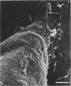
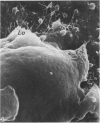
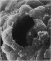
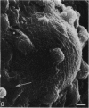
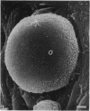
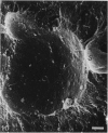
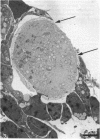
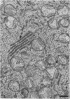
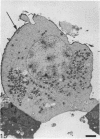
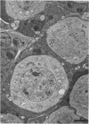
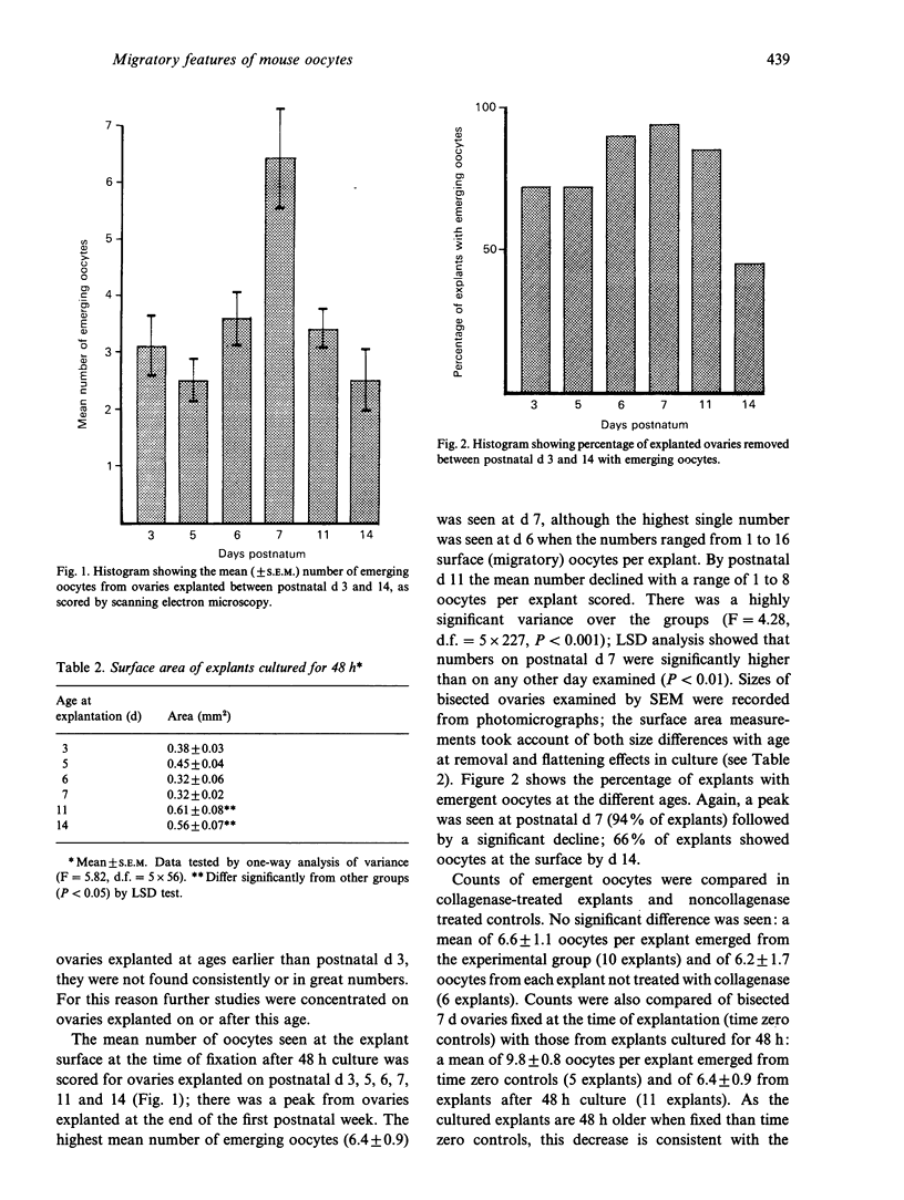
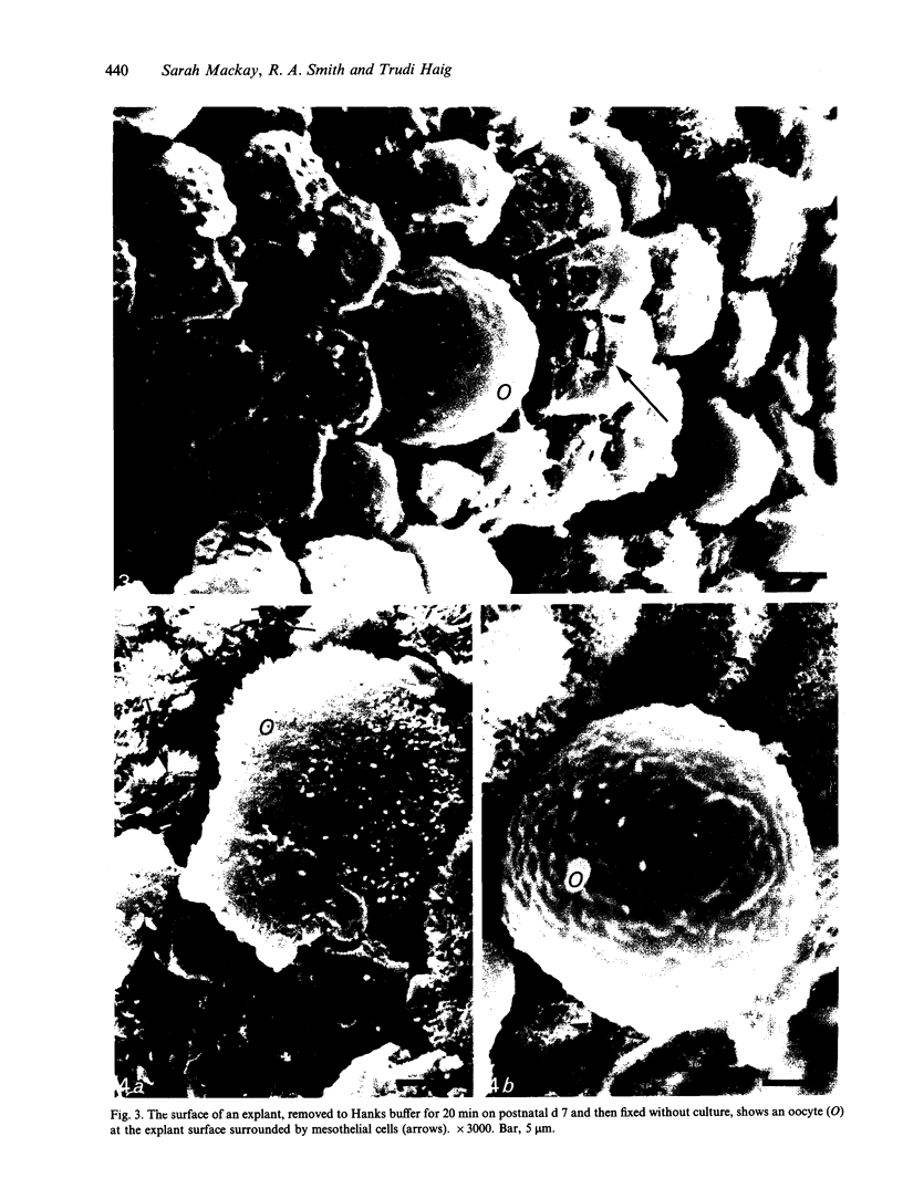
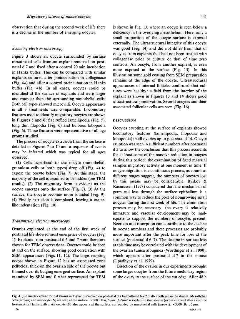
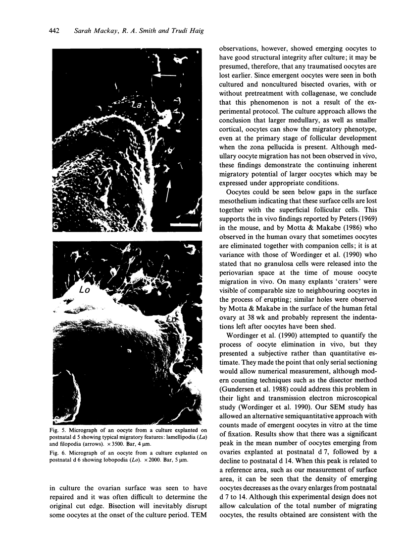
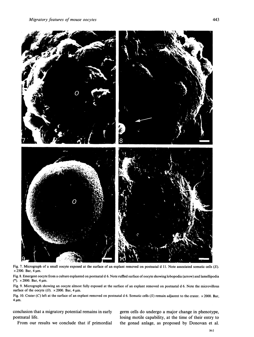
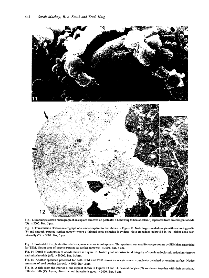
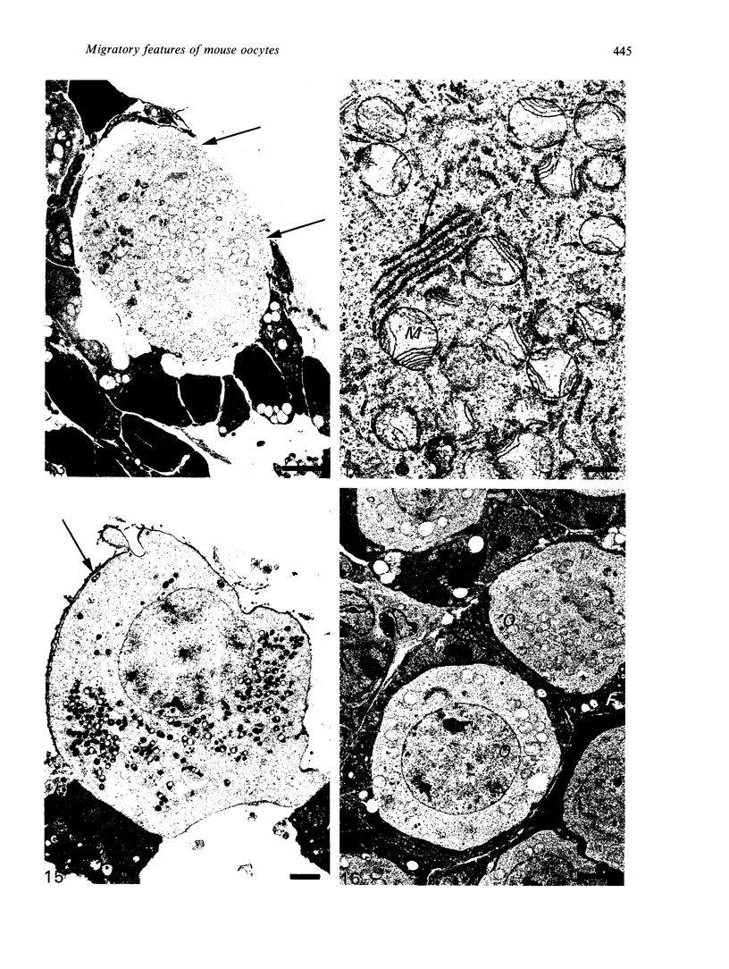
This investigation was undertaken to determine the potential of mouse oocytes for migratory activity using bisected ovaries in vitro. Bisection allowed larger medullary oocytes to be brought nearer to the surface; in this way the migratory potential of all oocytes could be studied. Observations were made following 48 h culture to allow for recovery from any initial traumatic effects resulting from bisection. Ovaries were explanted from fetuses at d 15 postcoitum and from neonatal and postnatal mice (d 1-7, 11, 12 and 14 of life) and examined by scanning and transmission electron microscopy. Oocytes were extruded from the surface and a sequence of events was inferred. Cells superficial to the oocyte sloughed off, exposing the oocytes which showed the migratory phenotype as they emerged onto the surface. Here each oocyte became rounder and was finally extruded, leaving a 'crater'. Scanning electron microscopy of the explant surface allowed counts to be made of emergent oocytes. The number of explants showing emergent oocytes was at a maximum when ovaries were removed at the end of the first week postnatum; the mean number of oocytes emerging from each also peaked at this time. Numbers of migratory oocytes declined in ovaries aged 11 d at explantation and by d 14 only 66% of explants showed oocytes at the surface. The distribution of oocytes of various sizes at the surface suggests that both small cortical oocytes and larger medullary oocytes can express the migratory phenotype. Transmission electron microscopy verified structural integrity of the emerging oocytes and revealed their relationship to underlying cells.
Full text
PDF
Images in this article
Selected References
These references are in PubMed. This may not be the complete list of references from this article.
- BLANDAU R. J., WHITE B. J., RUMERY R. E. OBSERVATIONS ON THE MOVEMENTS OF THE LIVING PRIMORDIAL GERM CELLS IN THE MOUSE. Fertil Steril. 1963 Sep-Oct;14:482–489. doi: 10.1016/s0015-0282(16)34981-0. [DOI] [PubMed] [Google Scholar]
- Donovan P. J., Stott D., Cairns L. A., Heasman J., Wylie C. C. Migratory and postmigratory mouse primordial germ cells behave differently in culture. Cell. 1986 Mar 28;44(6):831–838. doi: 10.1016/0092-8674(86)90005-x. [DOI] [PubMed] [Google Scholar]
- Gosden R. G., Telfer E., Faddy M. J., Brook D. J. Ovarian cyclicity and follicular recruitment in unilaterally ovariectomized mice. J Reprod Fertil. 1989 Sep;87(1):257–264. doi: 10.1530/jrf.0.0870257. [DOI] [PubMed] [Google Scholar]
- Gundersen H. J., Bagger P., Bendtsen T. F., Evans S. M., Korbo L., Marcussen N., Møller A., Nielsen K., Nyengaard J. R., Pakkenberg B. The new stereological tools: disector, fractionator, nucleator and point sampled intercepts and their use in pathological research and diagnosis. APMIS. 1988 Oct;96(10):857–881. doi: 10.1111/j.1699-0463.1988.tb00954.x. [DOI] [PubMed] [Google Scholar]
- JONES E. C., KROHN P. L. The relationships between age, numbers of ocytes and fertility in virgin and multiparous mice. J Endocrinol. 1961 Feb;21:469–495. doi: 10.1677/joe.0.0210469. [DOI] [PubMed] [Google Scholar]
- Mackay S., Smith R. A. Mouse gonadal differentiation in vitro in the presence of fetal calf serum. Cell Differ Dev. 1989 Jun;27(1):19–28. doi: 10.1016/0922-3371(89)90041-5. [DOI] [PubMed] [Google Scholar]
- Motta P. M., Makabe S. Elimination of germ cells during differentiation of the human ovary: an electron microscopic study. Eur J Obstet Gynecol Reprod Biol. 1986 Sep;22(5-6):271–286. doi: 10.1016/0028-2243(86)90115-2. [DOI] [PubMed] [Google Scholar]
- Peters H. The development of the mouse ovary from birth to maturity. Acta Endocrinol (Copenh) 1969 Sep;62(1):98–116. doi: 10.1530/acta.0.0620098. [DOI] [PubMed] [Google Scholar]
- Wordinger R., Sutton J., Brun-Zinkernagel A. M. Ultrastructure of oocyte migration through the mouse ovarian surface epithelium during neonatal development. Anat Rec. 1990 Jun;227(2):187–198. doi: 10.1002/ar.1092270207. [DOI] [PubMed] [Google Scholar]


