Abstract
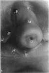
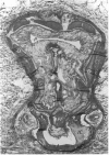
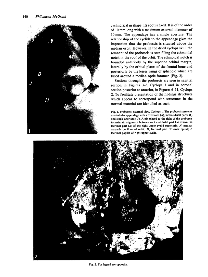
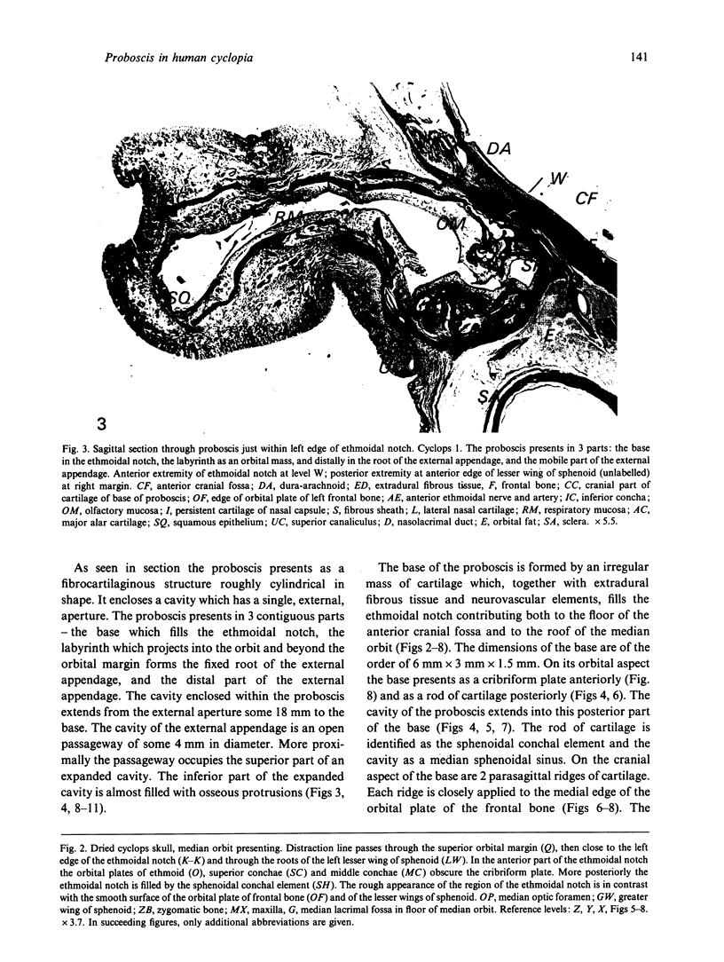
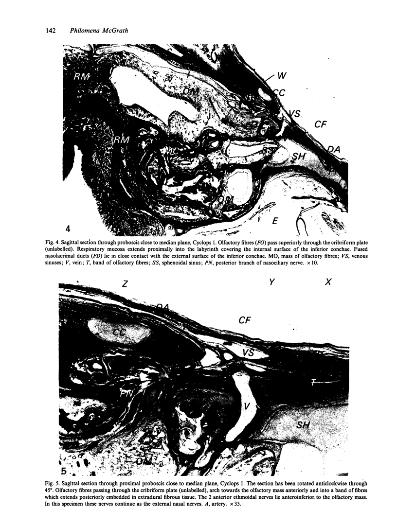
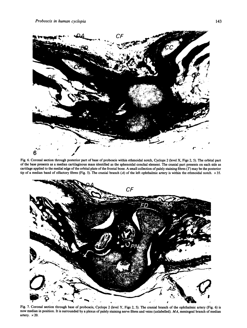
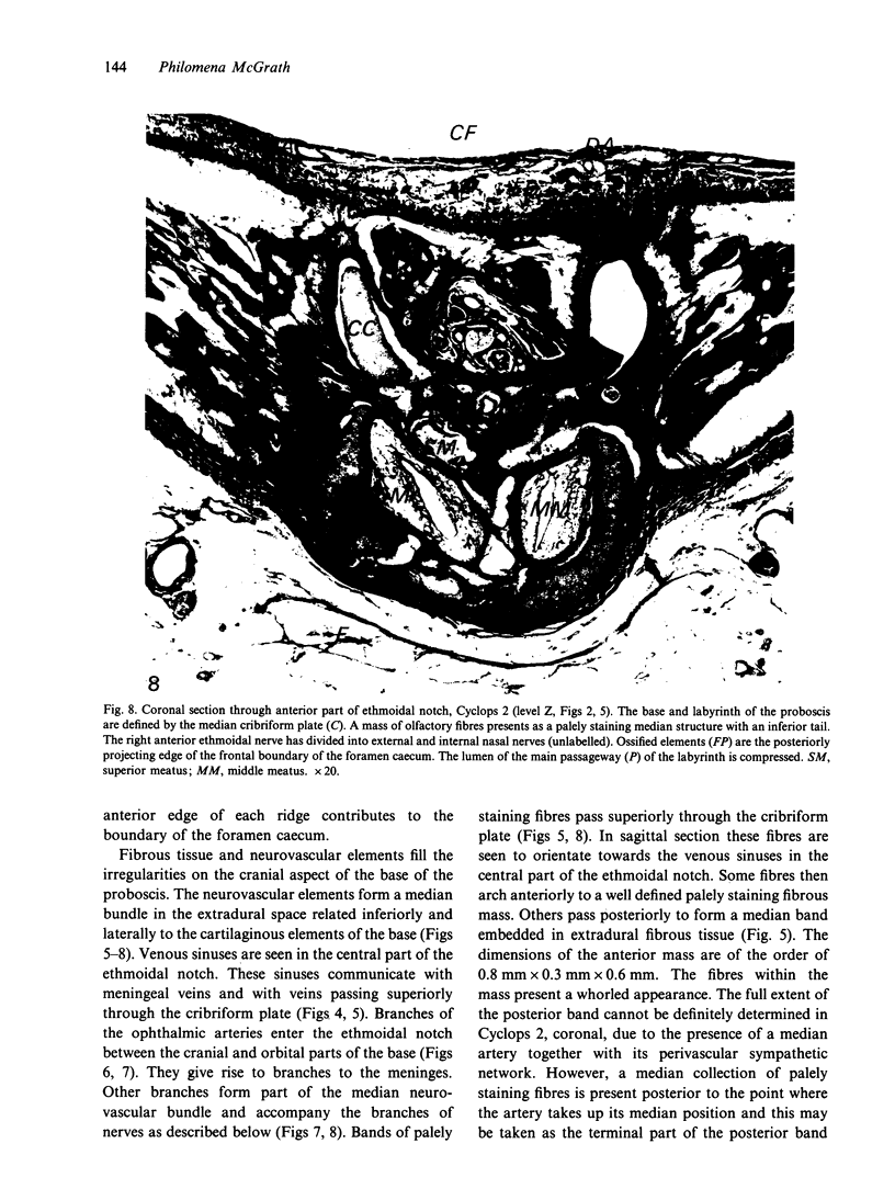
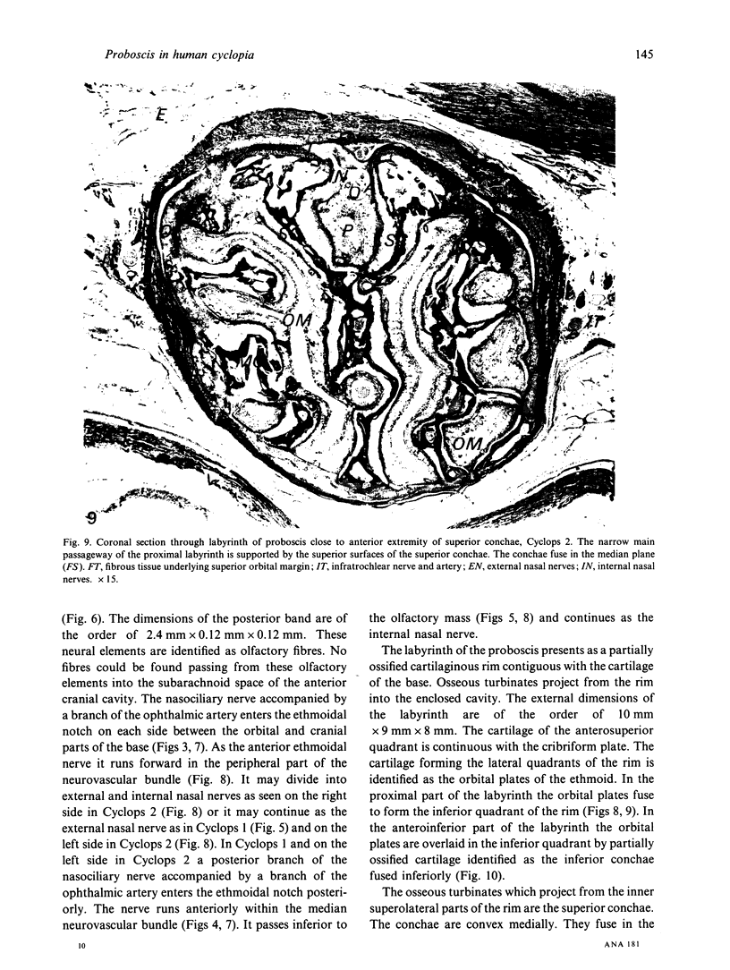
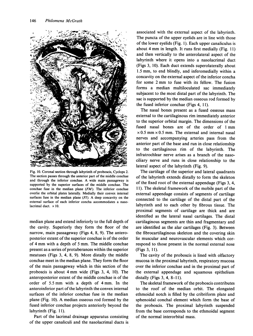
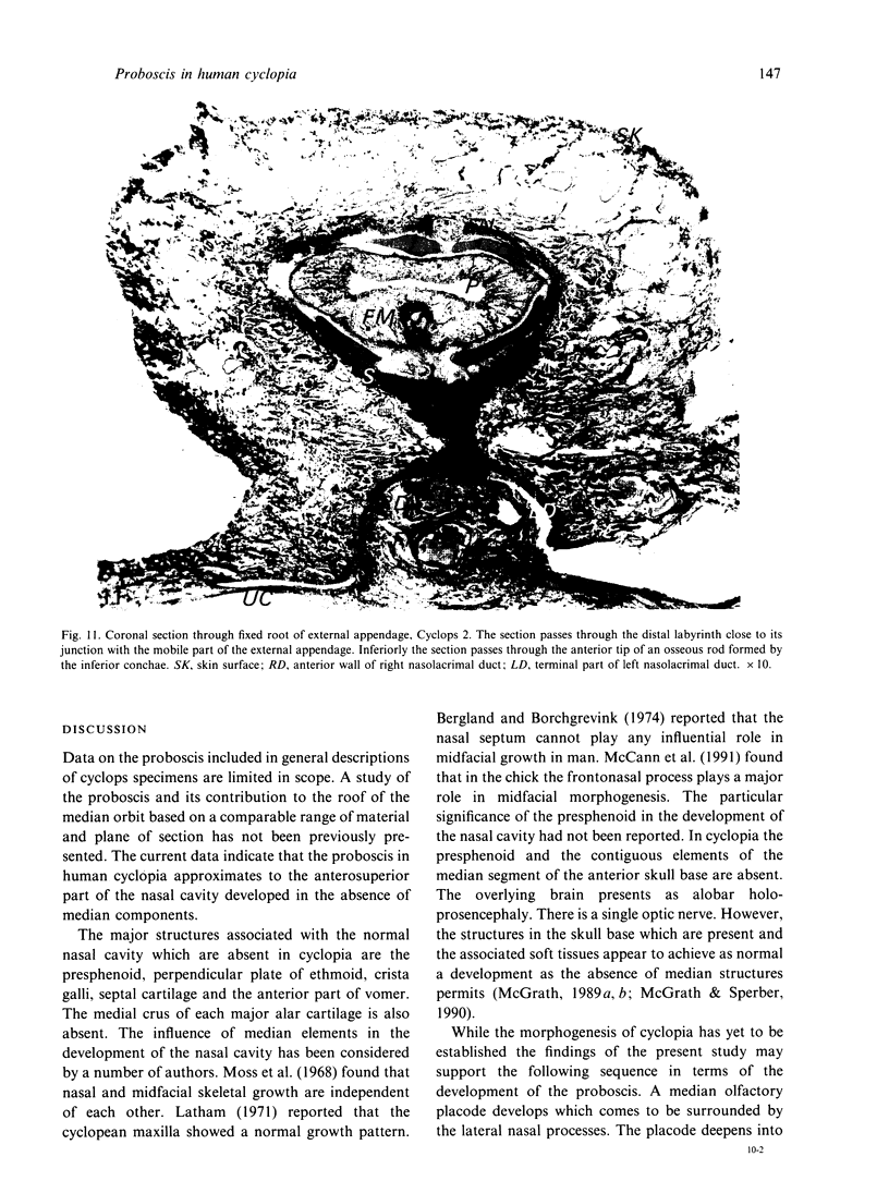
A 2-dimensional anatomical study has been undertaken of the proboscis and its contribution to the roof of the median orbit in human cyclopia. The cyclops material consists of 4 sectioned fetal heads and a dried cyclops skull. The skeleton of the proboscis is formed by the nasal capsule. The base of the proboscis lies in the floor of the anterior cranial fossa filling an extended ethmoidal notch and contributing to the roof of the median orbit anterior to the fused lesser wings of sphenoid. The cavity of the proboscis is lined with squamous epithelium, respiratory and olfactory mucosa. Olfactory fibres pass from the proboscis into the extradural space of the ethmoidal notch forming a collection of tissue similar to the inferior layer of the normal olfactory bulb. The data indicate that the proboscis represents the anterosuperior part of the normal nasal cavity developed in the absence of median components. It is suggested that the cyclops face constitutes a model for the study of the development of the normal face.
Full text
PDF
Images in this article
Selected References
These references are in PubMed. This may not be the complete list of references from this article.
- Bergland O., Borchgrevink H. The role of the nasal septum in midfacial growth in man elucidated by the maxillary development in certain types of facial clefts. A preliminary report. Scand J Plast Reconstr Surg. 1974;8(1-2):42–48. doi: 10.3109/02844317409084369. [DOI] [PubMed] [Google Scholar]
- Deligdisch L., Legum C., Peyser M. R., Toaff R. Cyclopia associated with triploidy and hydatidiform mole: a case report. Teratology. 1978 Oct;18(2):257–261. doi: 10.1002/tera.1420180212. [DOI] [PubMed] [Google Scholar]
- Kokich V. G., Ngim C. H., Siebert J. R., Clarren S. K., Cohen M. M., Jr Cyclopia: an anatomic and histologic study of two specimens. Teratology. 1982 Oct;26(2):105–113. doi: 10.1002/tera.1420260202. [DOI] [PubMed] [Google Scholar]
- Latham R. A. Mechanism of maxillary growth in the human cyclops. J Dent Res. 1971 Jul-Aug;50(4):929–933. doi: 10.1177/00220345710500042401. [DOI] [PubMed] [Google Scholar]
- McCann J. P., Owens P. D., Wilson D. J. Chick frontonasal process excision significantly affects mid-facial development. Anat Embryol (Berl) 1991;184(2):171–178. doi: 10.1007/BF00942748. [DOI] [PubMed] [Google Scholar]
- McGrath P. Palatine bone in human cyclopia. Acta Anat (Basel) 1989;135(2):149–157. doi: 10.1159/000146745. [DOI] [PubMed] [Google Scholar]
- McGrath P., Sperber G. H. Floor of the median orbit in human cyclopia: an anatomical study in three dimensions. J Anat. 1990 Apr;169:125–138. [PMC free article] [PubMed] [Google Scholar]
- McGrath P. Upper jaw in human cyclopia. Acta Anat (Basel) 1989;136(3):235–241. doi: 10.1159/000146892. [DOI] [PubMed] [Google Scholar]
- Moss M. L., Bromberg B. E., Song I. C., Eisenman G. The passive role of nasal septal cartilage in mid-facial growth. Plast Reconstr Surg. 1968 Jun;41(6):536–542. doi: 10.1097/00006534-196806000-00004. [DOI] [PubMed] [Google Scholar]
- SARMA V. OCULAR ABNORMALITIES OF THE FOETUS WITH SPECIAL REFERENCE TO CYCLOPIA. Br J Ophthalmol. 1963 Apr;47:193–202. doi: 10.1136/bjo.47.4.193. [DOI] [PMC free article] [PubMed] [Google Scholar]
- Torczynski E., Jacobiec F. A., Johnston M. C., Font R. L., Madewell J. A. Synophthalmia and cyclopia: a histopathologic, radiographic, and organogenetic analysis. Doc Ophthalmol. 1977 Dec 30;44(2):311–378. doi: 10.1007/BF00230088. [DOI] [PubMed] [Google Scholar]


