

Abstract
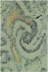
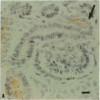
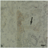
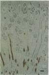
The human kidney develops from 2 embryonic tissues, the ureteric bud and the metanephric blastema. The site in the adult renal distal tubule corresponding to the junction between these tissues has never been established unequivocally and is usually said to be the union between the collecting duct and the connecting piece, based on microdissection evidence. We have examined kidneys from 21 human fetuses of various ages using an immunohistological method for substances related to the ABO blood group system, various cytokeratins including those detected by the monoclonal antibody PKK2, and Tamm-Horsfall protein. The ureteric bud and connecting piece expressed the type 1 precursor chain of ABO antigens mostly early in gestation, the H antigen of the ABO system mostly later in gestation, and cytokeratins detected by PKK2. The induced nephrons after the S-shaped body stage expressed Tamm-Horsfall protein. In the adult renal tubule, distal from the macula densa, it was already known that there is a sharp junction between the segment expressing Tamm-Horsfall protein and the more distal segment that expresses the H antigen and cytokeratins detected by PKK2. The finding that the ureteric bud and connecting piece express the same antigens as this segment while the S-shaped body eventually expresses Tamm-Horsfall protein is consistent with the concept that (1) the connecting piece arises from the ureteric bud, not the S-shaped body, and (2) the junction of ureteric bud derivatives and metanephric blastema derivatives is on the distal side of the macula densa at the distal end of Tamm-Horsfall staining.
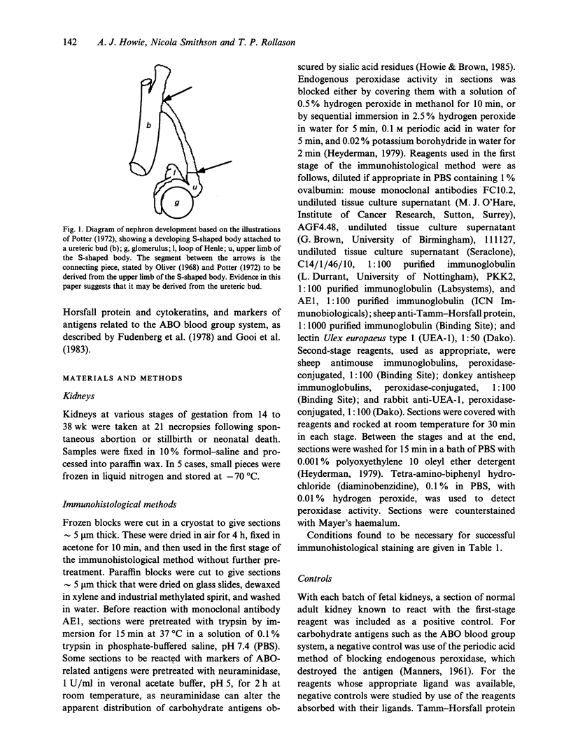
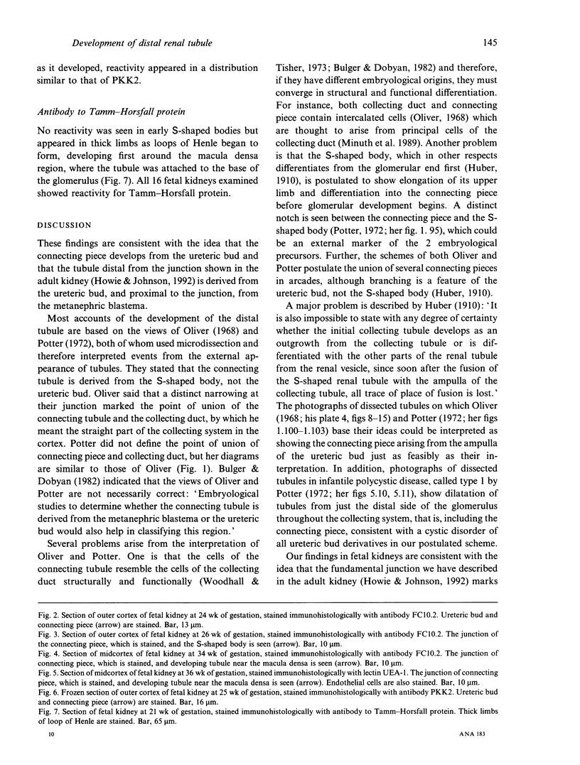
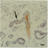
Full text
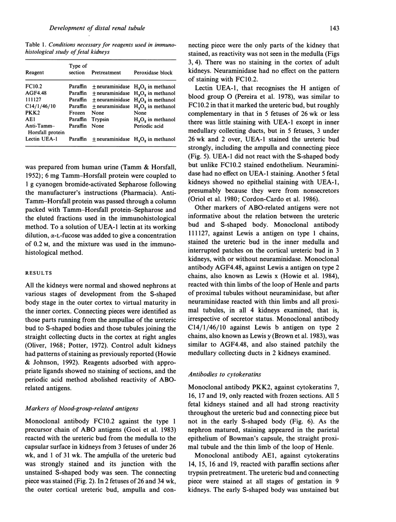
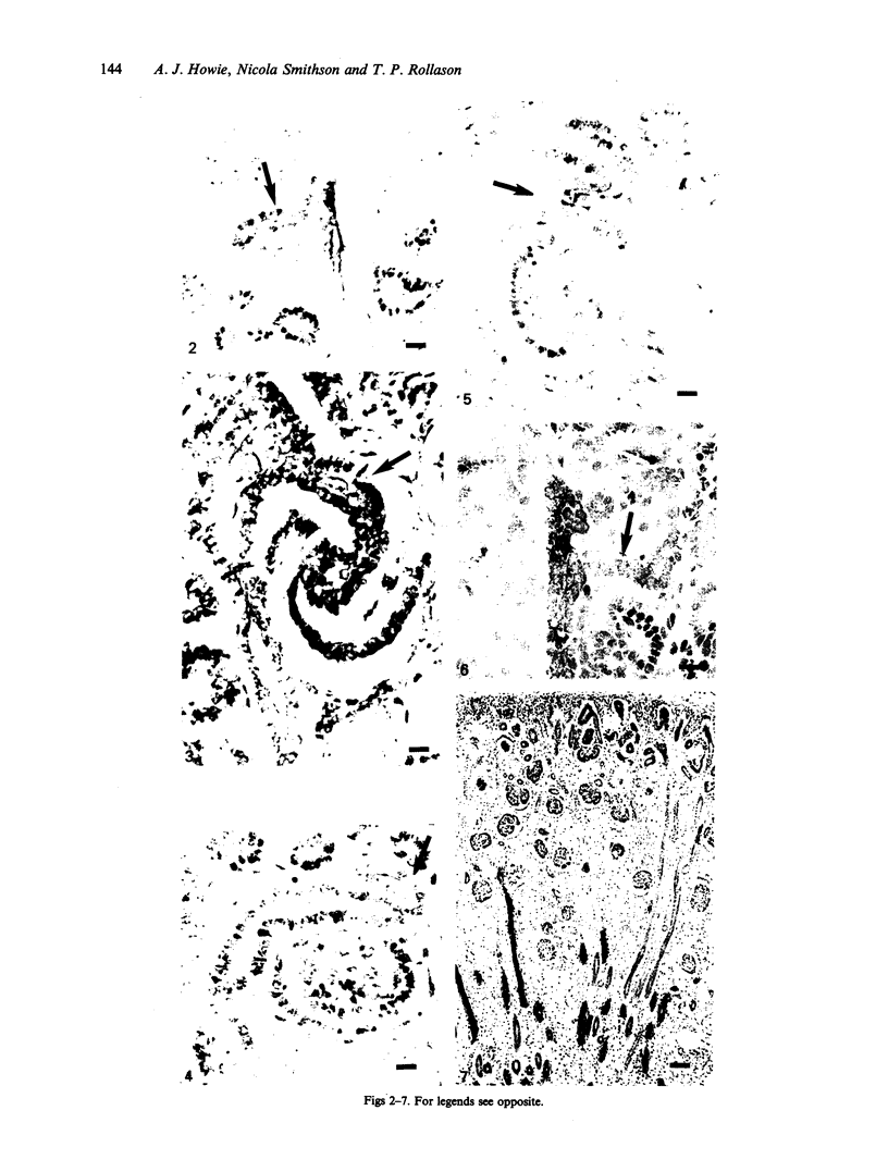
PDF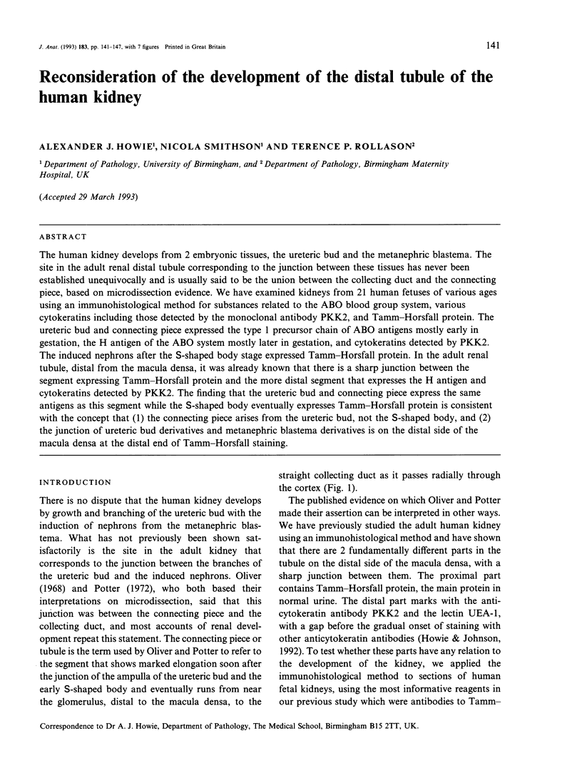
Images in this article

Selected References
These references are in PubMed. This may not be the complete list of references from this article.
- Brown A., Feizi T., Gooi H. C., Embleton M. J., Picard J. K., Baldwin R. W. A monoclonal antibody against human colonic adenoma recognizes difucosylated Type-2-blood-group chains. Biosci Rep. 1983 Feb;3(2):163–170. doi: 10.1007/BF01121947. [DOI] [PubMed] [Google Scholar]
- Bulger R. E., Dobyan D. C. Recent advances in renal morphology. Annu Rev Physiol. 1982;44:147–179. doi: 10.1146/annurev.ph.44.030182.001051. [DOI] [PubMed] [Google Scholar]
- Cordon-Cardo C., Lloyd K. O., Finstad C. L., McGroarty M. E., Reuter V. E., Bander N. H., Old L. J., Melamed M. R. Immunoanatomic distribution of blood group antigens in the human urinary tract. Influence of secretor status. Lab Invest. 1986 Oct;55(4):444–454. [PubMed] [Google Scholar]
- Ekblom P., Miettinen A., Virtanen I., Wahlström T., Dawnay A., Saxén L. In vitro segregation of the metanephric nephron. Dev Biol. 1981 May;84(1):88–95. doi: 10.1016/0012-1606(81)90373-0. [DOI] [PubMed] [Google Scholar]
- Gooi H. C., Williams L. K., Uemura K., Hounsell E. F., McIlhinney R. A., Feizi T. A marker of human foetal endoderm defined by a monoclonal antibody involves Type 1 blood group chains. Mol Immunol. 1983 Jun;20(6):607–613. doi: 10.1016/0161-5890(83)90005-6. [DOI] [PubMed] [Google Scholar]
- Heyderman E. Immunoperoxidase technique in histopathology: applications, methods, and controls. J Clin Pathol. 1979 Oct;32(10):971–978. doi: 10.1136/jcp.32.10.971. [DOI] [PMC free article] [PubMed] [Google Scholar]
- Howie A. J., Brown G. Effect of neuraminidase on the expression of the 3-fucosyl-N-acetyllactosamine antigen in human tissues. J Clin Pathol. 1985 Apr;38(4):409–416. doi: 10.1136/jcp.38.4.409. [DOI] [PMC free article] [PubMed] [Google Scholar]
- Howie A. J., Brown G., Fisher A. G., Khan M. Widespread distribution in human tissues of an antigenic determinant of granulocytes. J Clin Pathol. 1984 May;37(5):555–559. doi: 10.1136/jcp.37.5.555. [DOI] [PMC free article] [PubMed] [Google Scholar]
- Howie A. J., Johnson G. D. Confocal microscopic and other observations on the distal end of the thick limb of the human loop of Henle. Cell Tissue Res. 1992 Jan;267(1):11–16. doi: 10.1007/BF00318686. [DOI] [PubMed] [Google Scholar]
- Howie A. J., Lote C. J., Hillis A. N. Immunoreactive Tamm-Horsfall protein in the kidney and skin of the frog Rana temporaria. Cell Tissue Res. 1991 Mar;263(3):585–587. doi: 10.1007/BF00327292. [DOI] [PubMed] [Google Scholar]
- Minuth W. W., Gilbert P., Rudolph U., Spielman W. S. Successive histochemical differentiation steps during postnatal development of the collecting duct in rabbit kidney. Histochemistry. 1989;93(1):19–25. doi: 10.1007/BF00266842. [DOI] [PubMed] [Google Scholar]
- Oriol R., Cartron J. P., Cartron J., Mulet C. Biosynthesis of ABH and Lewis antigens in normal and transplanted kidneys. Transplantation. 1980 Mar;29(3):184–188. doi: 10.1097/00007890-198003000-00003. [DOI] [PubMed] [Google Scholar]
- Pereira M. E., Kisailus E. C., Gruezo F., Kabat E. A. Immunochemical studies on the combining site of the blood group H-specific lectin 1 from Ulex europeus seeds. Arch Biochem Biophys. 1978 Jan 15;185(1):108–115. doi: 10.1016/0003-9861(78)90149-2. [DOI] [PubMed] [Google Scholar]
- TAMM I., HORSFALL F. L., Jr A mucoprotein derived from human urine which reacts with influenza, mumps, and Newcastle disease viruses. J Exp Med. 1952 Jan;95(1):71–97. doi: 10.1084/jem.95.1.71. [DOI] [PMC free article] [PubMed] [Google Scholar]
- Williams L. K., Sullivan A., McIlhinney R. A., Neville A. M. A monoclonal antibody marker of human primitive endoderm. Int J Cancer. 1982 Dec 15;30(6):731–738. doi: 10.1002/ijc.2910300609. [DOI] [PubMed] [Google Scholar]
- Woodhall P. B., Tisher C. C. Response of the distal tubule and cortical collecting duct to vasopressin in the rat. J Clin Invest. 1973 Dec;52(12):3095–3108. doi: 10.1172/JCI107509. [DOI] [PMC free article] [PubMed] [Google Scholar]