Abstract
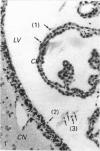
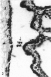
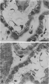
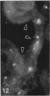
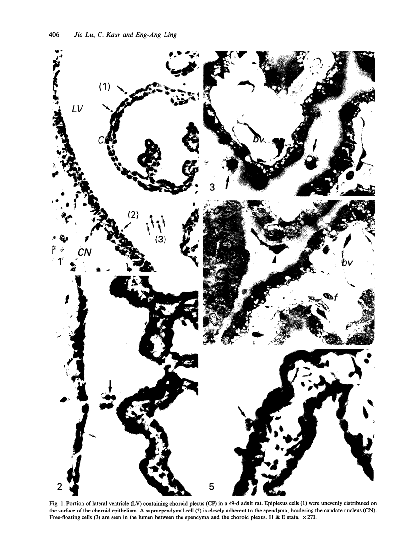
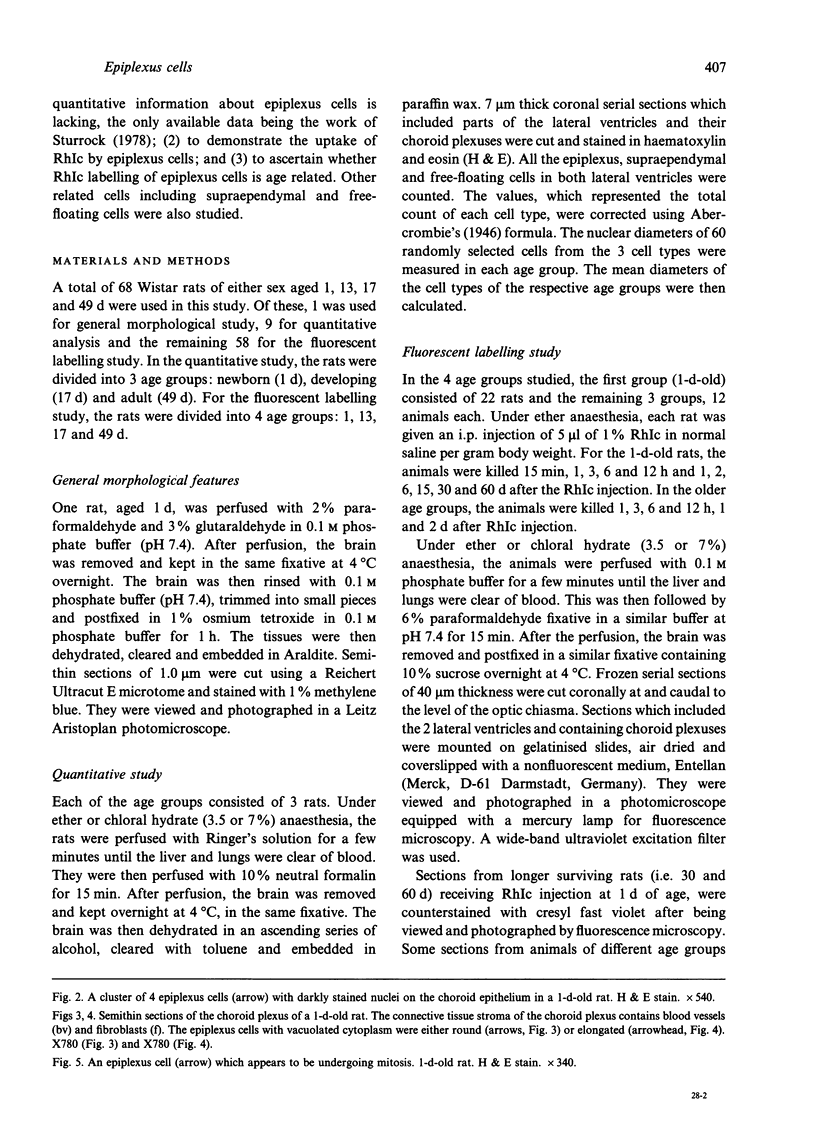
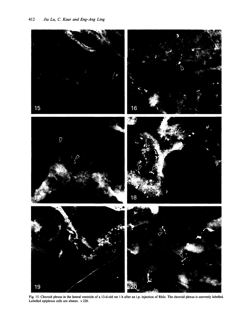
The labelling of epiplexus cells associated with the choroid plexus in the lateral ventricles was examined in rats of different ages with the fluorescent dye, rhodamine isothiocyanate (RhIc). A quantitative study was also attempted; this showed that the number of epiplexus cells and their related cells, namely supraependymal and free-floating cells, increased with age. The mean absolute number of epiplexus cells ranged from approximately 700 in the newborn to approximately 2200 in rats of 17 d of age; thereafter it remained unchanged. The number of free-floating cells also increased substantially but showed considerable individual variation. Following i.p. injection, the tracer was rapidly taken up by the epiplexus cells. This provided strong support for their phagocytic nature. In the newborn (1 d) and developing (13 d, 17 d) rats, RhIc-labelled epiplexus cells were first observed 3 h after the injection. In adult rats, labelled cells were not observed until 12 h after injection. In either case, the fluorescence in the epiplexus cells gradually increased with time. It is suggested from this study that the blood-CSF barrier in the choroid plexus in postnatal rats is incomplete, thereby allowing a rapid transvascular diffusion of the injected RhIc into the blood circulation. The fluorescent dye which enters the ventricle by way of the choroid epithelium is subsequently taken up by the epiplexus cells. Such an unimpeded passage, however, is reduced in the adult rats, probably due to the maturation of the blood capillaries as well as the choroid epithelium.
Full text
PDF
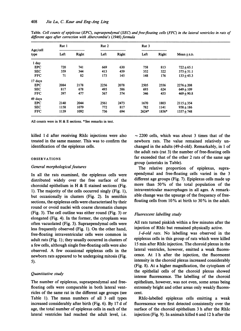
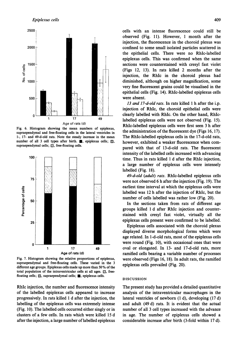
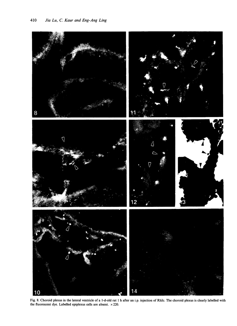
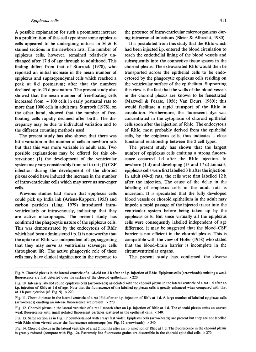
Images in this article
Selected References
These references are in PubMed. This may not be the complete list of references from this article.
- Allen D. J. Scanning electron microscopy of epiplexus macrophages (Kolmer cells) in the dog. J Comp Neurol. 1975 May 15;161(2):197–213. doi: 10.1002/cne.901610205. [DOI] [PubMed] [Google Scholar]
- Bleier R., Albrecht R. Supraependymal macrophages of third ventricle of hamster: morphological, functional and histochemical characterization in situ and in culture. J Comp Neurol. 1980 Aug 1;192(3):489–504. doi: 10.1002/cne.901920308. [DOI] [PubMed] [Google Scholar]
- Carpenter S. J., McCarthy L. E., Borison H. L. Electron microscopic study of the epiplexus (Kolmer) cells of the cat choroid plexus. Z Zellforsch Mikrosk Anat. 1970;110(4):471–486. doi: 10.1007/BF00330099. [DOI] [PubMed] [Google Scholar]
- Hosoya Y., Fujita T. Scanning electron microscope observation of intraventricular macrophages (Kolmer cells) in the rat brain. Arch Histol Jpn. 1973 Jan;35(2):133–140. doi: 10.1679/aohc1950.35.133. [DOI] [PubMed] [Google Scholar]
- KAPPERS J. A. Beitrag zur experimentellen Untersuchung von Funktion und Herkunft der Kolmerschen Zellen des Plexus chorioideus beim Axolotl und Meerschweinchen. Z Anat Entwicklungsgesch. 1953;117(1):1–19. [PubMed] [Google Scholar]
- Kaur C., Ling E. A., Gopalakrishnakone P., Wong W. C. Response of intraventricular macrophages to crotoxin-coated microcarrier beads injected into the lateral ventricle of postnatal rats. J Anat. 1990 Feb;168:63–72. [PMC free article] [PubMed] [Google Scholar]
- Leong S. K., Ling E. A. Amoeboid and ramified microglia: their interrelationship and response to brain injury. Glia. 1992;6(1):39–47. doi: 10.1002/glia.440060106. [DOI] [PubMed] [Google Scholar]
- Ling E. A., Gopalakrishnakone P., Tan C. K. Electron-microscopical study of the choroid plexus and epiplexus cells in cats following a cisternal injection of crotoxin complex. Acta Anat (Basel) 1988;131(3):241–248. doi: 10.1159/000146523. [DOI] [PubMed] [Google Scholar]
- Ling E. A., Gopalakrishnakone P., Voon F. C. Epiplexus cells in the embryos of the turtle, Trionyx sinensis. Arch Histol Jpn. 1985 Oct;48(4):355–361. doi: 10.1679/aohc.48.355. [DOI] [PubMed] [Google Scholar]
- Ling E. A., Kaur C., Wong W. C. Light and electron microscopic demonstration of non-specific esterase in amoeboid microglial cells in the corpus callosum in postnatal rats: a cytochemical link to monocytes. J Anat. 1982 Sep;135(Pt 2):385–394. [PMC free article] [PubMed] [Google Scholar]
- Ling E. A. Scanning electron microscopic study of epiplexus cells in the lateral ventricles of the monkey (Macaca fascicularis). J Anat. 1983 Dec;137(Pt 4):645–652. [PMC free article] [PubMed] [Google Scholar]
- Ling E. A. Ultrastruct and origin of epiplexus cells in the telencephalic choroid plexus of postnatal rats studied by intravenous injection of carbon particles. J Anat. 1979 Oct;129(Pt 3):479–492. [PMC free article] [PubMed] [Google Scholar]
- Ling E. A. Ultrastructure and mode of formation of epiplexus cells in the choroid plexus in the lateral ventricles of the monkey (Macaca fascicularis). J Anat. 1981 Dec;133(Pt 4):555–569. [PMC free article] [PubMed] [Google Scholar]
- MAXWELL D. S., PEASE D. C. The electron microscopy of the choroid plexus. J Biophys Biochem Cytol. 1956 Jul 25;2(4):467–474. doi: 10.1083/jcb.2.4.467. [DOI] [PMC free article] [PubMed] [Google Scholar]
- Maxwell W. L., Hardy I. G., Watt C., McGadey J., Graham D. I., Adams J. H., Gennarelli T. A. Changes in the choroid plexus, responses by intrinsic epiplexus cells and recruitment from monocytes after experimental head acceleration injury in the non-human primate. Acta Neuropathol. 1992;84(1):78–84. doi: 10.1007/BF00427218. [DOI] [PubMed] [Google Scholar]
- Maxwell W. L., McGadey J. Response of intraventricular macrophages after a penetrant cerebral lesion. J Anat. 1988 Oct;160:145–155. [PMC free article] [PubMed] [Google Scholar]
- Mitchell J. A. Morphology and distribution of type II supraependymal cells in the third ventricle of the guinea pig. J Morphol. 1979 Jan;159(1):67–80. doi: 10.1002/jmor.1051590106. [DOI] [PubMed] [Google Scholar]
- Peters A., Swan R. C. The choroid plexus of the mature and aging rat: the choroidal epithelium. Anat Rec. 1979 Jul;194(3):325–353. doi: 10.1002/ar.1091940303. [DOI] [PubMed] [Google Scholar]
- Peters A. The surface fine structure of the choroid plexus and ependymal lining of the rat lateral ventricle. J Neurocytol. 1974 Mar;3(1):99–108. doi: 10.1007/BF01111935. [DOI] [PubMed] [Google Scholar]
- Sturrock R. R. A developmental study of epiplexus cells and supraependymal cells and their possible relationship to microglia. Neuropathol Appl Neurobiol. 1978 Sep-Oct;4(5):307–322. doi: 10.1111/j.1365-2990.1978.tb01345.x. [DOI] [PubMed] [Google Scholar]
- Sturrock R. R. A light microscopic and scanning electron microscopic study of intraventricular macrophages in the brains of aged mice. J Anat. 1983 Jun;136(Pt 4):761–771. [PMC free article] [PubMed] [Google Scholar]
- Sturrock R. R. A semithin light microscopic, transmission electron microscopic and scanning electron microscopic study of macrophages in the lateral ventricle of mice from embryonic to adult life. J Anat. 1979 Aug;129(Pt 1):31–44. [PMC free article] [PubMed] [Google Scholar]
- Sturrock R. R. An ultrastructural study of intraventricular macrophages in the brains of aged mice. Anat Anz. 1988;165(4):283–290. [PubMed] [Google Scholar]
- van Deurs B. Structural aspects of brain barriers, with special reference to the permeability of the cerebral endothelium and choroidal epithelium. Int Rev Cytol. 1980;65:117–191. doi: 10.1016/s0074-7696(08)61960-9. [DOI] [PubMed] [Google Scholar]


