Abstract
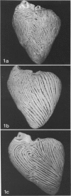
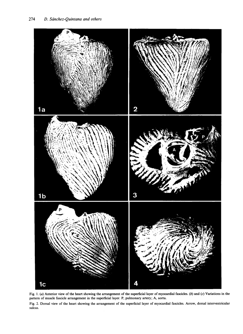
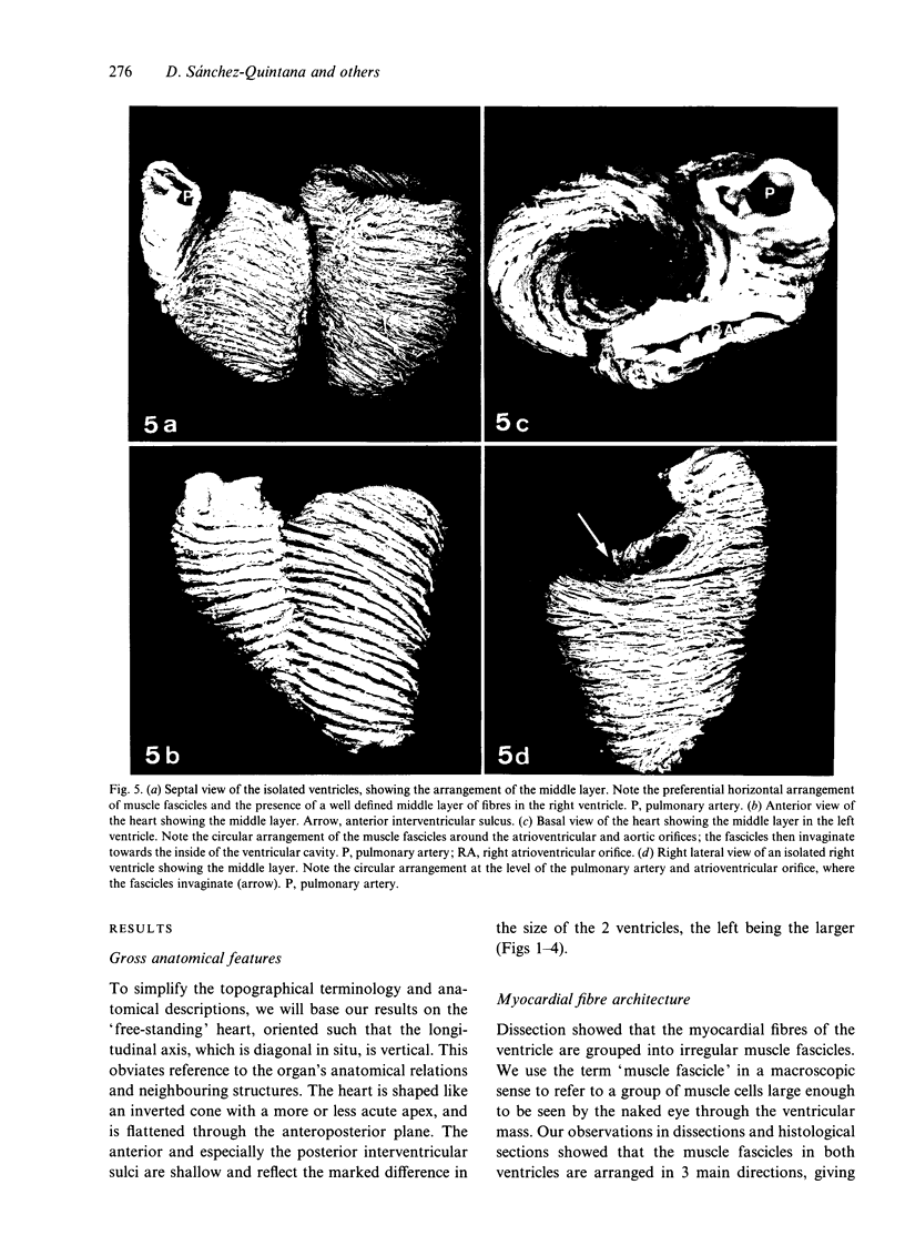
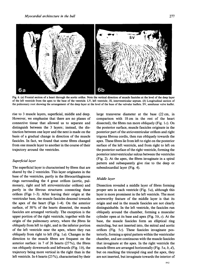
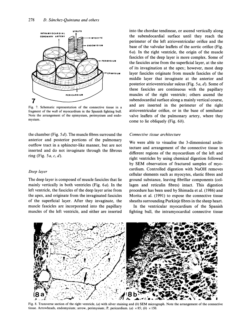
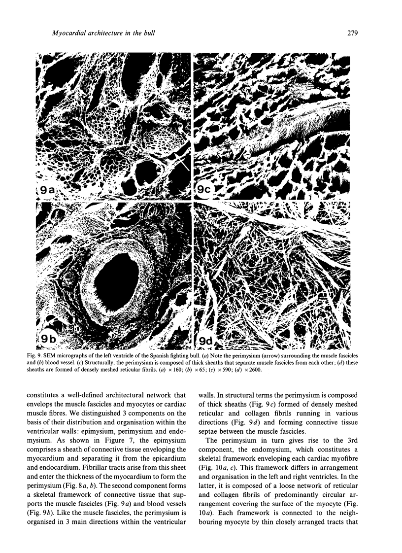
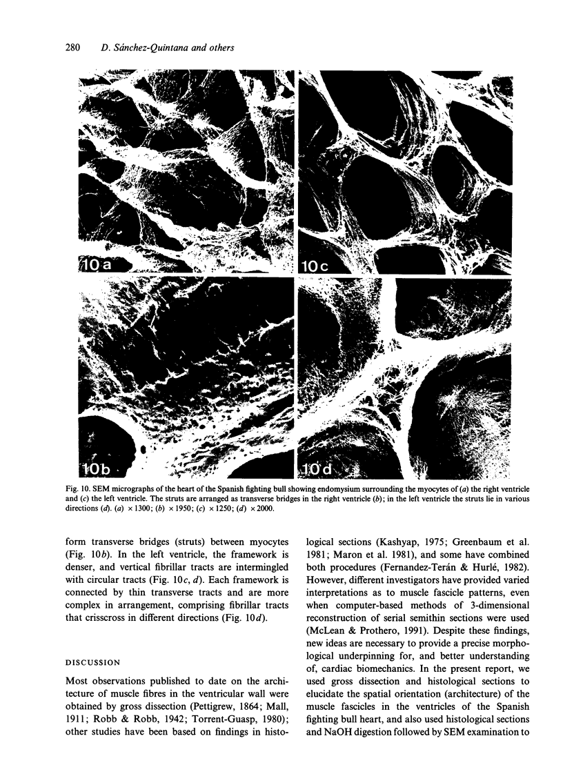
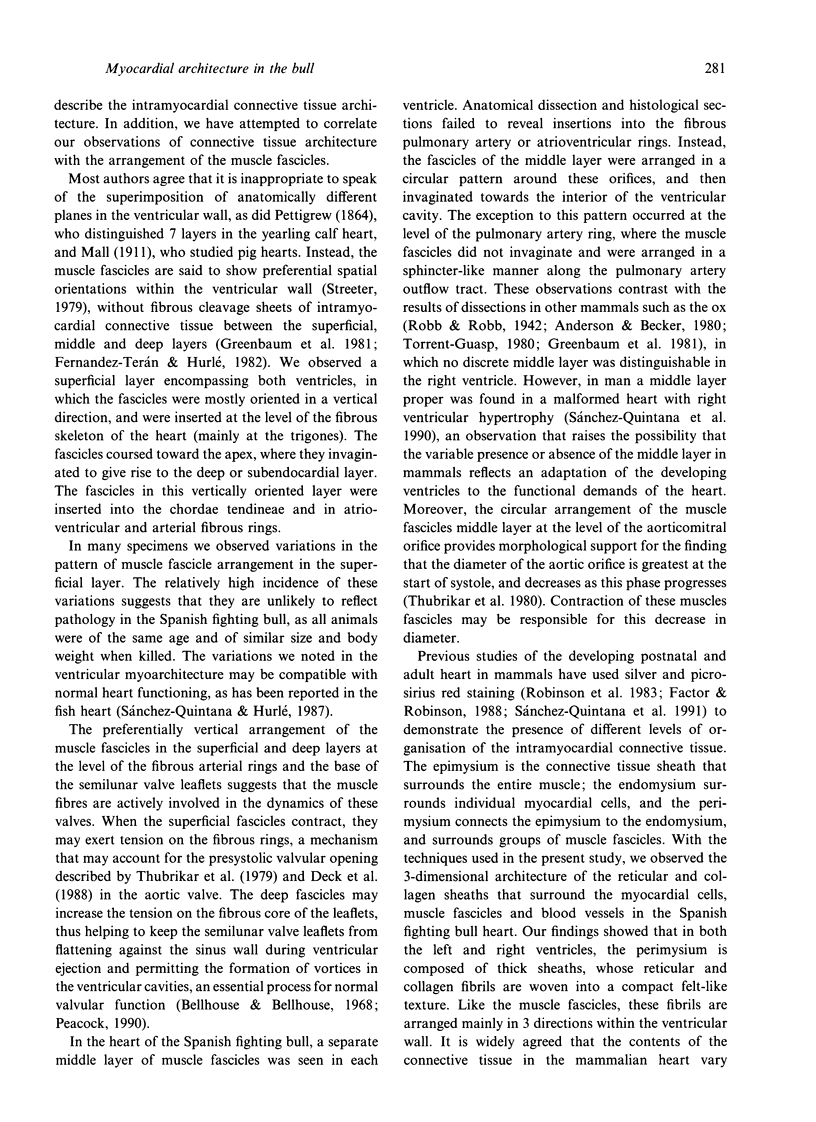
The spatial arrangement of the muscle fascicles and intramyocardial connective tissue was examined in the ventricles of the heart of the Spanish fighting bull (Bos taurus). In both ventricles, the muscle fascicles of the myocardium are arranged in 3 main directions, forming 3 muscle layers within the ventricular wall. The preferentially vertical arrangement of the muscle fascicles in the superficial and deep layers at the level of the fibrous aortic rings and the base of the semilunar valve leaflets suggests that these fascicles are actively involved in valvular dynamics. After controlled digestion of myocytes and elastic fibres with NaOH, a 3-dimensional arrangement of the scaffolding of connective tissue that supports the muscle fascicles and myocytes was observed. The arrangement and structure of this scaffolding may influence the order of contraction of muscle fascicles in different layers of the ventricle. In addition, differences were observed between the connective tissue scaffolding surrounding the myocytes of the 2 ventricles; these variations were correlated with the different biomechanical properties.
Full text
PDF
Images in this article
Selected References
These references are in PubMed. This may not be the complete list of references from this article.
- Bellhouse B. J., Bellhouse F. H. Mechanism of closure of the aortic valve. Nature. 1968 Jan 6;217(5123):86–87. doi: 10.1038/217086b0. [DOI] [PubMed] [Google Scholar]
- Borg T. K., Caulfield J. B. The collagen matrix of the heart. Fed Proc. 1981 May 15;40(7):2037–2041. [PubMed] [Google Scholar]
- Deck J. D., Thubrikar M. J., Schneider P. J., Nolan S. P. Structure, stress, and tissue repair in aortic valve leaflets. Cardiovasc Res. 1988 Jan;22(1):7–16. doi: 10.1093/cvr/22.1.7. [DOI] [PubMed] [Google Scholar]
- Dolber P. C., Spach M. S. Thin collagenous septa in cardiac muscle. Anat Rec. 1987 May;218(1):45–55. doi: 10.1002/ar.1092180109. [DOI] [PubMed] [Google Scholar]
- Factor S. M., Robinson T. F. Comparative connective tissue structure-function relationships in biologic pumps. Lab Invest. 1988 Feb;58(2):150–156. [PubMed] [Google Scholar]
- Fernandez-Teran M. A., Hurle J. M. Myocardial fiber architecture of the human heart ventricles. Anat Rec. 1982 Oct;204(2):137–147. doi: 10.1002/ar.1092040207. [DOI] [PubMed] [Google Scholar]
- GRANT R. P. NOTES ON THE MUSCULAR ARCHITECTURE OF THE LEFT VENTRICLE. Circulation. 1965 Aug;32:301–308. doi: 10.1161/01.cir.32.2.301. [DOI] [PubMed] [Google Scholar]
- Greenbaum R. A., Ho S. Y., Gibson D. G., Becker A. E., Anderson R. H. Left ventricular fibre architecture in man. Br Heart J. 1981 Mar;45(3):248–263. doi: 10.1136/hrt.45.3.248. [DOI] [PMC free article] [PubMed] [Google Scholar]
- JONES D. B. Inflammation and repair of glomerulus. Am J Pathol. 1951 Nov-Dec;27(6):991–1009. [PMC free article] [PubMed] [Google Scholar]
- Macías-Rodríguez D., Sánchez Quintana D., Hurle J. M. Arquitectura del miocardio ventricular en la zona de unión ventriculoarterial. Bases anatómicas del posible papel del miocardio en la dinámica de las válvulas semilunares. Rev Esp Cardiol. 1986 Nov-Dec;39(6):441–445. [PubMed] [Google Scholar]
- Maron B. J., Anan T. J., Roberts W. C. Quantitative analysis of the distribution of cardiac muscle cell disorganization in the left ventricular wall of patients with hypertrophic cardiomyopathy. Circulation. 1981 Apr;63(4):882–894. doi: 10.1161/01.cir.63.4.882. [DOI] [PubMed] [Google Scholar]
- McLean M., Prothero J. Myofiber orientation in the weanling mouse heart. Am J Anat. 1991 Dec;192(4):425–441. doi: 10.1002/aja.1001920410. [DOI] [PubMed] [Google Scholar]
- Morita T., Shimada T., Kitamura H., Nakamura M. Demonstration of connective tissue sheaths surrounding working myocardial cells and Purkinje cells of the sheep moderator band. Arch Histol Cytol. 1991 Dec;54(5):539–550. doi: 10.1679/aohc.54.539. [DOI] [PubMed] [Google Scholar]
- Peacock J. A. An in vitro study of the onset of turbulence in the sinus of Valsalva. Circ Res. 1990 Aug;67(2):448–460. doi: 10.1161/01.res.67.2.448. [DOI] [PubMed] [Google Scholar]
- Price M. G. Molecular analysis of intermediate filament cytoskeleton--a putative load-bearing structure. Am J Physiol. 1984 Apr;246(4 Pt 2):H566–H572. doi: 10.1152/ajpheart.1984.246.4.H566. [DOI] [PubMed] [Google Scholar]
- Robinson T. F., Cohen-Gould L., Factor S. M. Skeletal framework of mammalian heart muscle. Arrangement of inter- and pericellular connective tissue structures. Lab Invest. 1983 Oct;49(4):482–498. [PubMed] [Google Scholar]
- Sanchez-Quintana D., Garcia-Martinez V., Hurle J. M. Myocardial fiber architecture in the human heart. Anatomical demonstration of modifications in the normal pattern of ventricular fiber architecture in a malformed adult specimen. Acta Anat (Basel) 1990;138(4):352–358. [PubMed] [Google Scholar]
- Sanchez-Quintana D., Garcia-Martinez V., Macias D., Hurle J. M. Structural arrangement of the extracellular matrix network during myocardial development in the chick embryo heart. Anat Embryol (Berl) 1991;184(5):451–460. doi: 10.1007/BF01236051. [DOI] [PubMed] [Google Scholar]
- Sanchez-Quintana D., Hurle J. M. Ventricular myocardial architecture in marine fishes. Anat Rec. 1987 Mar;217(3):263–273. doi: 10.1002/ar.1092170307. [DOI] [PubMed] [Google Scholar]
- Shimada T., Noguchi T., Asami I., Campbell G. R. Functional morphology of the conduction system and the myocardium in the sheep heart as revealed by scanning and transmission electron microscopic analyses. Arch Histol Jpn. 1986 Aug;49(3):283–295. doi: 10.1679/aohc.49.283. [DOI] [PubMed] [Google Scholar]
- THOMAS C. E. The muscular architecture of the ventricles of hog and dog hearts. Am J Anat. 1957 Jul;101(1):17–57. doi: 10.1002/aja.1001010103. [DOI] [PubMed] [Google Scholar]
- Thubrikar M., Bosher L. P., Nolan S. P. The mechanism of opening of the aortic valve. J Thorac Cardiovasc Surg. 1979 Jun;77(6):863–870. [PubMed] [Google Scholar]
- Thubrikar M., Nolan S. P., Bosher L. P., Deck J. D. The cyclic changes and structure of the base of the aortic valve. Am Heart J. 1980 Feb;99(2):217–224. doi: 10.1016/0002-8703(80)90768-1. [DOI] [PubMed] [Google Scholar]
- Torrent Guasp F. La estructuración macroscópica del miocardio ventricular. Rev Esp Cardiol. 1980;33(3):265–287. [PubMed] [Google Scholar]
- Weber K. T. Cardiac interstitium in health and disease: the fibrillar collagen network. J Am Coll Cardiol. 1989 Jun;13(7):1637–1652. doi: 10.1016/0735-1097(89)90360-4. [DOI] [PubMed] [Google Scholar]


