Abstract
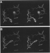
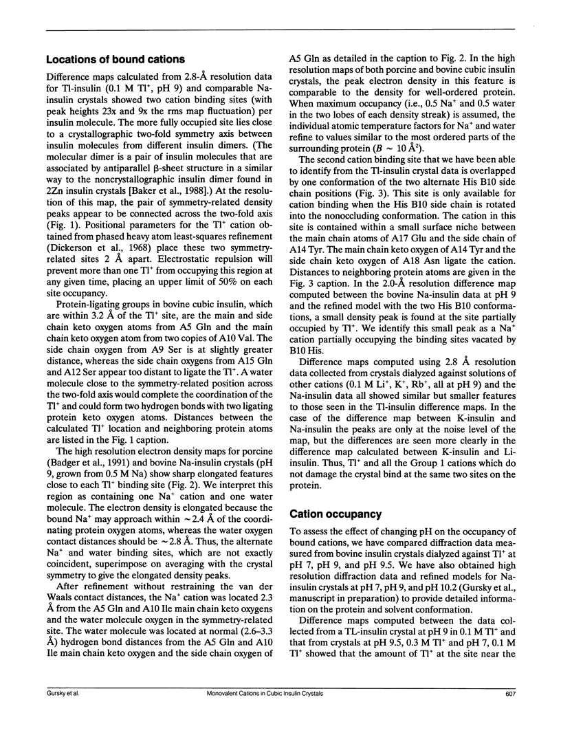
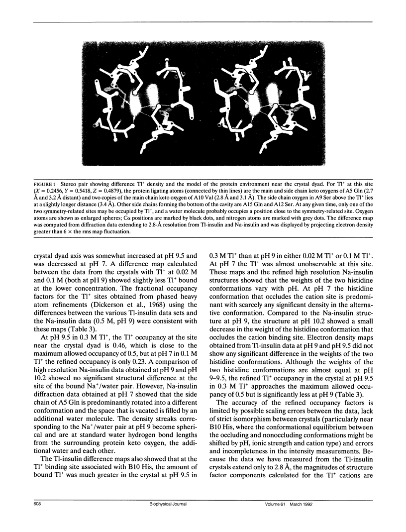
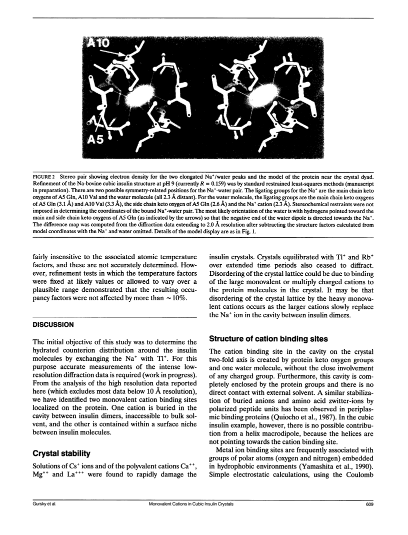
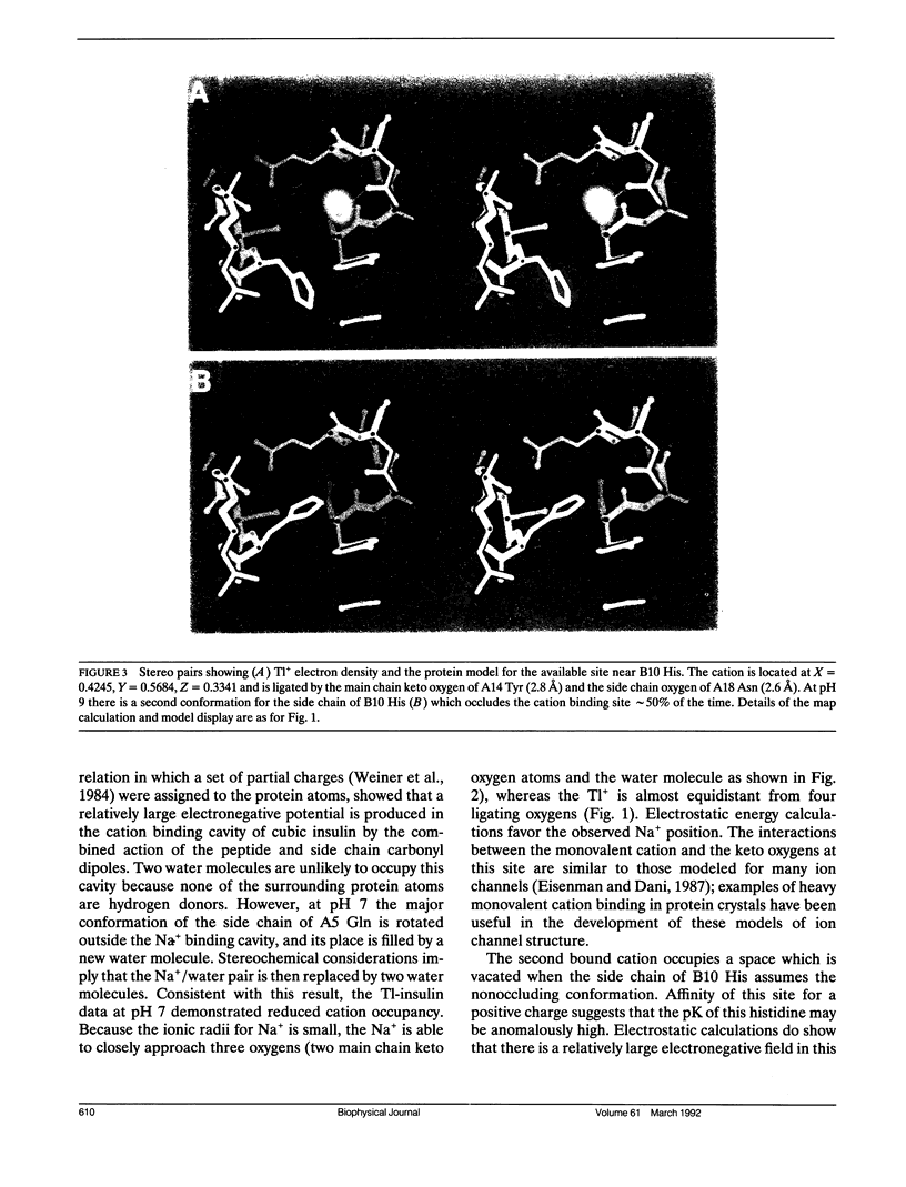
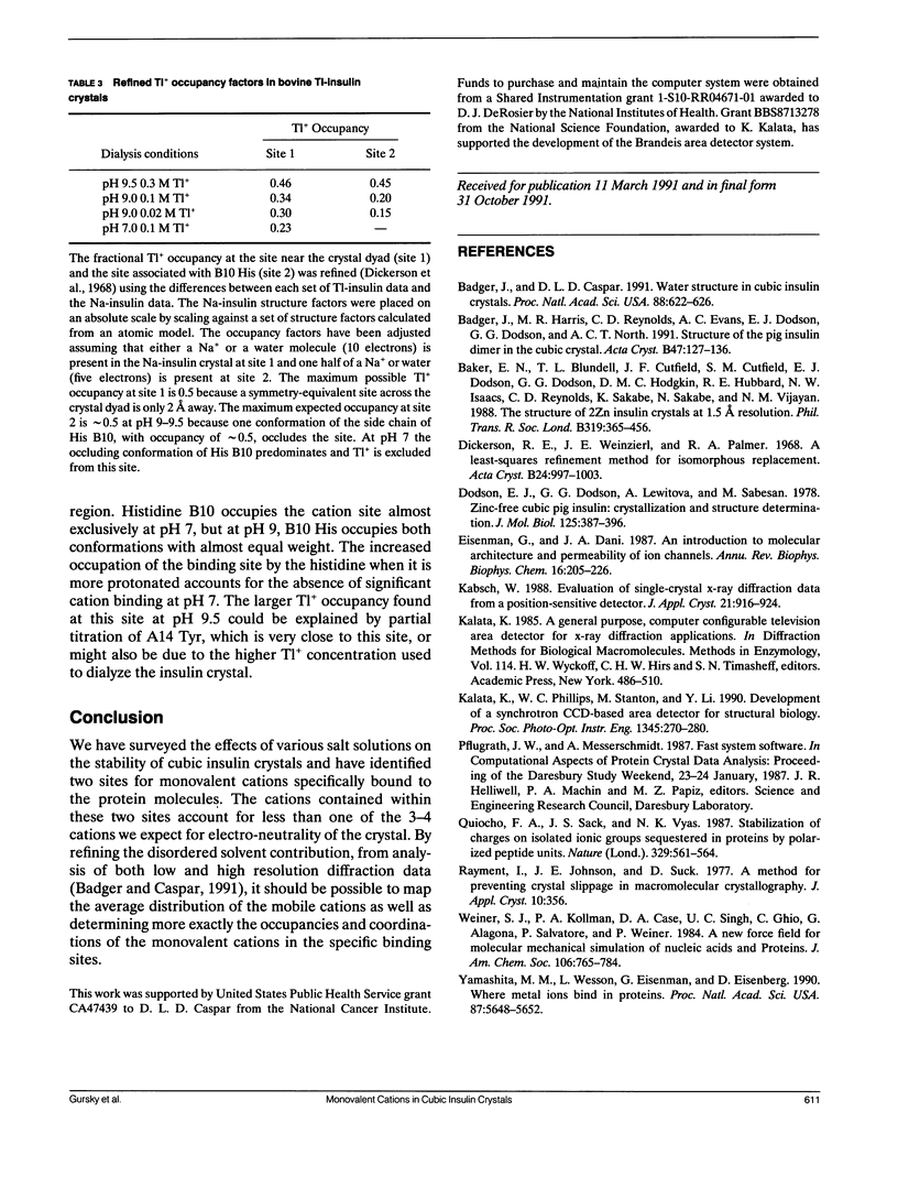
Two localized monovalent cation binding sites have been identified in cubic insulin from 2.8 A-resolution difference electron density maps comparing crystals in which the Na+ ions have been replaced by Tl+. One cation is buried in a closed cavity between insulin dimers and is stabilized by interaction with protein carbonyl dipoles in two juxtaposed alternate positions related by the crystal dyad. The second cation binding site, which also involves ligation with carbonyl dipoles, is competitively occupied by one position of two alternate His B10 side chain conformations. The cation occupancy in both sites depends on the net charge on the protein which was varied by equilibrating crystals in the pH range 7-10. Detailed structures of the cation binding sites were inferred from the refined 2-A resolution map of the sodium-insulin crystal at pH 9. At pH 9, the localized monovalent cations account for less than one of the three to four positive counterion charges necessary to neutralize the negative charge on each protein molecule. The majority of the monovalent counterions are too mobile to show up in the electron density maps calculated using data only at resolution higher than 10 A. Monovalent cations of ionic radius less than 1.5 A are required for crystal stability. Replacing Na+ with Cs+, Mg++, Ca++ or La+++ disrupts the lattice order, but crystals at pH 9 with 0.1 M Li+, K+, NH4+, Rb+ or Tl+ diffract to at least 2.8 A resolution.
Full text
PDF
Images in this article
Selected References
These references are in PubMed. This may not be the complete list of references from this article.
- Badger J., Caspar D. L. Water structure in cubic insulin crystals. Proc Natl Acad Sci U S A. 1991 Jan 15;88(2):622–626. doi: 10.1073/pnas.88.2.622. [DOI] [PMC free article] [PubMed] [Google Scholar]
- Badger J., Harris M. R., Reynolds C. D., Evans A. C., Dodson E. J., Dodson G. G., North A. C. Structure of the pig insulin dimer in the cubic crystal. Acta Crystallogr B. 1991 Feb 1;47(Pt 1):127–136. doi: 10.1107/s0108768190009570. [DOI] [PubMed] [Google Scholar]
- Baker E. N., Blundell T. L., Cutfield J. F., Cutfield S. M., Dodson E. J., Dodson G. G., Hodgkin D. M., Hubbard R. E., Isaacs N. W., Reynolds C. D. The structure of 2Zn pig insulin crystals at 1.5 A resolution. Philos Trans R Soc Lond B Biol Sci. 1988 Jul 6;319(1195):369–456. doi: 10.1098/rstb.1988.0058. [DOI] [PubMed] [Google Scholar]
- Dodson E. J., Dodson G. G., Lewitova A., Sabesan M. Zinc-free cubic pig insulin: crystallization and structure determination. J Mol Biol. 1978 Nov 5;125(3):387–396. doi: 10.1016/0022-2836(78)90409-6. [DOI] [PubMed] [Google Scholar]
- Eisenman G., Dani J. A. An introduction to molecular architecture and permeability of ion channels. Annu Rev Biophys Biophys Chem. 1987;16:205–226. doi: 10.1146/annurev.bb.16.060187.001225. [DOI] [PubMed] [Google Scholar]
- Quiocho F. A., Sack J. S., Vyas N. K. Stabilization of charges on isolated ionic groups sequestered in proteins by polarized peptide units. Nature. 1987 Oct 8;329(6139):561–564. doi: 10.1038/329561a0. [DOI] [PubMed] [Google Scholar]
- Yamashita M. M., Wesson L., Eisenman G., Eisenberg D. Where metal ions bind in proteins. Proc Natl Acad Sci U S A. 1990 Aug;87(15):5648–5652. doi: 10.1073/pnas.87.15.5648. [DOI] [PMC free article] [PubMed] [Google Scholar]


