Abstract
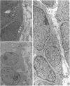
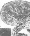
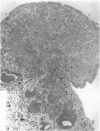
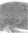
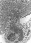
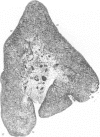
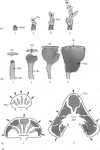
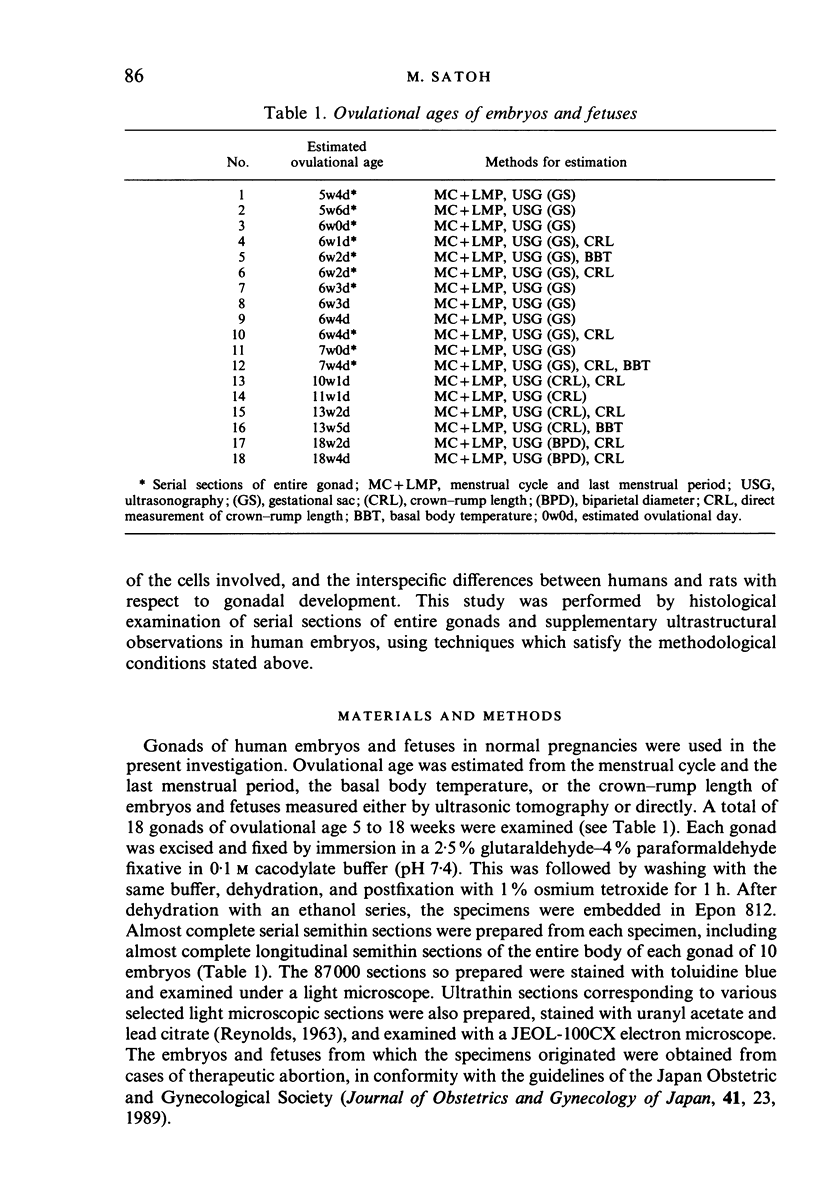
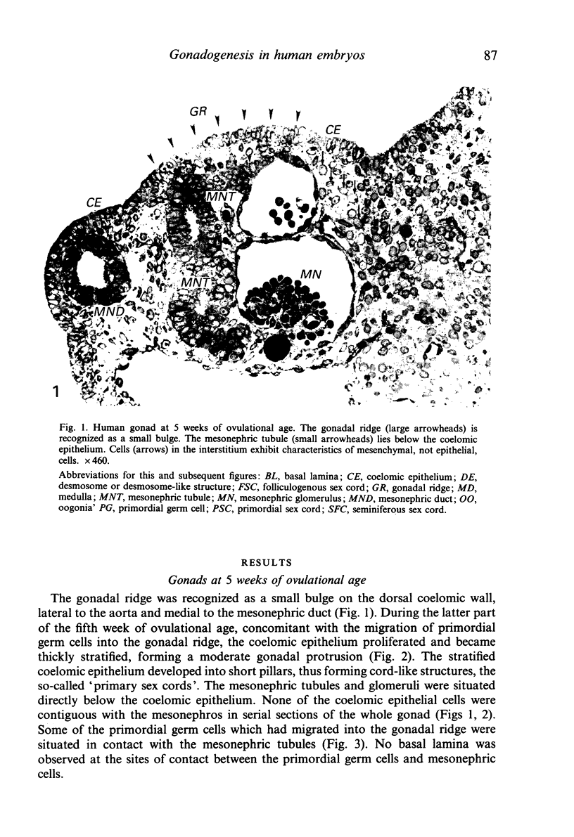
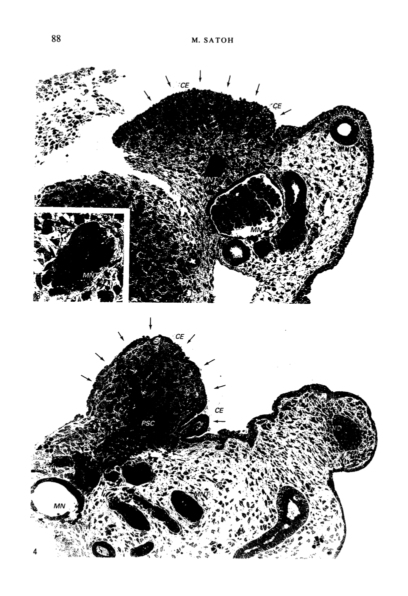
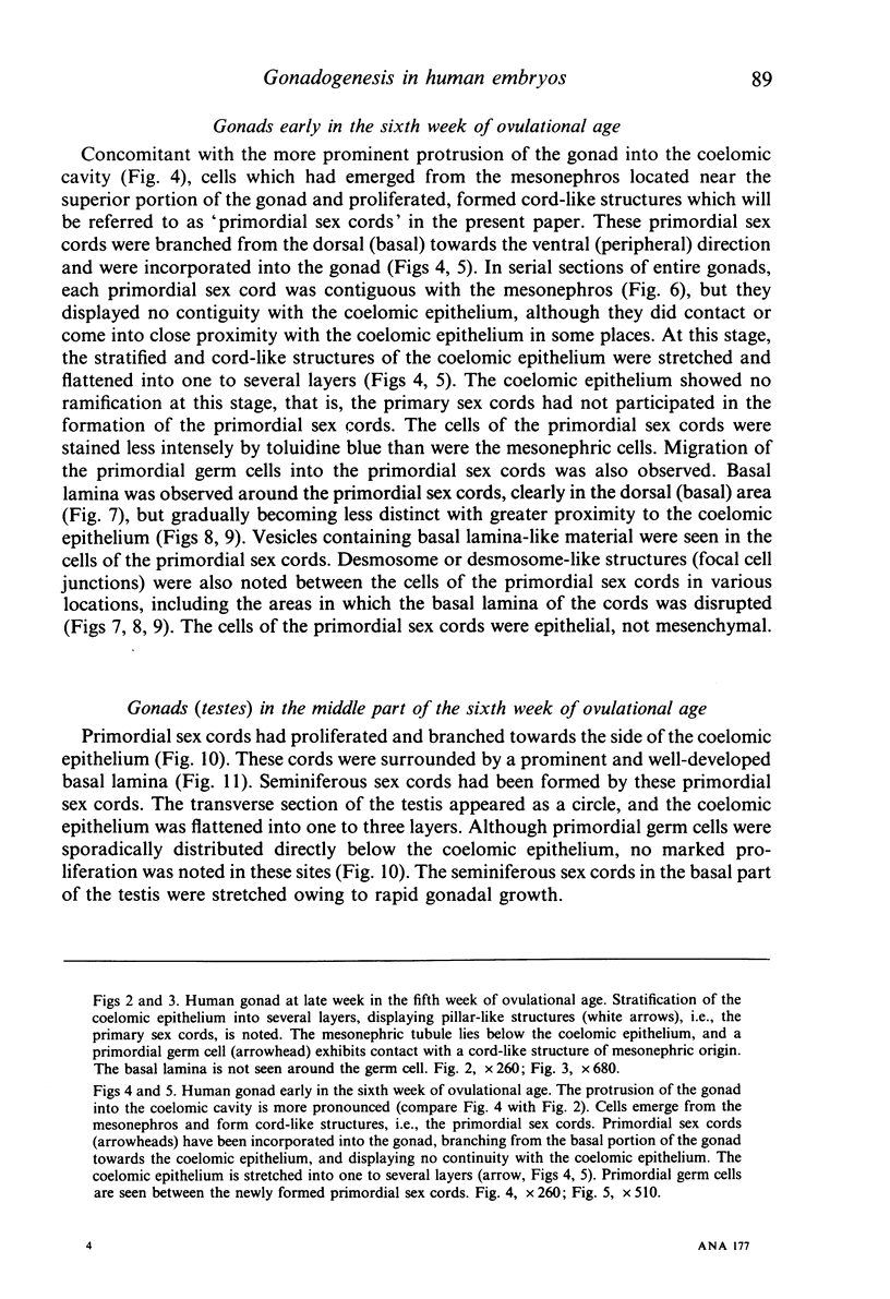
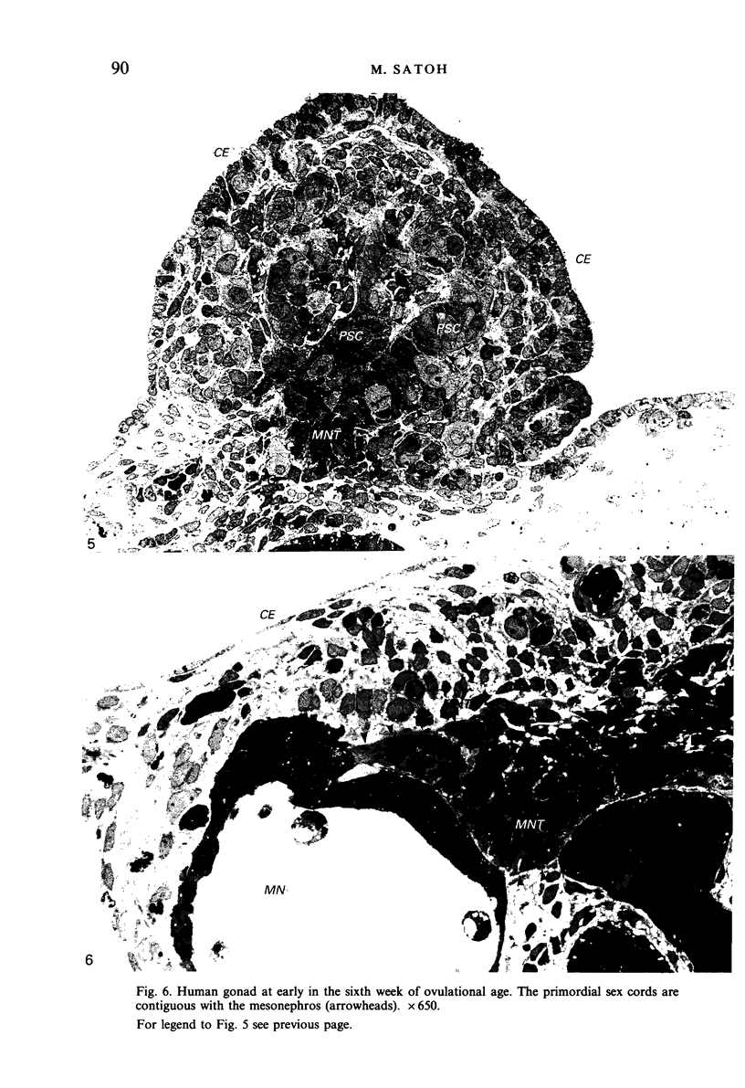
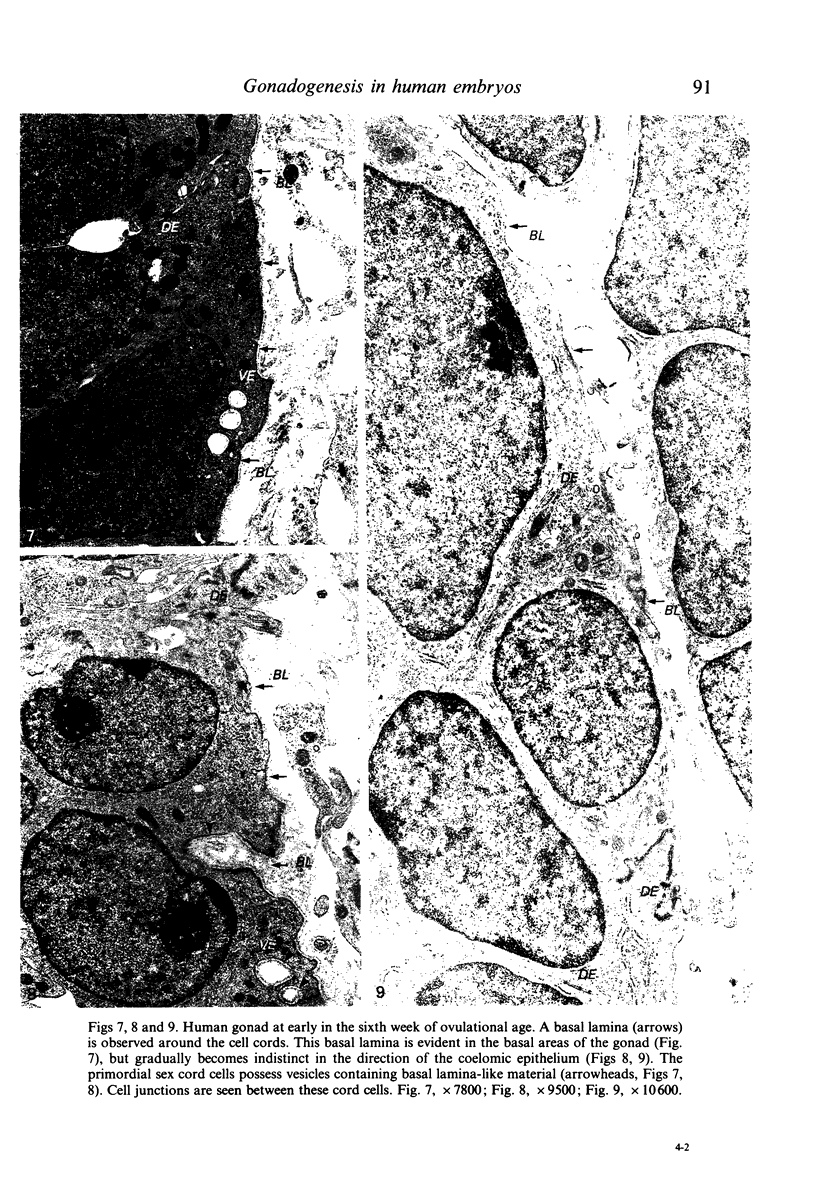
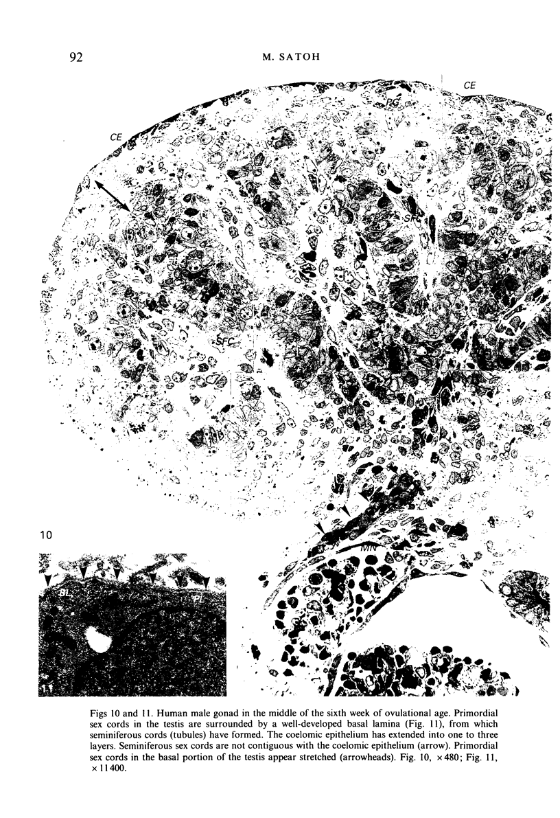
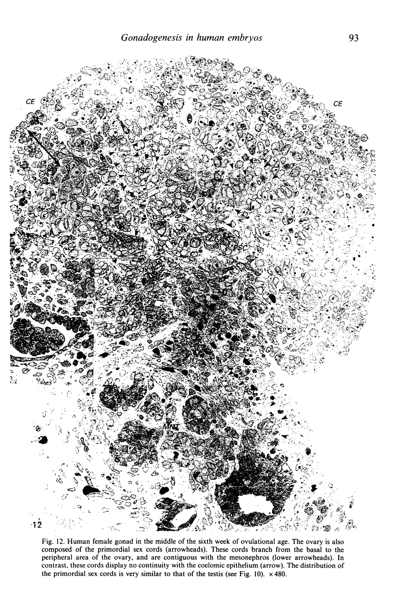
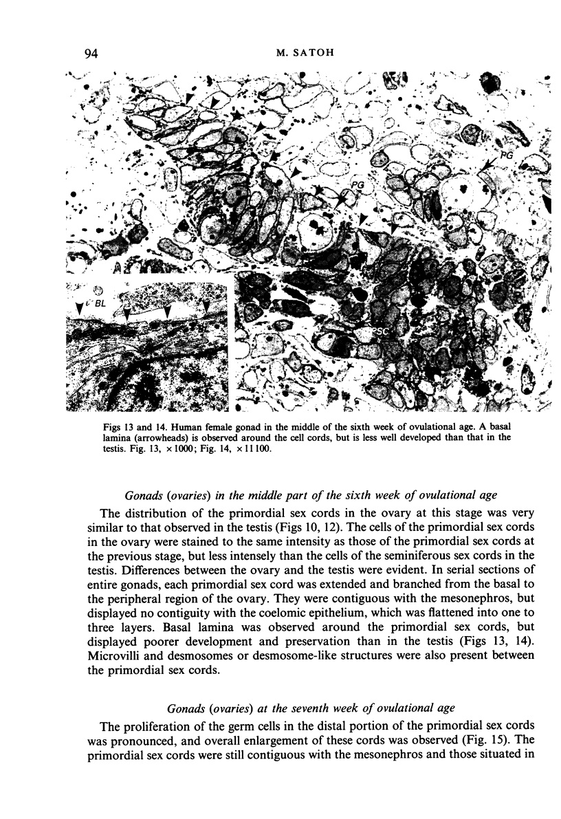
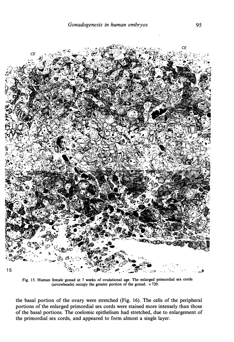
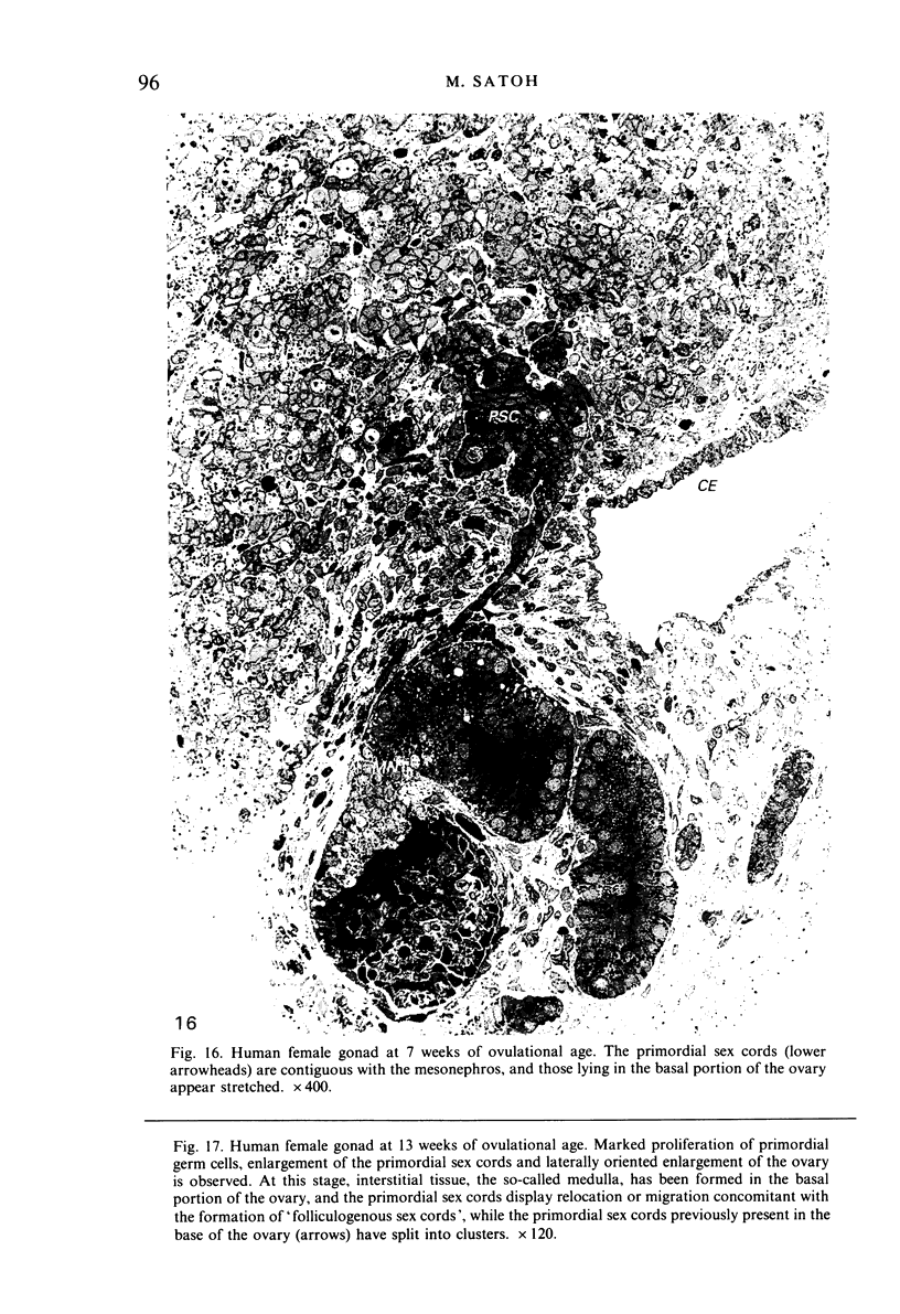
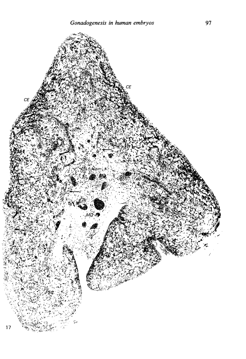
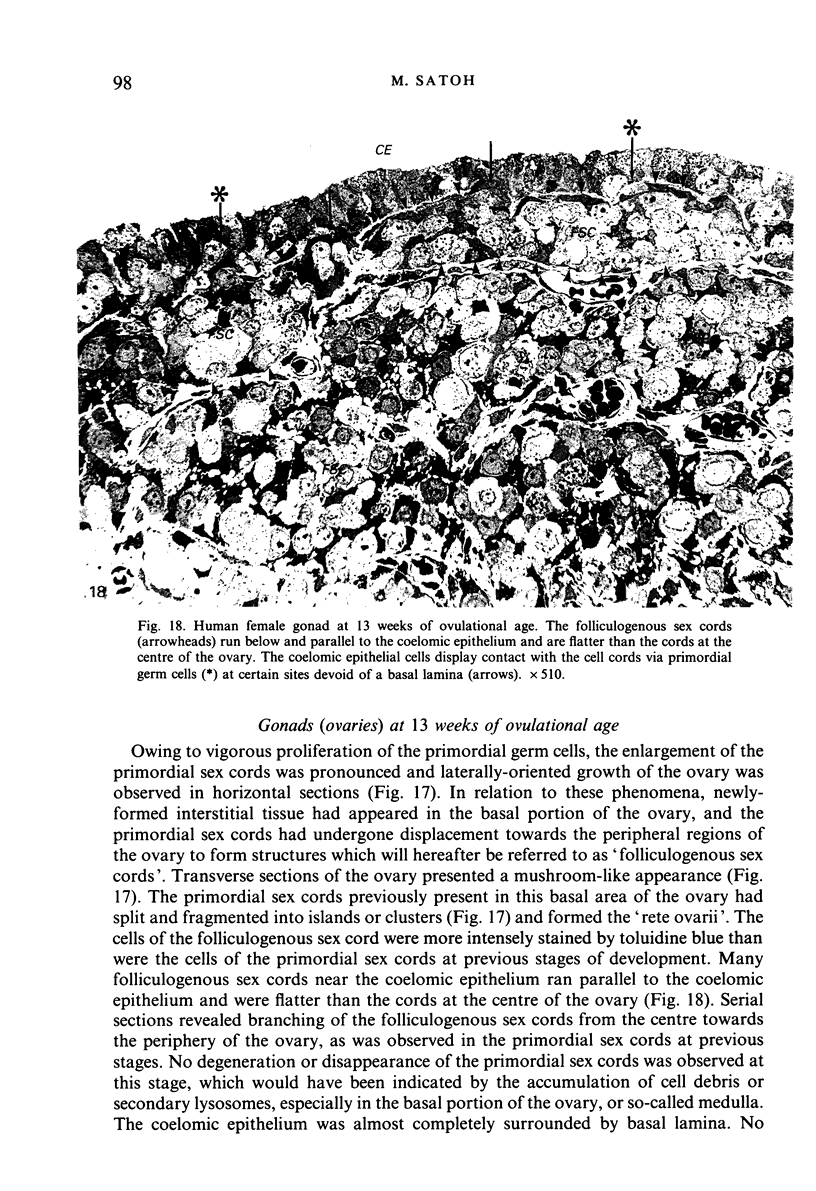
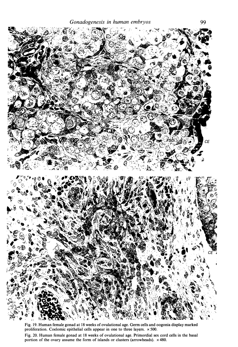
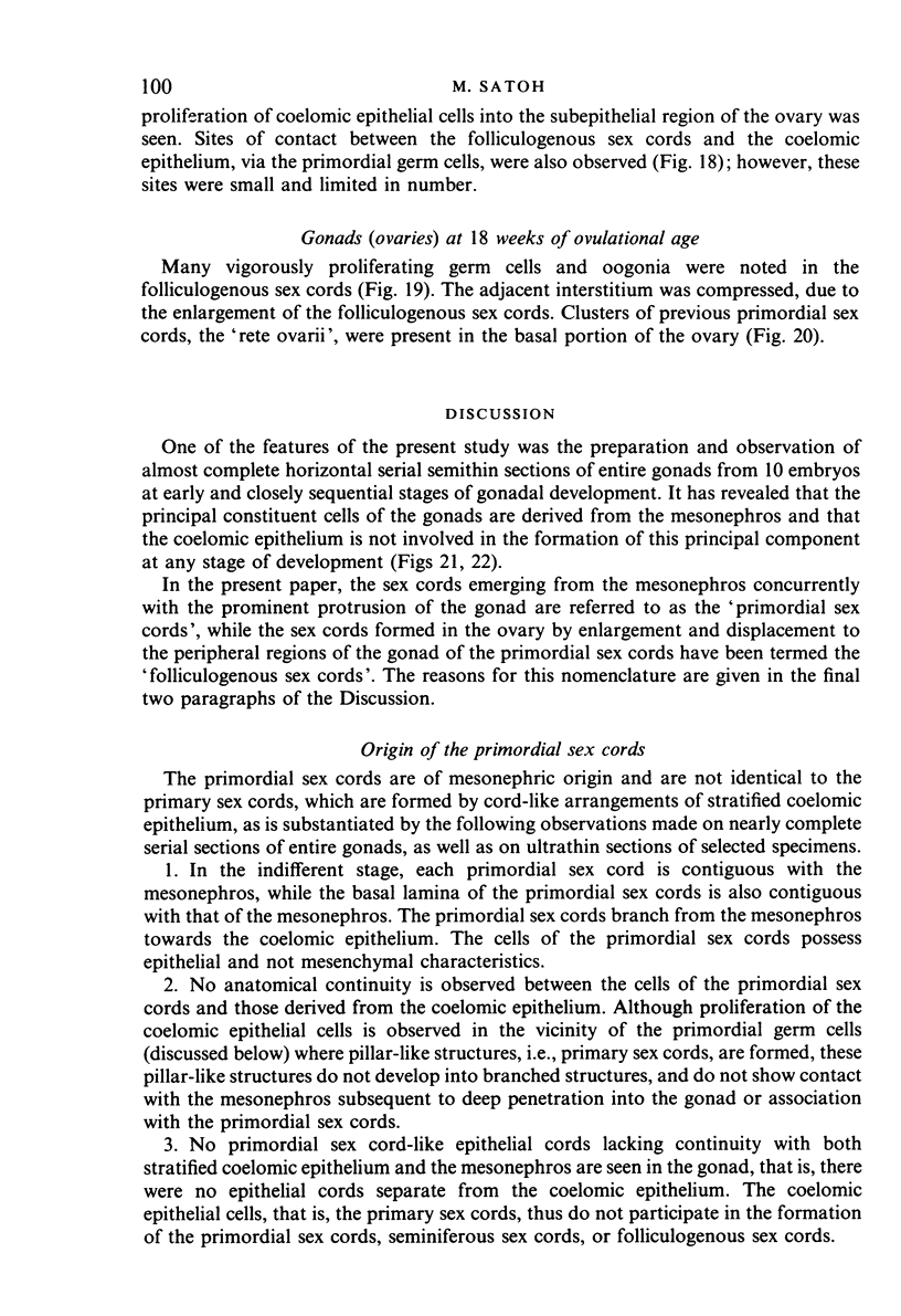
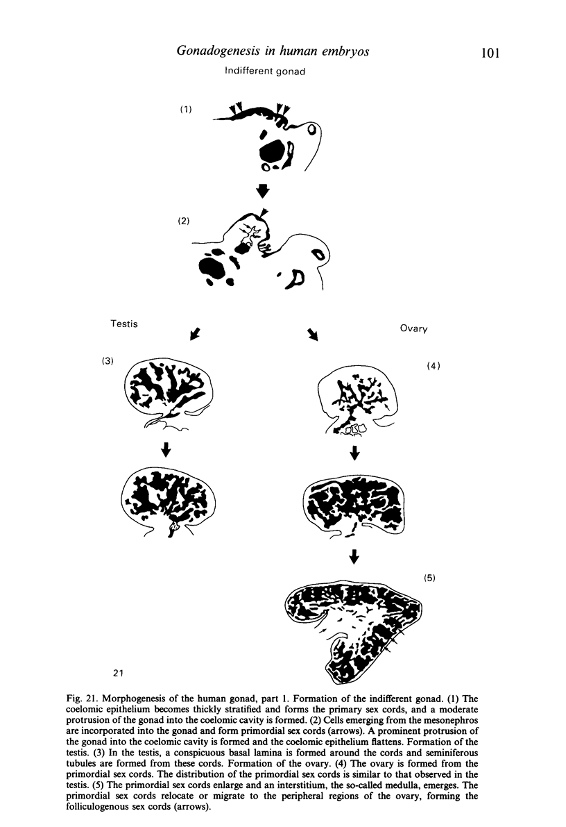
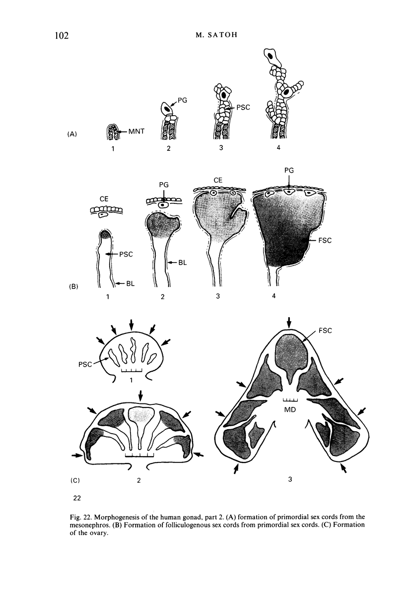
The histogenesis and organogenesis of the human gonad in 12 embryos and 6 fetuses of ovulational ages 5 to 18 weeks was investigated by histological and ultrastructural examination, including observation of almost complete serial Epon-embedded sections of entire gonads of 10 embryos. This investigation revealed that the main constituent cells of the gonads are derived from the mesonephros, and that the coelomic epithelium is not involved in the formation of the main component at any stage. With the migration of the primordial germ cells into the gonadal ridge, the coelomic epithelium becomes stratified to form a moderate protrusion of the gonad into the coelomic cavity and the coelomic epithelial cells develop into short pillars which form cord-like structures, the so-called primary sex cords. Shortly afterwards, concomitantly with the development into the subsequent prominent protrusion of the gonad into the coelomic cavity, cells emerging from the mesonephros are incorporated into the gonad to form 'primordial sex cords'. At this stage, a stratified, pile-like arrangement of coelomic epithelium flattens into monolaminar or oligolaminar structures. In the testis, the 'primordial sex cords' differentiate into seminiferous sex cords by elaborating a surrounding basal lamina. In the ovary, these 'primordial sex cords' become displaced towards the peripheral regions of the gonad by the enlargement of these cords, as well as by the formation of the interstitium, or so-called medulla, at the base of the ovary; they differentiate into 'folliculogenous sex cords' which give rise to follicular cells.
Full text
PDF
Images in this article
Selected References
These references are in PubMed. This may not be the complete list of references from this article.
- Byskov A. G., Lintern-Moore S. Follicle formation in the immature mouse ovary: the role of the rete ovarii. J Anat. 1973 Nov;116(Pt 2):207–217. [PMC free article] [PubMed] [Google Scholar]
- Fouquet J. P., Dang D. C. A comparative study of the development of the fetal testis and ovary in the monkey (Macaca fascicularis). Reprod Nutr Dev. 1980;20(5A):1439–1459. doi: 10.1051/rnd:19800804. [DOI] [PubMed] [Google Scholar]
- PINKERTON J. H., McKAY D. G., ADAMS E. C., HERTIG A. T. Development of the human ovary--a study using histochemical technics. Obstet Gynecol. 1961 Aug;18:152–181. [PubMed] [Google Scholar]
- Pelliniemi L. J. Ultrastructure of the indifferent gonad in male and female pig embryos. Tissue Cell. 1976;8(1):163–174. doi: 10.1016/0040-8166(76)90028-8. [DOI] [PubMed] [Google Scholar]
- REYNOLDS E. S. The use of lead citrate at high pH as an electron-opaque stain in electron microscopy. J Cell Biol. 1963 Apr;17:208–212. doi: 10.1083/jcb.17.1.208. [DOI] [PMC free article] [PubMed] [Google Scholar]
- Satoh M. The histogenesis of the gonad in rat embryos. J Anat. 1985 Dec;143:17–37. [PMC free article] [PubMed] [Google Scholar]
- Wartenberg H. Development of the early human ovary and role of the mesonephros in the differentiation of the cortex. Anat Embryol (Berl) 1982;165(2):253–280. doi: 10.1007/BF00305481. [DOI] [PubMed] [Google Scholar]
- Yoshinaga K., Hess D. L., Hendrickx A. G., Zamboni L. The development of the sexually indifferent gonad in the prosimian, Galago crassicaudatus crassicaudatus. Am J Anat. 1988 Jan;181(1):89–105. doi: 10.1002/aja.1001810110. [DOI] [PubMed] [Google Scholar]


