

Abstract
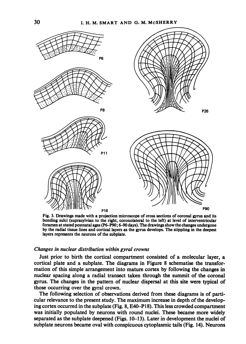
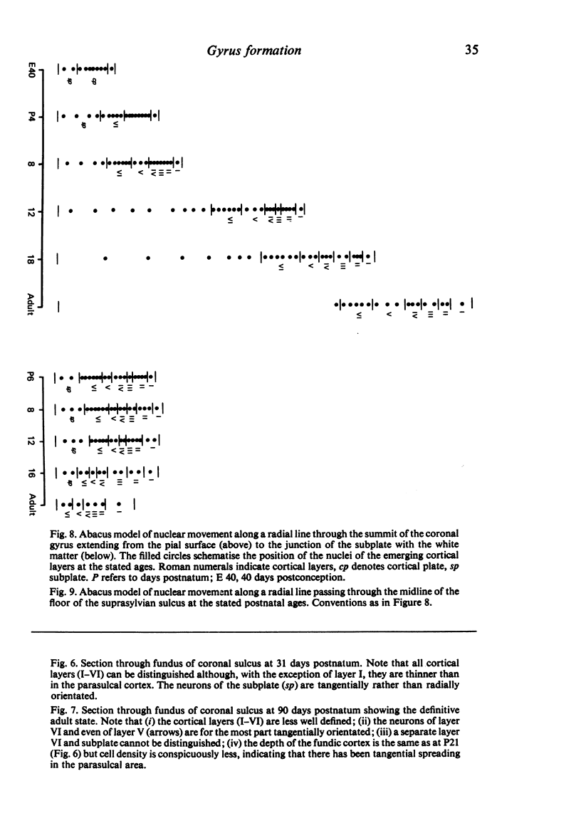
The internal changes within a developing gyrus of the ferret cerebral cortex were studied by recording (i) the changing length and direction of the radial tissue lines and (ii) the emergence of the tangential banding of the classical six cortical layers. Together these lines provided a coordinate net whose deformations during development gave an indication of the differential growth occurring within a gyrus. The changes in these features suggested that a gyrus was initiated by an area of local growth appearing in the subplate and then in the suprajacent segment of cortical plate. During subsequent growth there was tangential spreading of the more mature tissue at the gyral crown while at the site of the future sulci the cortical plate remained immature and growth was retarded. During later stages the majority of tangential growth occurred in the parasulcal area. At this site a very much thinner cortex was generated from a segment of cortical plate of the same depth and degree of nuclear crowding as elsewhere, implying that growth here was resolved into tangential spreading. The cells and fibres of the deeper cortical layers of the sulcal cortex eventually became tangentially orientated suggesting that they subserved a commissural function between the columnar systems of adjacent gyri. At the scale prevailing in the ferret, gyrus formation was seen as a configuration which tended to conserve both the total length of the cortical columns and the depth of the individual cortical layers.
Full text
PDF


Images in this article


Selected References
These references are in PubMed. This may not be the complete list of references from this article.
- Angevine J. B., Jr, Sidman R. L. Autoradiographic study of cell migration during histogenesis of cerebral cortex in the mouse. Nature. 1961 Nov 25;192:766–768. doi: 10.1038/192766b0. [DOI] [PubMed] [Google Scholar]
- Fernández V., Bravo H. Autoradiographic study of development of the cerebral cortex in the rabbit. Brain Behav Evol. 1974;9(5):317–332. doi: 10.1159/000123674. [DOI] [PubMed] [Google Scholar]
- Marin-Padilla M. Dual origin of the mammalian neocortex and evolution of the cortical plate. Anat Embryol (Berl) 1978 Feb 20;152(2):109–126. doi: 10.1007/BF00315920. [DOI] [PubMed] [Google Scholar]
- Marin-Padilla M. Early prenatal ontogenesis of the cerebral cortex (neocortex) of the cat (Felis domestica). A Golgi study. I. The primordial neocortical organization. Z Anat Entwicklungsgesch. 1971;134(2):117–145. doi: 10.1007/BF00519296. [DOI] [PubMed] [Google Scholar]
- Nieuwenhuys R. Topological analysis of the brain stem: a general introduction. J Comp Neurol. 1974 Aug 1;156(3):255–276. doi: 10.1002/cne.901560302. [DOI] [PubMed] [Google Scholar]
- Rakic P. Neurons in rhesus monkey visual cortex: systematic relation between time of origin and eventual disposition. Science. 1974 Feb 1;183(4123):425–427. doi: 10.1126/science.183.4123.425. [DOI] [PubMed] [Google Scholar]
- Richman D. P., Stewart R. M., Hutchinson J. W., Caviness V. S., Jr Mechanical model of brain convolutional development. Science. 1975 Jul 4;189(4196):18–21. doi: 10.1126/science.1135626. [DOI] [PubMed] [Google Scholar]
- Rickmann M., Chronwall B. M., Wolff J. R. On the development of non-pyramidal neurons and axons outside the cortical plate: the early marginal zone as a pallial anlage. Anat Embryol (Berl) 1977 Dec 2;151(3):285–307. doi: 10.1007/BF00318931. [DOI] [PubMed] [Google Scholar]
- Smart I. H., McSherry G. M. Gyrus formation in the cerebral cortex in the ferret. I. Description of the external changes. J Anat. 1986 Jun;146:141–152. [PMC free article] [PubMed] [Google Scholar]
- Todd P. H. A geometric model for the cortical folding pattern of simple folded brains. J Theor Biol. 1982 Aug 7;97(3):529–538. doi: 10.1016/0022-5193(82)90380-0. [DOI] [PubMed] [Google Scholar]
- WELKER W. I., CAMPOS G. B. Physiological significance of sulci in somatic sensory cerebral cortex in mammals of the family procyonidae. J Comp Neurol. 1963 Feb;120:19–36. doi: 10.1002/cne.901200103. [DOI] [PubMed] [Google Scholar]