Abstract
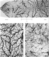
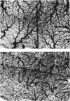
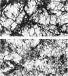
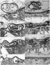
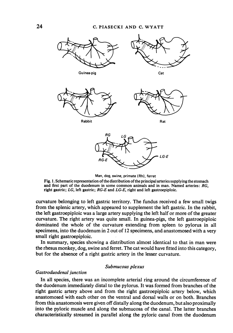
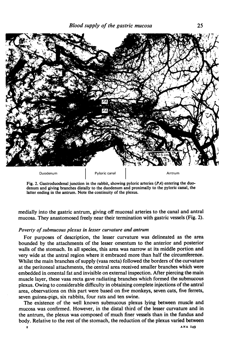
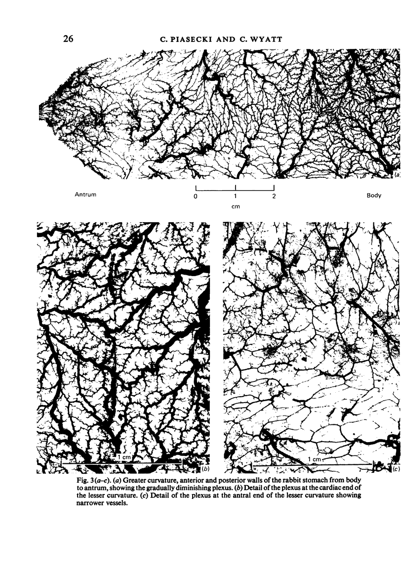
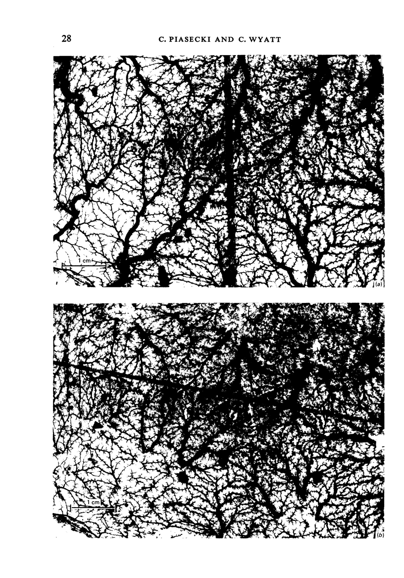
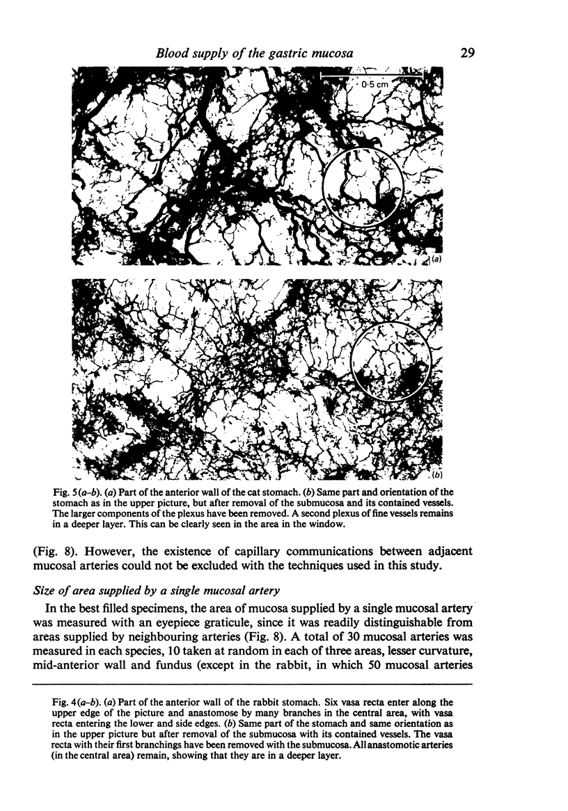
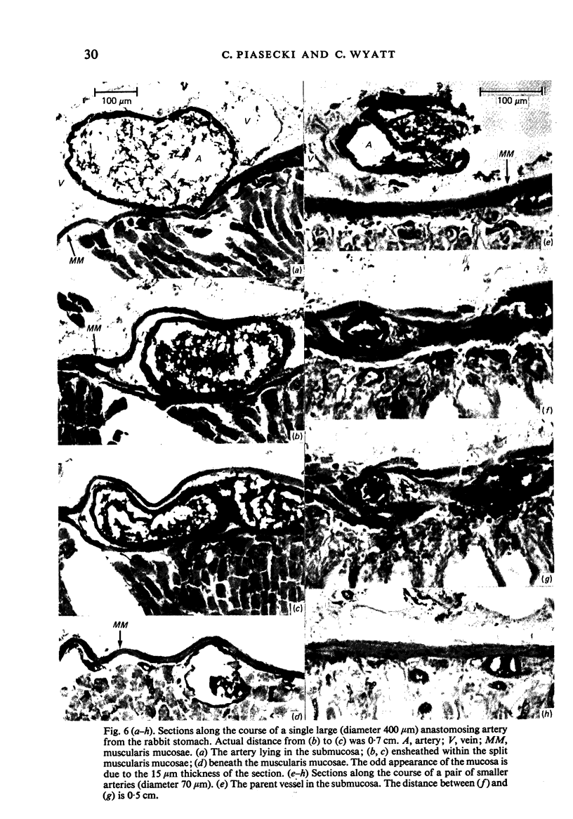
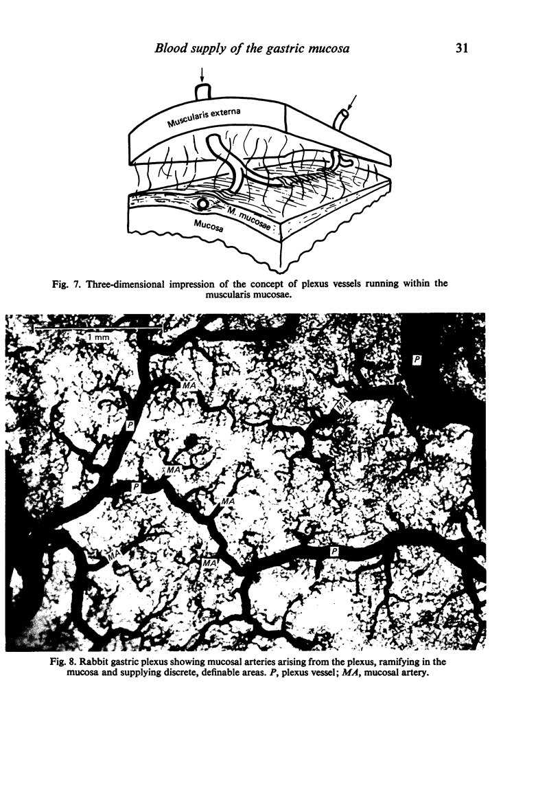
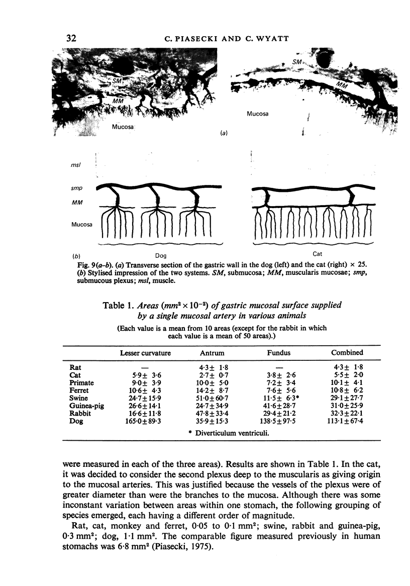
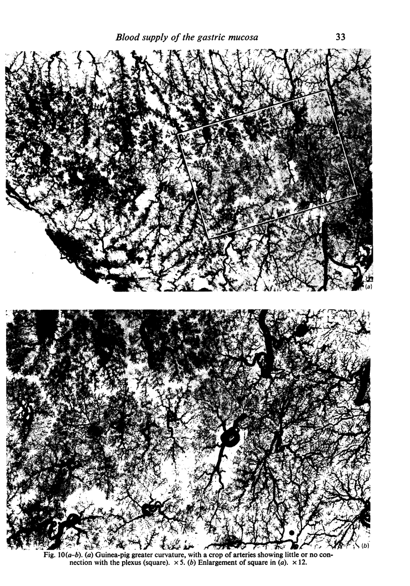
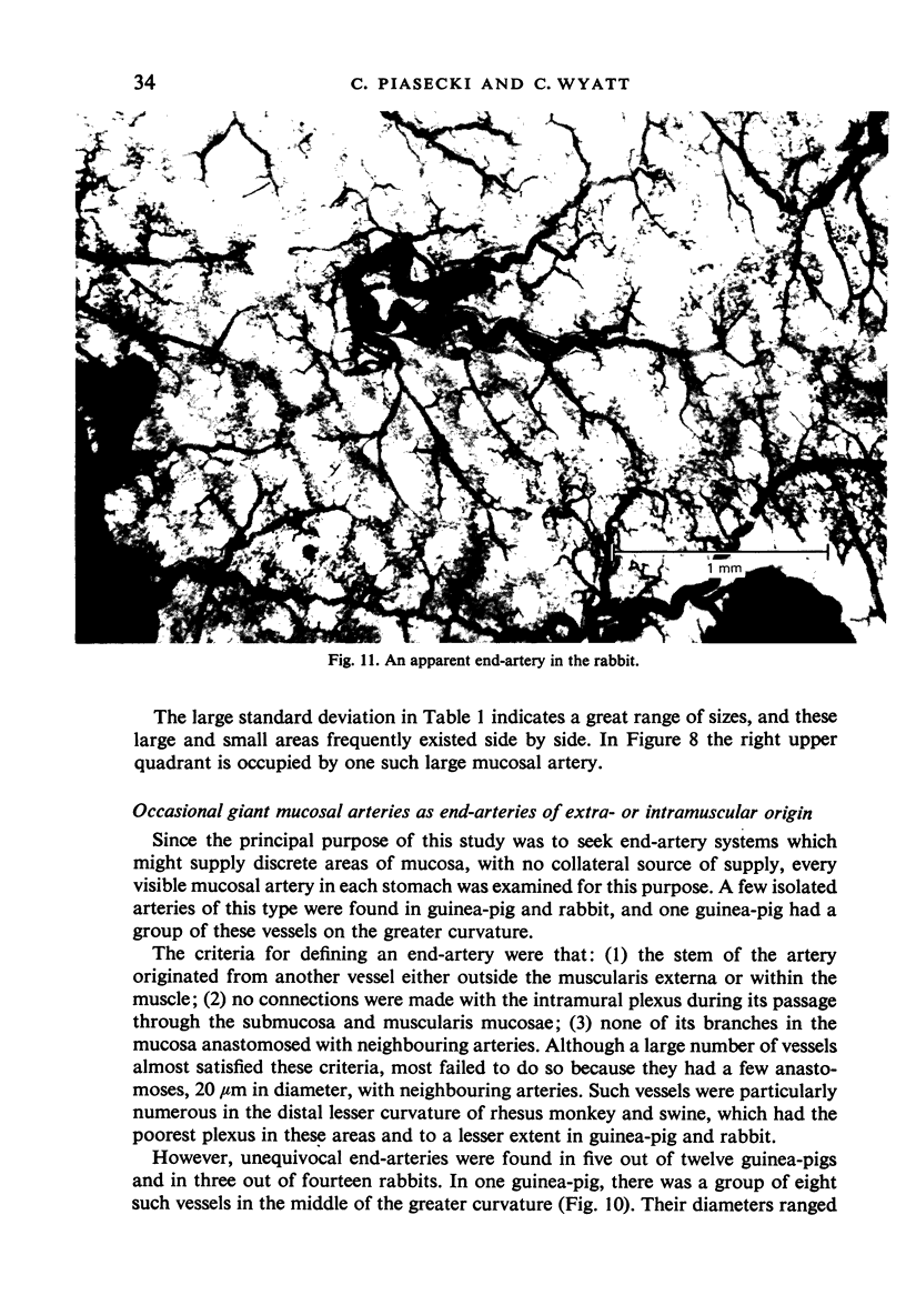
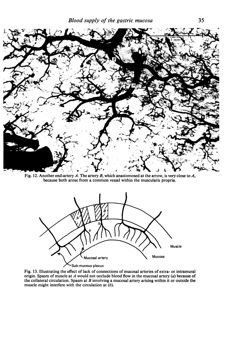
The form of the gastric arterial supply to the mucosa has been studied in dog, swine, ferret, cat, guinea-pig, rabbit and rhesus monkey. In all these species, the bore of vessels in the submucous plexus diminished from body to pylorus, though this was most marked in the guinea-pig and rabbit. The plexus was also continuous across the pylorus with duodenal vessels. Thus the well known poverty of vascularity in distal parts of the human stomach is shared by other species and is unlikely to be a contributory factor to the initiation of peptic ulcer, a disease limited to man. In dog, swine, ferret and cat, as in man, the primary (largest) and secondary (smaller) components of the plexus lay entirely in the submucosa. In the cat, there was a secondary plexus of much smaller vessels deep to the muscularis mucosae. In the guinea-pig, rat, rabbit and monkey, both plexuses were mostly embedded within the muscularis mucosae. As a result, mucosal arteries had two modes of origin: (a) the first, in which they did not pass through the muscularis mucosae as exemplified in the cat, and (b) the second, where they did pass through muscularis mucosae as exemplified by the dog, ferret and swine; in other species, they passed through part of the muscularis mucosae. Areas of mucosa supplied by a single mucosal artery were measured, and ranged widely from the smallest in the cat to the largest in the dog. These features do not seem to have been reported previously, and may be associated with as yet undiscovered functional mechanisms of the muscularis mucosae. Mucosal arteries of extramural origin were found to occur occasionally in the guinea-pig and rabbit, and hence these may provide an experimental model of the pattern existing in man.
Full text
PDF
Images in this article
Selected References
These references are in PubMed. This may not be the complete list of references from this article.
- ARABEHETY J. T., DOLCINI H., GRAY S. J. Sympathetic influences on circulation of the gastric mucosa of the rat. Am J Physiol. 1959 Oct;197:915–922. doi: 10.1152/ajplegacy.1959.197.4.915. [DOI] [PubMed] [Google Scholar]
- BARLOW T. E., BENTLEY F. H., WALDER D. N. Arteries, veins, and arteriovenous anastomoses in the human stomach. Surg Gynecol Obstet. 1951 Dec;93(6):657–671. [PubMed] [Google Scholar]
- DE BUSSCHER G. Les anastomoses artérioveineuses de l'estomac. Acta Neerl Morphol Norm Pathol. 1948;6(1-2):87–105. [PubMed] [Google Scholar]
- Guth P. H., Rosenberg A. In vivo microscopy of the gastric microcirculation. Am J Dig Dis. 1972 May;17(5):391–398. doi: 10.1007/BF02231290. [DOI] [PubMed] [Google Scholar]
- Hase T., Moss B. J. Microvascular changes of gastric mucosa in the development of stress ulcer in rats. Gastroenterology. 1973 Aug;65(2):224–234. [PubMed] [Google Scholar]
- Kirk R. M. Factors determining the site of chronic gastroduodenal ulcers. Hepatogastroenterology. 1982 Apr;29(2):75–85. [PubMed] [Google Scholar]
- Kirk R. M. Site and localization of duodenal ulcers: a study at operation. Gut. 1968 Aug;9(4):414–419. doi: 10.1136/gut.9.4.414. [DOI] [PMC free article] [PubMed] [Google Scholar]
- MALLIK K. C. B. An experimental study of the pathogenesis of gastric ulcers produced by pilocarpine. J Pathol Bacteriol. 1955 Oct;70(2):315–324. doi: 10.1002/path.1700700207. [DOI] [PubMed] [Google Scholar]
- NYLANDER G., OLERUD S. The vascular pattern of the gastric mucosa of the rat following vagotomy. Surg Gynecol Obstet. 1961 Apr;112:475–480. [PubMed] [Google Scholar]
- Oi M., Ito Y., Kumagai F., Yoshida K., Tanaka Y., Yoshikawa K., Miho O., Kijima M. A possible dual control mechanism in the origin of peptic ulcer. A study on ulcer location as affected by mucosa and musculature. Gastroenterology. 1969 Sep;57(3):280–293. [PubMed] [Google Scholar]
- Oka S. [Microcirculation of the gastro-intestinal mucosa and the architecture of the blood vessels]. Saishin Igaku. 1970 Aug;25(8):1705–1713. [PubMed] [Google Scholar]
- Piasecki C. A possible vascular factor in the aetiology of duodenal ulceration. Br J Surg. 1971 Sep;58(9):660–662. doi: 10.1002/bjs.1800580907. [DOI] [PubMed] [Google Scholar]
- Piasecki C. Blood supply to the human gastroduodenal mucosa with special reference to the ulcer-bearing areas. J Anat. 1974 Nov;118(Pt 2):295–335. [PMC free article] [PubMed] [Google Scholar]
- Piasecki C. Observations on the submucous plexus and mucosal arteries of the dog's stomach and first part of the duodenum. J Anat. 1975 Feb;119(Pt 1):133–148. [PMC free article] [PubMed] [Google Scholar]
- Piasecki C. Role of ischaemia in the initiation of peptic ulcer. Ann R Coll Surg Engl. 1977 Nov;59(6):476–478. [PMC free article] [PubMed] [Google Scholar]
- SCHNITZLEIN H. N. Regulation of blood flow through the stomach of the rat. Anat Rec. 1957 Apr;127(4):735–753. doi: 10.1002/ar.1091270409. [DOI] [PubMed] [Google Scholar]


