Abstract
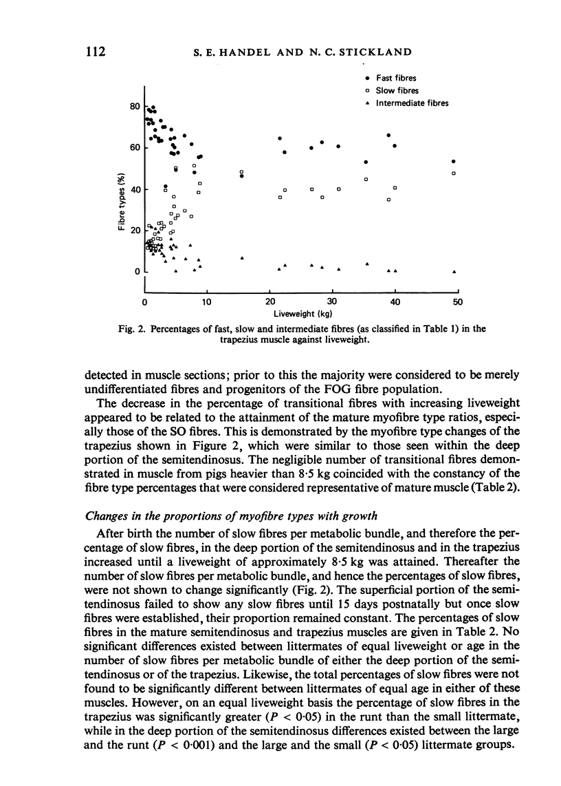
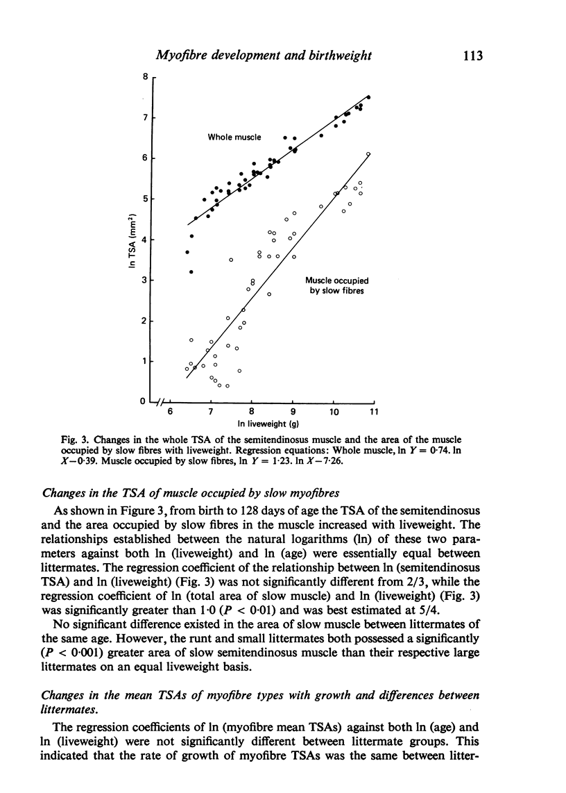
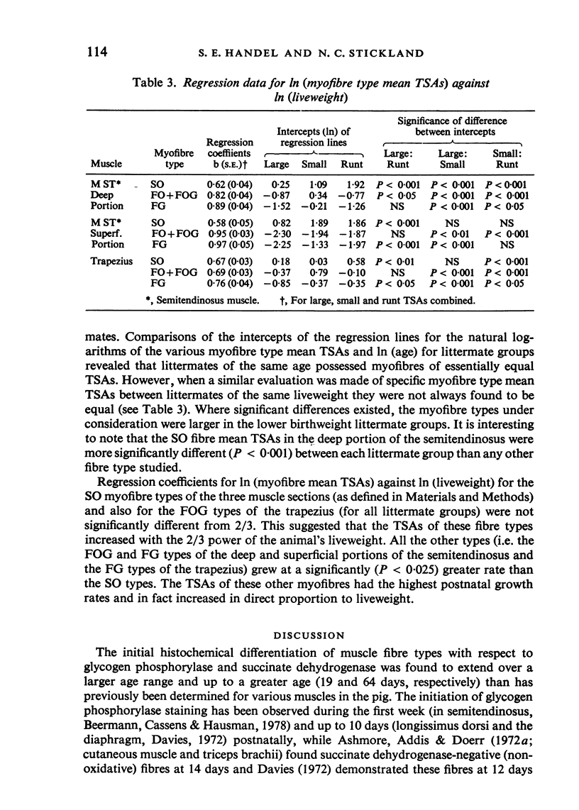
Muscle growth and development was studied in 49 Large White pigs from a total of 17 litters. Representative large (mean birthweight of 1544 g), small (1144 g) and runt (776 g) littermates were selected and slaughtered at the same age, ages ranging from birth to 128 days. Fresh frozen, serial transverse sections taken from the semi-tendinosus and trapezius muscles of these animals were stained for the histochemical demonstration of acid and alkaline pre-incubated adenosine triphosphatase, succinate dehydrogenase and glycogen phosphorylase. Profiles of the muscle fibre types were compiled for each animal. In both muscles the number of slow oxidative (SO) fibres, that were arranged together in groups within 'metabolic bundles', increased with growth. The transverse sectional area (TSA) of the semitendinosus muscle increased with the 2/3 power of liveweight whereas the area occupied by SO fibres increased at a rate significantly greater than 1.0 (P less than 0.01). Regression analysis revealed that the area of this muscle occupied by SO fibres was greater (P less than 0.001) in runt and small littermates relative to their large littermates when they were compared at an equal liveweight. This greater TSA of the semitendinosus classified as 'SO' in lower birthweight pigs was the result of a combination of higher percentages (P less than 0.05) of SO fibres and significantly greater (P less than 0.001) SO fibre mean TSAs. The mean TSAs of all myofibre types were similar between littermates of the same age but most types were of greater TSA in the lower birthweight littermates when compared (by regression analysis) at the same liveweight suggesting that fibre TSA was age- rather than weight-related. The higher percentage of SO fibres in the low birthweight pigs, when compared at an equivalent liveweight to their large littermates, appeared to be related to their affected secondary/primary fibre number ratio. This phenomenon, plus the data on the number of slow fibres per metabolic bundle, indicated that it was apparently the number of slow fibres per metabolic bundle which was regulated with liveweight gain rather than the resultant percentage of slow fibres within the muscle.
Full text
PDF
Images in this article
Selected References
These references are in PubMed. This may not be the complete list of references from this article.
- Ashmore C. R., Addis P. B., Doerr L. Development of muscle fibers in the fetal pig. J Anim Sci. 1973 Jun;36(6):1088–1093. doi: 10.2527/jas1973.3661088x. [DOI] [PubMed] [Google Scholar]
- Ashmore C. R., Robinson D. W., Rattray P., Doerr L. Biphasic development of muscle fibers in the fetal lamb. Exp Neurol. 1972 Nov;37(2):241–255. doi: 10.1016/0014-4886(72)90071-4. [DOI] [PubMed] [Google Scholar]
- Beermann D. H., Cassens R. G., Hausman G. J. A second look at fiber type differentiation in porcine skeletal muscle. J Anim Sci. 1978 Jan;46(1):125–132. doi: 10.2527/jas1978.461125x. [DOI] [PubMed] [Google Scholar]
- Burke R. E., Levine D. N., Zajac F. E., 3rd Mammalian motor units: physiological-histochemical correlation in three types in cat gastrocnemius. Science. 1971 Nov 12;174(4010):709–712. doi: 10.1126/science.174.4010.709. [DOI] [PubMed] [Google Scholar]
- Close R. Effects of cross-union of motor nerves to fast and slow skeletal muscles. Nature. 1965 May 22;206(4986):831–832. doi: 10.1038/206831a0. [DOI] [PubMed] [Google Scholar]
- Davies A. S. Postnatal changes in the histochemical fibre types of procine skeletal muscle. J Anat. 1972 Nov;113(Pt 2):213–240. [PMC free article] [PubMed] [Google Scholar]
- Edström L., Kugelberg E. Histochemical composition, distribution of fibres and fatiguability of single motor units. Anterior tibial muscle of the rat. J Neurol Neurosurg Psychiatry. 1968 Oct;31(5):424–433. doi: 10.1136/jnnp.31.5.424. [DOI] [PMC free article] [PubMed] [Google Scholar]
- Guth L., Samaha F. J. Procedure for the histochemical demonstration of actomyosin ATPase. Exp Neurol. 1970 Aug;28(2):365–367. [PubMed] [Google Scholar]
- Hegarty P. V., Allen C. E. Effect of pre-natal runting on the post-natal development of skeletal muscles in swine and rats. J Anim Sci. 1978 Jun;46(6):1634–1640. doi: 10.2527/jas1978.4661634x. [DOI] [PubMed] [Google Scholar]
- Holmes J. H., Ashmore C. R. A histochemical study of development of muscle fiber type and size in normal and "double muscled" cattle. Growth. 1972 Dec;36(4):351–372. [PubMed] [Google Scholar]
- Kugelberg E. Adaptive transformation of rat soleus motor units during growth. J Neurol Sci. 1976 Mar;27(3):269–289. doi: 10.1016/0022-510x(76)90001-0. [DOI] [PubMed] [Google Scholar]
- NACHLAS M. M., TSOU K. C., DE SOUZA E., CHENG C. S., SELIGMAN A. M. Cytochemical demonstration of succinic dehydrogenase by the use of a new p-nitrophenyl substituted ditetrazole. J Histochem Cytochem. 1957 Jul;5(4):420–436. doi: 10.1177/5.4.420. [DOI] [PubMed] [Google Scholar]
- Oldfors A., Mair W. G., Sourander P. Muscle changes in protein-deprived young rats. A morphometrical, histochemical and ultrastructural study. J Neurol Sci. 1983 May;59(2):291–302. doi: 10.1016/0022-510x(83)90046-1. [DOI] [PubMed] [Google Scholar]
- Peter J. B., Barnard R. J., Edgerton V. R., Gillespie C. A., Stempel K. E. Metabolic profiles of three fiber types of skeletal muscle in guinea pigs and rabbits. Biochemistry. 1972 Jul 4;11(14):2627–2633. doi: 10.1021/bi00764a013. [DOI] [PubMed] [Google Scholar]
- Powell S. E., Aberle E. D. Skeletal muscle and adipose tissue cellularity in runt and normal birth weight swine. J Anim Sci. 1981 Apr;52(4):748–756. doi: 10.2527/jas1981.524748x. [DOI] [PubMed] [Google Scholar]
- Salmons S., Sréter F. A. Significance of impulse activity in the transformation of skeletal muscle type. Nature. 1976 Sep 2;263(5572):30–34. doi: 10.1038/263030a0. [DOI] [PubMed] [Google Scholar]
- Sivachelvan M. N., Davies A. S. Antenatal anticipation of postnatal muscle function. J Anat. 1981 Jun;132(Pt 4):545–555. [PMC free article] [PubMed] [Google Scholar]
- Sreter F. A., Seidel J. C., Gergely J. Studies on myosin from red and white skeletal muscles of the rabbit. I. Adenosine triphosphatase activity. J Biol Chem. 1966 Dec 25;241(24):5772–5776. [PubMed] [Google Scholar]
- Suzuki A., Cassens R. G. A histochemical study of myofiber types in muscle of the growing pig. J Anim Sci. 1980 Dec;51(6):1449–1461. doi: 10.2527/jas1981.5161449x. [DOI] [PubMed] [Google Scholar]
- Suzuki A., Cassens R. G. A histochemical study of myofiber types in the serratus ventralis thoracis muscle of sheep during growth. J Anim Sci. 1983 Jun;56(6):1447–1458. doi: 10.2527/jas1983.5661447x. [DOI] [PubMed] [Google Scholar]
- TAKEUCHI T., KURIAKI H. Histochemical detection of phosphorylase in animal tissues. J Histochem Cytochem. 1955 May;3(3):153–160. doi: 10.1177/3.3.153. [DOI] [PubMed] [Google Scholar]


