Abstract
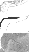
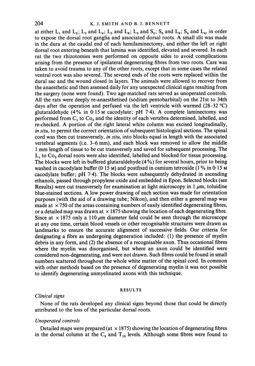
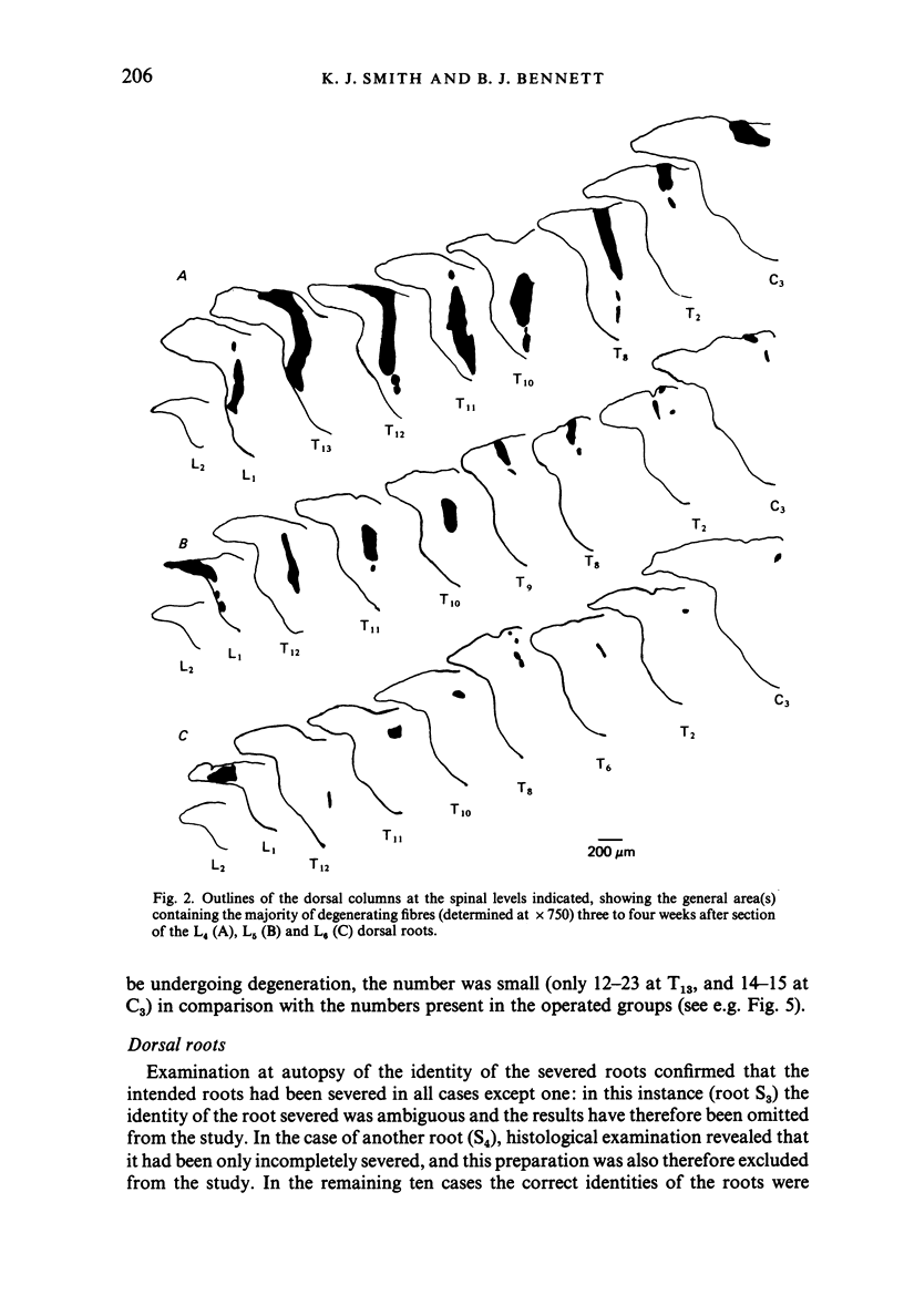
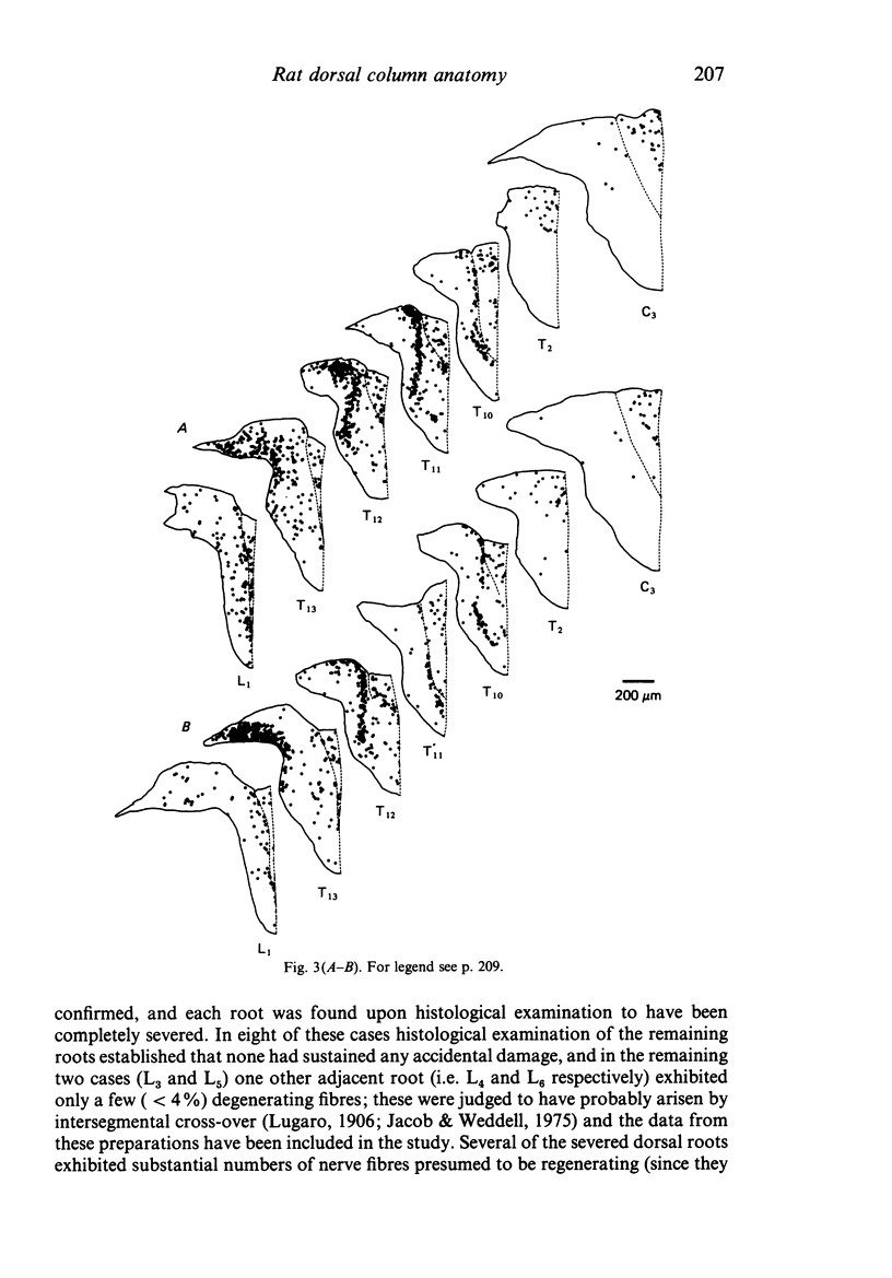
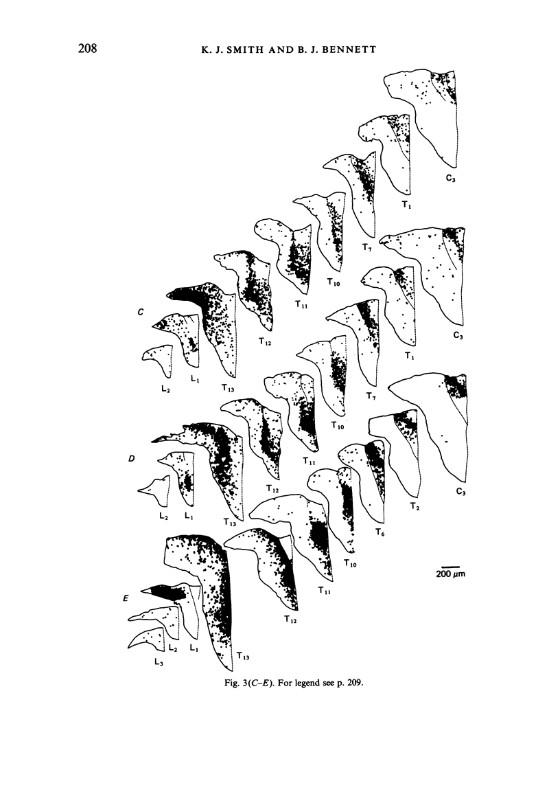
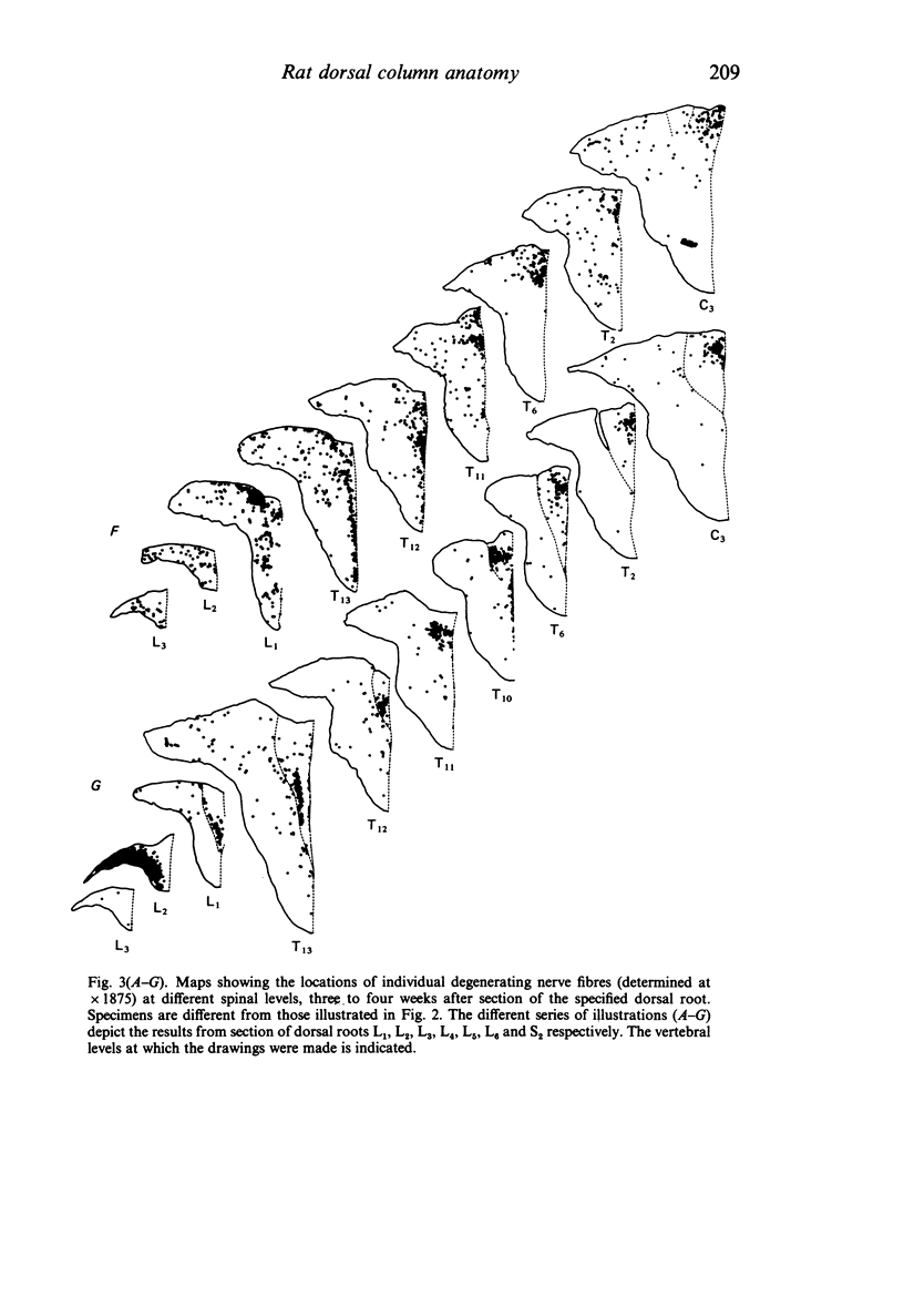
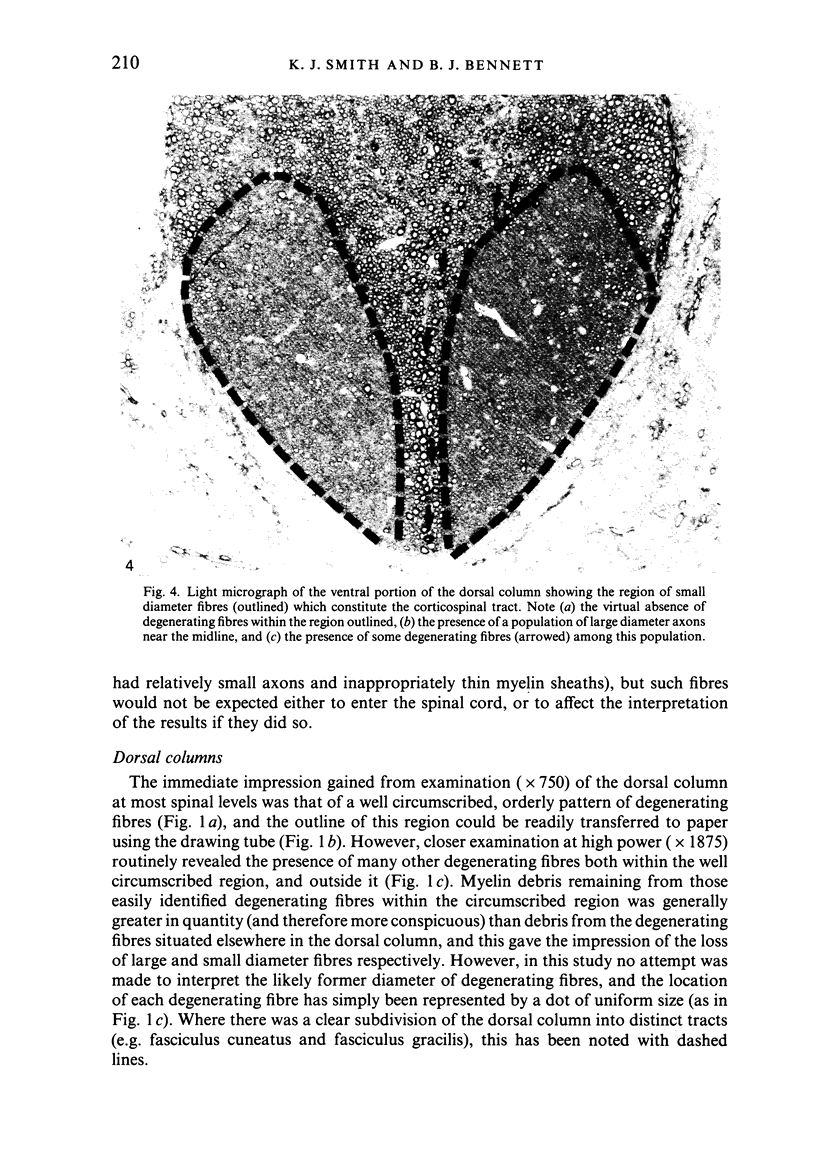
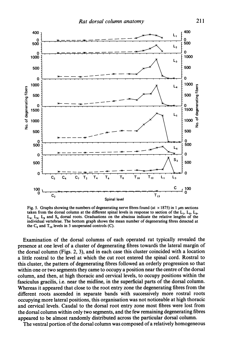
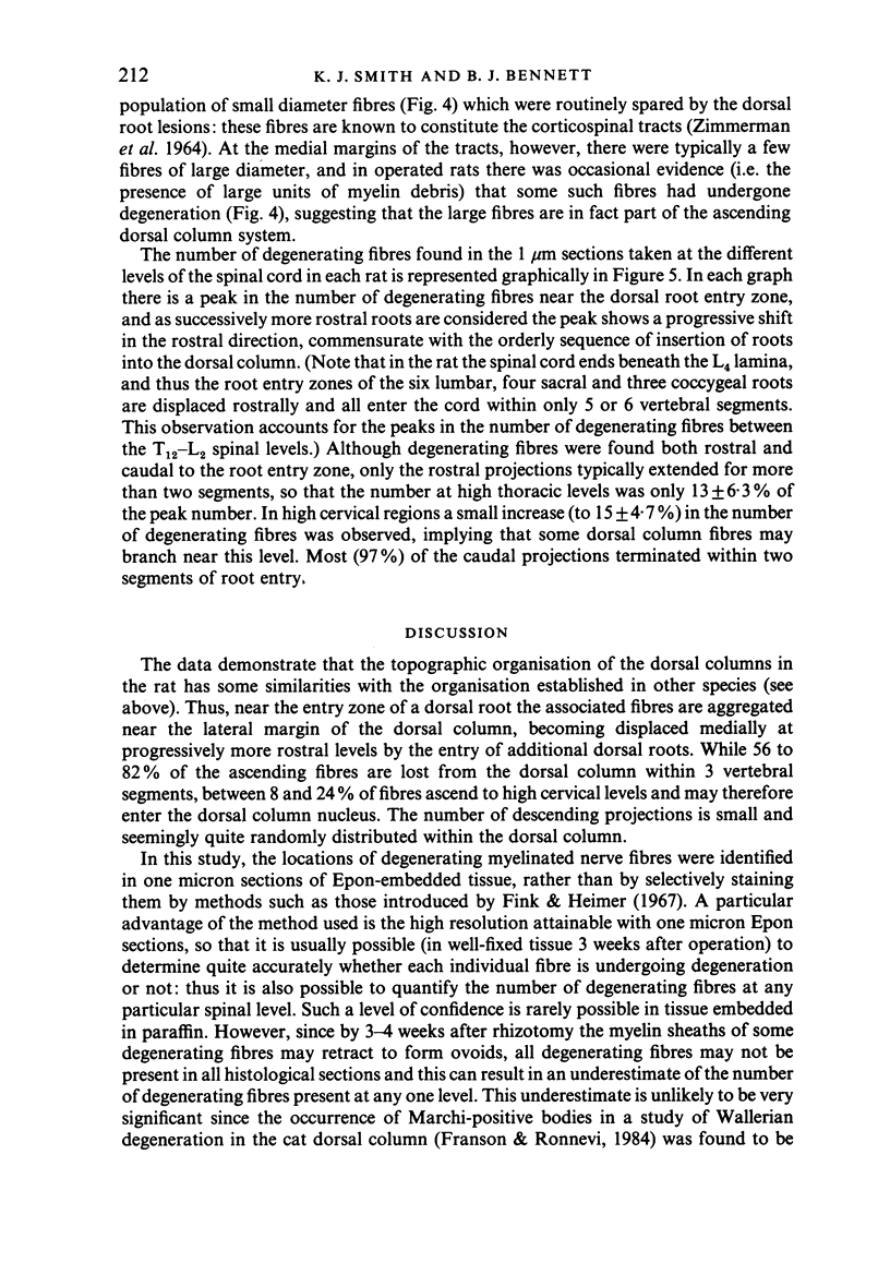
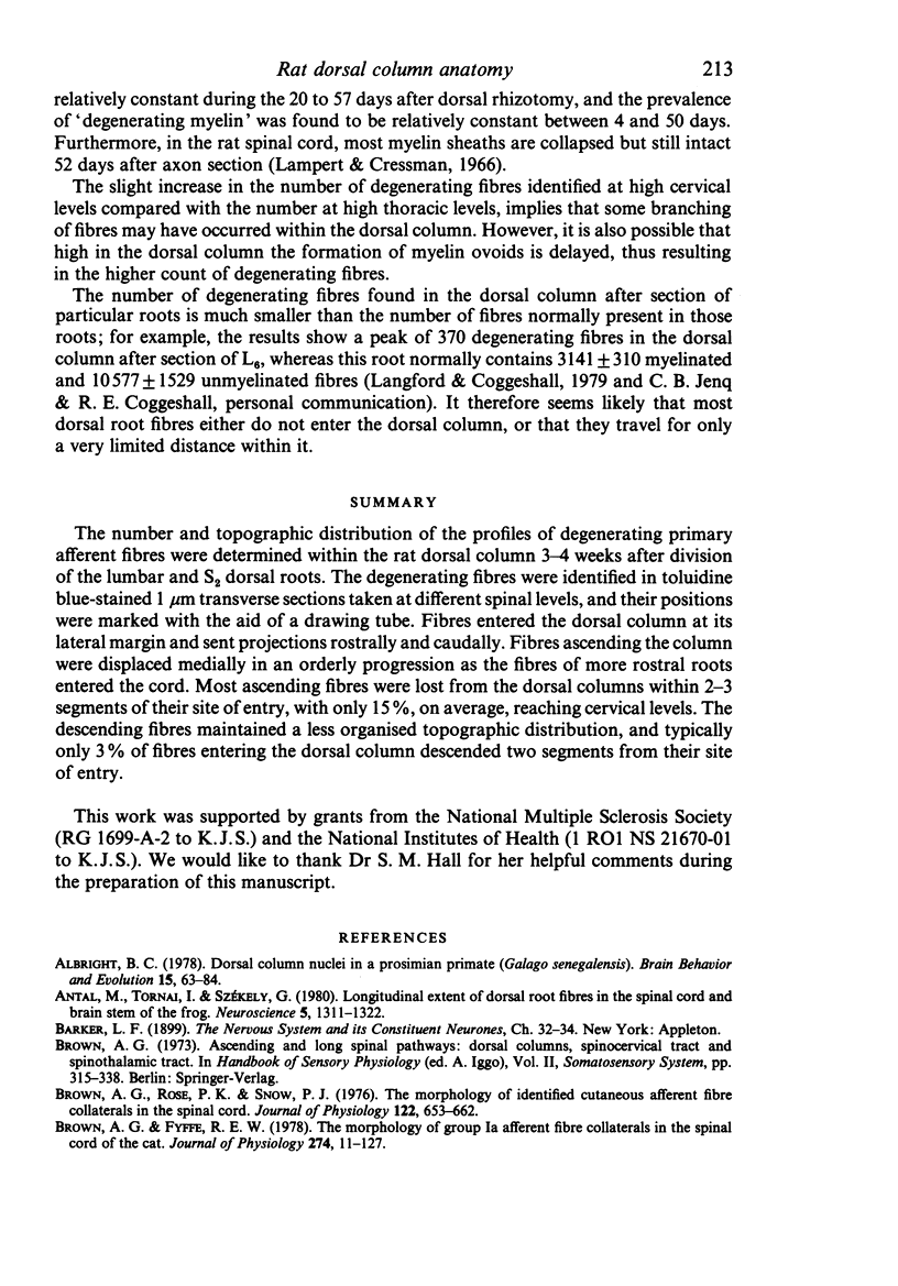
The number and topographic distribution of the profiles of degenerating primary afferent fibres were determined within the rat dorsal column 3-4 weeks after division of the lumbar and S2 dorsal roots. The degenerating fibres were identified in toluidine blue-stained 1 micron transverse sections taken at different spinal levels, and their positions were marked with the aid of a drawing tube. Fibres entered the dorsal column at its lateral margin and sent projections rostrally and caudally. Fibres ascending the column were displaced medially in an orderly progression as the fibres of more rostral roots entered the cord. Most ascending fibres were lost from the dorsal columns within 2-3 segments of their site of entry, with only 15%, on average, reaching cervical levels. The descending fibres maintained a less organised topographic distribution, and typically only 3% of fibres entering the dorsal column descended two segments from their site of entry.
Full text
PDF

Images in this article
Selected References
These references are in PubMed. This may not be the complete list of references from this article.
- Albright B. C. Dorsal column nuclei in a prosimian primate (Galago senegalensis). I. Gracile nucleus: morphology and primary afferent fibers from low thoracic, lumbosacral and coccygeal spinal segments. Brain Behav Evol. 1978;15(1):63–84. doi: 10.1159/000123772. [DOI] [PubMed] [Google Scholar]
- Antal M., Tornai I., Székely G. Longitudinal extent of dorsal root fibres in the spinal cord and brain stem of the frog. Neuroscience. 1980;5(7):1311–1322. doi: 10.1016/0306-4522(80)90203-1. [DOI] [PubMed] [Google Scholar]
- Brown A. G., Fyffe R. E. The morphology of group Ia afferent fibre collaterals in the spinal cord of the cat. J Physiol. 1978 Jan;274:111–127. doi: 10.1113/jphysiol.1978.sp012137. [DOI] [PMC free article] [PubMed] [Google Scholar]
- Carpenter M. B., Stein B. M., Shriver J. E. Central projections of spinal dorsal roots in the monkey. II. Lower thoracic, lumbosarcral and coccygeal dorsal roots. Am J Anat. 1968 Jul;123(1):75–118. doi: 10.1002/aja.1001230104. [DOI] [PubMed] [Google Scholar]
- Culberson J. L., Brown P. B. Projections of hindlimb dorsal roots to lumbosacral spinal cord of cat. J Neurophysiol. 1984 Mar;51(3):516–528. doi: 10.1152/jn.1984.51.3.516. [DOI] [PubMed] [Google Scholar]
- Culberson J. L., Haines D. E., Kimmel D. L., Brown P. B. Contralateral projection of primary afferent fibers to mammalian spinal cord. Exp Neurol. 1979 Apr;64(1):83–97. doi: 10.1016/0014-4886(79)90007-4. [DOI] [PubMed] [Google Scholar]
- Culberson J. L., Kimmel D. L. Primary afferent fiber distribution at brachial and lumbosacral spinal cord levels in the opossum (Didelphis marsupialis virigniana). Brain Behav Evol. 1975;12(4-6):229–246. doi: 10.1159/000124437. [DOI] [PubMed] [Google Scholar]
- Fink R. P., Heimer L. Two methods for selective silver impregnation of degenerating axons and their synaptic endings in the central nervous system. Brain Res. 1967 Apr;4(4):369–374. doi: 10.1016/0006-8993(67)90166-7. [DOI] [PubMed] [Google Scholar]
- Franson P., Ronnevi L. O. Myelin breakdown and elimination in the posterior funiculus of the adult cat after dorsal rhizotomy: a light and electron microscopic qualitative and quantitative study. J Comp Neurol. 1984 Feb 10;223(1):138–151. doi: 10.1002/cne.902230111. [DOI] [PubMed] [Google Scholar]
- GLEES P., SOLER J. Fibre content of the posterior column and synaptic connections of nucleus gracilis. Z Zellforsch Mikrosk Anat. 1951;36(4):381–400. doi: 10.1007/BF00335070. [DOI] [PubMed] [Google Scholar]
- GOLDBY F., ROBINSON L. R. The central connexions of dorsal spinal nerve roots and the ascending tracts in the spinal cord of Lacerta viridis. J Anat. 1962 Apr;96:153–170. [PMC free article] [PubMed] [Google Scholar]
- Imai Y., Kusama T. Distribution of the dorsal root fibers in the cat. An experimental study with the Nauta method. Brain Res. 1969 Apr;13(2):338–359. doi: 10.1016/0006-8993(69)90292-3. [DOI] [PubMed] [Google Scholar]
- Jacob M., Weddell G. Neural intersegmental connection in the spinal root and ganglion region of the rat. J Comp Neurol. 1975 May 1;161(1):115–123. doi: 10.1002/cne.901610109. [DOI] [PubMed] [Google Scholar]
- Joseph B. S., Whitlock D. G. Central projections of brachial and lumbar dorsal roots in reptiles. J Comp Neurol. 1968 Mar;132(3):469–484. doi: 10.1002/cne.901320308. [DOI] [PubMed] [Google Scholar]
- Joseph B. S., Whitlock D. G. Central projections of selected spinal dorsal roots in anuran amphibians. Anat Rec. 1968 Feb;160(2):279–288. doi: 10.1002/ar.1091600214. [DOI] [PubMed] [Google Scholar]
- Kusuma A., ten Donkelaar H. J. Dorsal root projections in various types of reptiles. Brain Behav Evol. 1980;17(4):291–309. doi: 10.1159/000121805. [DOI] [PubMed] [Google Scholar]
- Kusuma A., ten Donkelaar H. J. Staining of the dorsal root primary afferent fibers by anterograde movement of horseradish peroxidase and retrograde labelling of motoneurons and preganglionic autonomic cells in the turtle spinal cord. Neurosci Lett. 1979 Oct;14(2-3):141–146. doi: 10.1016/0304-3940(79)96138-x. [DOI] [PubMed] [Google Scholar]
- LIU C. N. Afferent nerves to Clarke's and the lateral cuneate nuclei in the cat. AMA Arch Neurol Psychiatry. 1956 Jan;75(1):67–77. doi: 10.1001/archneurpsyc.1956.02330190083009. [DOI] [PubMed] [Google Scholar]
- Lampert P. W., Cressman M. R. Fine-structural changes of myelin sheaths after axonal degeneration in the spinal cord of rats. Am J Pathol. 1966 Dec;49(6):1139–1155. [PMC free article] [PubMed] [Google Scholar]
- Langford L. A., Coggeshall R. E. Branching of sensory axons in the dorsal root and evidence for the absence of dorsal root efferent fibers. J Comp Neurol. 1979 Mar 1;184(1):193–204. doi: 10.1002/cne.901840111. [DOI] [PubMed] [Google Scholar]
- Leonard R. B., Cohen D. H. Spinal terminal fields of dorsal root fibers in the pigeon (Columba livia). J Comp Neurol. 1975 Sep 15;163(2):181–192. doi: 10.1002/cne.901630204. [DOI] [PubMed] [Google Scholar]
- Nikundiwe A. M., de Boer-van Huizen R., ten Donkelaar H. J. Dorsal root projections in the clawed toad (Xenopus laevis) as demonstrated by anterograde labeling with horseradish peroxidase. Neuroscience. 1982;7(9):2089–2103. doi: 10.1016/0306-4522(82)90121-x. [DOI] [PubMed] [Google Scholar]
- Petras J. M. Afferent fibres to the spinal cord. The terminal distribution of dorsal root and encephalospinal axons. Med Serv J Can. 1966 Jul-Aug;22(7):668–694. [PubMed] [Google Scholar]
- SPRAGUE J. M., HONGCHIEN H. A. THE TERMINAL FIELDS OF DORSAL ROOT FIBERS IN THE LUMBOSACRAL SPINAL CORD OF THE CAT, AND THE DENDRITIC ORGANIZATION OF THE MOTOR NUCLEI. Prog Brain Res. 1964;11:120–154. doi: 10.1016/s0079-6123(08)64046-7. [DOI] [PubMed] [Google Scholar]
- SPRAGUE J. M. The distribution of dorsal root fibres on motor cells in the lumbosacral spinal cord of the cat, and the site of excitatory and inhibitory terminals in monosynaptic pathways. Proc R Soc Lond B Biol Sci. 1958 Dec 24;149(937):534–556. doi: 10.1098/rspb.1958.0091. [DOI] [PubMed] [Google Scholar]
- Shriver J. E., Stein B. M., Carpenter M. B. Central projections of spinal dorsal roots in the monkey. I. Cervical and upper thoracic dorasal roots. Am J Anat. 1968 Jul;123(1):27–74. doi: 10.1002/aja.1001230103. [DOI] [PubMed] [Google Scholar]
- Smith M. C., Deacon P. Topographical anatomy of the posterior columns of the spinal cord in man. The long ascending fibres. Brain. 1984 Sep;107(Pt 3):671–698. doi: 10.1093/brain/107.3.671. [DOI] [PubMed] [Google Scholar]
- Umetani T., Hasegawa M., Yoshida S., Takeda H. Spinal projections of the dorsal root fibers in the cat. Kobe J Med Sci. 1981 Oct;27(5):173–188. [PubMed] [Google Scholar]
- Wall P. D., Werman R. The physiology and anatomy of long ranging afferent fibres within the spinal cord. J Physiol. 1976 Feb;255(2):321–334. doi: 10.1113/jphysiol.1976.sp011282. [DOI] [PMC free article] [PubMed] [Google Scholar]
- Whitsel B. L., Petrucelli L. M., Sapiro G., Ha H. Fiber sorting in the fasciculus gracilis of squirrel monkeys. Exp Neurol. 1970 Nov;29(2):227–242. doi: 10.1016/0014-4886(70)90054-3. [DOI] [PubMed] [Google Scholar]
- Whitsel B. L., Petrucelli L. M., Sapiro G. Modality representation in the lumbar and cervical fasciculus gracilis of squirrel monkeys. Brain Res. 1969 Sep;15(1):67–78. doi: 10.1016/0006-8993(69)90310-2. [DOI] [PubMed] [Google Scholar]
- ZIMMERMAN E. A., CHAMBERS W. W., LIU C. N. AN EXPERIMENTAL STUDY OF THE ANATOMICAL ORGANIZATION OF THE CORTICO-BULBAR SYSTEM IN THE ALBINO RAT. J Comp Neurol. 1964 Dec;123:301–323. doi: 10.1002/cne.901230302. [DOI] [PubMed] [Google Scholar]


