Abstract
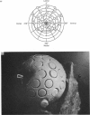
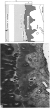
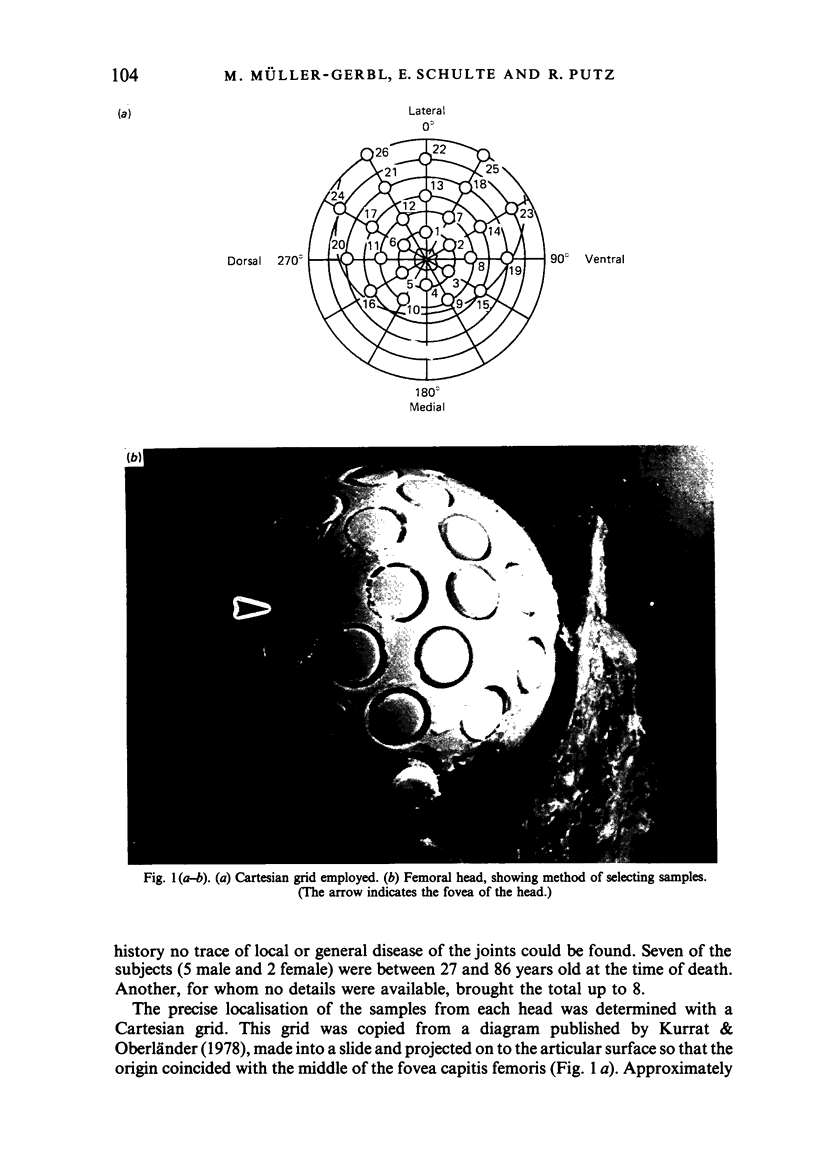
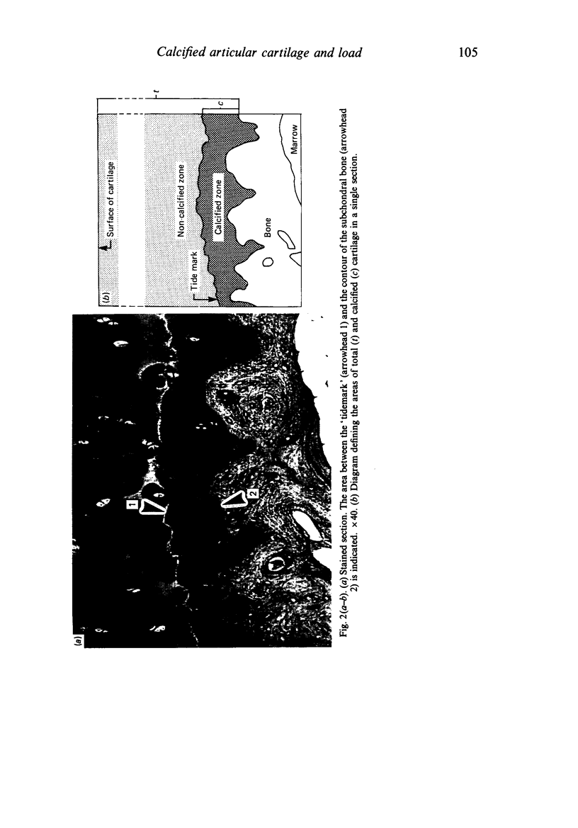
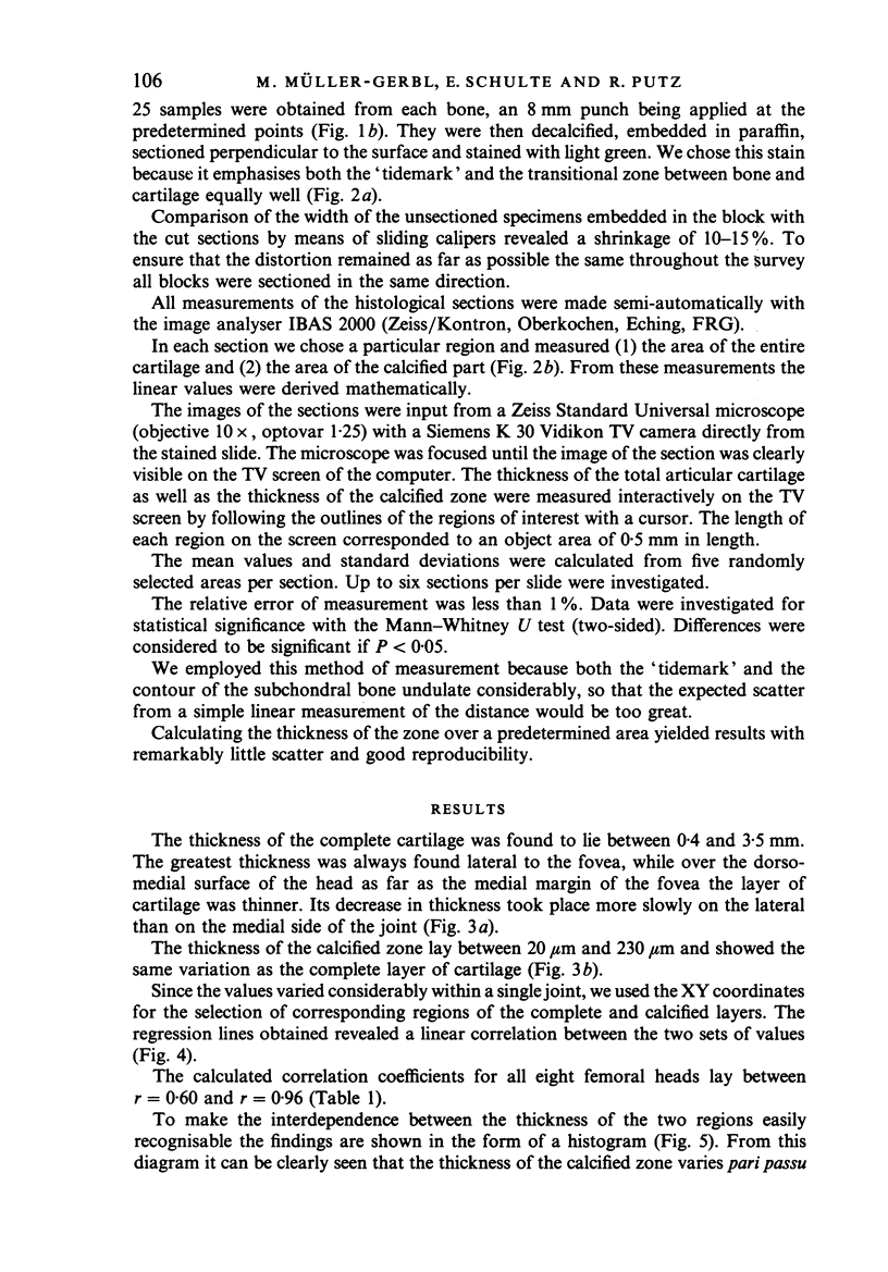
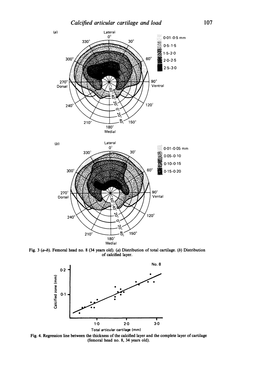
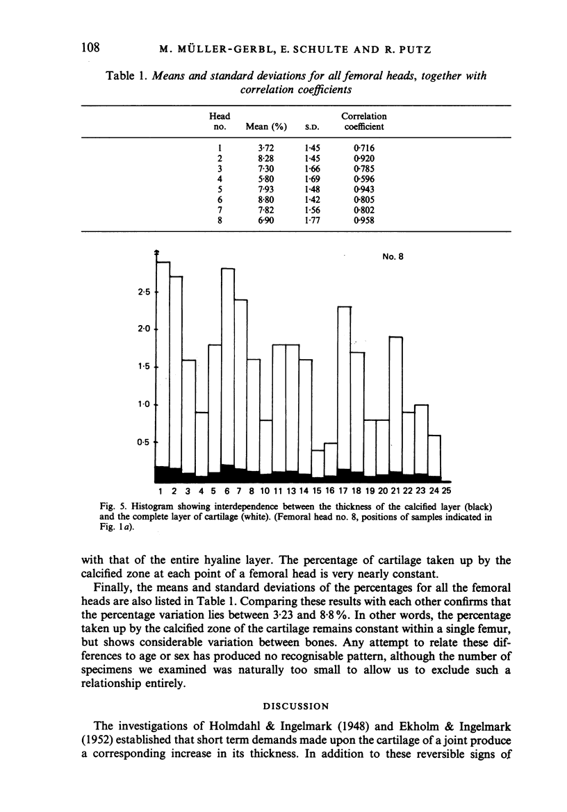
The thickness of both the articular cartilage and its calcified zone were measured at 25 carefully selected points in 8 human femoral heads, and the ratio of one to the other was found to be remarkably constant for each bone. The thickness of the calcified zone therefore shows the same distribution pattern as that of the total cartilage and, since the latter is dependent upon the distribution of the load, the thickness of the calcified region also appears to be related to mechanical stress. The volume of the calcified zone, however, expressed as a percentage of the total cartilage, varied considerably from one bone to another within the range from 3.23 to 8.8%. Too few specimens were examined to allow correlation with age or sex to be either refuted or confirmed.
Full text
PDF
Images in this article
Selected References
These references are in PubMed. This may not be the complete list of references from this article.
- EKHOLM R., INGELMARK B. E. Functional thickness variations of human articular cartilage. Acta Soc Med Ups. 1952;57(1-2):39–59. [PubMed] [Google Scholar]
- FAWNS H. T., LANDELLS J. W. Histochemical studies of rheumatic conditions. I. Observations on the fine structures of the matrix of normal bone and cartilage. Ann Rheum Dis. 1953 Jun;12(2):105–113. doi: 10.1136/ard.12.2.105. [DOI] [PMC free article] [PubMed] [Google Scholar]
- Green W. T., Jr, Martin G. N., Eanes E. D., Sokoloff L. Microradiographic study of the calcified layer of articular cartilage. Arch Pathol. 1970 Aug;90(2):151–158. [PubMed] [Google Scholar]
- Greenwald A. S., O'Connor J. J. The transmission of load through the human hip joint. J Biomech. 1971 Dec;4(6):507–528. doi: 10.1016/0021-9290(71)90041-8. [DOI] [PubMed] [Google Scholar]
- Jones I. L., Klämfeldt A., Sandström T. The effect of continuous mechanical pressure upon the turnover of articular cartilage proteoglycans in vitro. Clin Orthop Relat Res. 1982 May;(165):283–289. [PubMed] [Google Scholar]
- Kummer B. Die Beanspruchung des menschlichen Hüftgelenks. I. Allgemeine Problematik. Z Anat Entwicklungsgesch. 1968;127(4):277–285. [PubMed] [Google Scholar]
- Kurrat H. J. Die Beanspruchung des menschlichen Hüftgelenks. VI. Eine funktionelle Analyse der Knorpeldickenverteilung am menschlichen Femurkopf. Anat Embryol (Berl) 1977 Mar 30;150(2):129–140. doi: 10.1007/BF00316645. [DOI] [PubMed] [Google Scholar]
- Kurrat H. J., Oberländer W. The thickness of the cartilage in the hip joint. J Anat. 1978 May;126(Pt 1):145–155. [PMC free article] [PubMed] [Google Scholar]
- Lane L. B., Bullough P. G. Age-related changes in the thickness of the calcified zone and the number of tidemarks in adult human articular cartilage. J Bone Joint Surg Br. 1980 Aug;62(3):372–375. doi: 10.1302/0301-620X.62B3.7410471. [DOI] [PubMed] [Google Scholar]
- Oberländer W. Die Beanspruchung des menschlichen Hüftgelenks. VII. Die Verteilung der Knorpeldicke im Acetabulum und ihre funktionelle Deutung. Anat Embryol (Berl) 1977 Mar 30;150(2):141–153. doi: 10.1007/BF00316646. [DOI] [PubMed] [Google Scholar]
- Stougård J. The calcified cartilage and the subchondral bone under normal and abnormal conditions. Acta Pathol Microbiol Scand A. 1974 Mar;82(2):182–188. doi: 10.1111/j.1699-0463.1974.tb03842.x. [DOI] [PubMed] [Google Scholar]
- Tillmann B. Die Beanspruchung des menschlichen Hüftgelenks. 3. Die Form der Facies lunata. Z Anat Entwicklungsgesch. 1969;128(4):329–349. [PubMed] [Google Scholar]
- Tillmann B. Zur Lokalisation von degenerativen Veränderungen am Femurkopf bei der Coxarthrose. Z Orthop Ihre Grenzgeb. 1973 Feb;111(1):23–27. [PubMed] [Google Scholar]


