Abstract
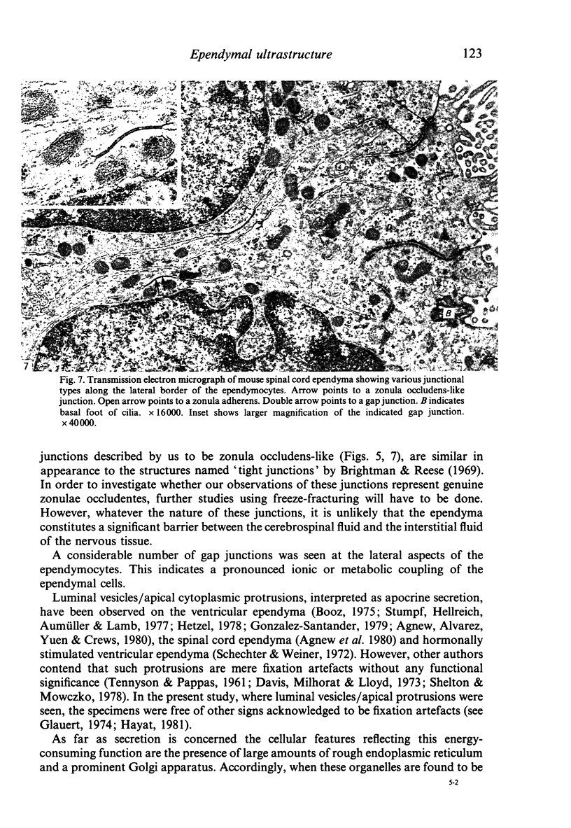
This study was done in order to investigate the normal ultrastructure of well-preserved mouse spinal canal ependyma using light, scanning and transmission electron microscopy. The ependymal lining was found to consist of a simple, cuboidal epithelium essentially similar to the unspecialized cuboidal ependyma of the brain ventricles. Apart from great variation in kinociliary density, no intracellular difference was noted between the ependymal cells. In contrast to earlier findings, indications of the existence of zonulae occludentes between the apical part of the ependymal cells were observed. Our findings do not support the hypothesis of secretion or intracellular transport by the ependyma, or that the ependyma constitutes a significant diffusion barrier.
Full text
PDF
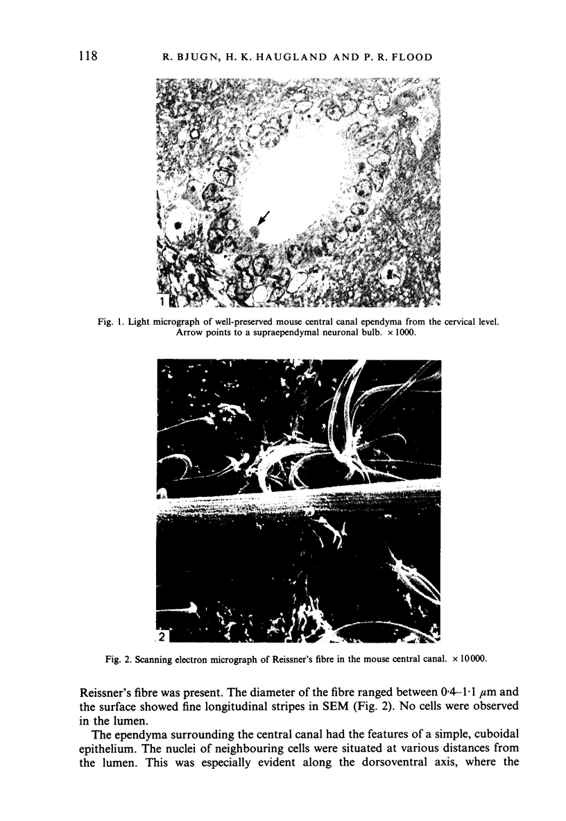
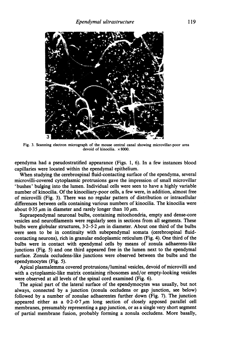
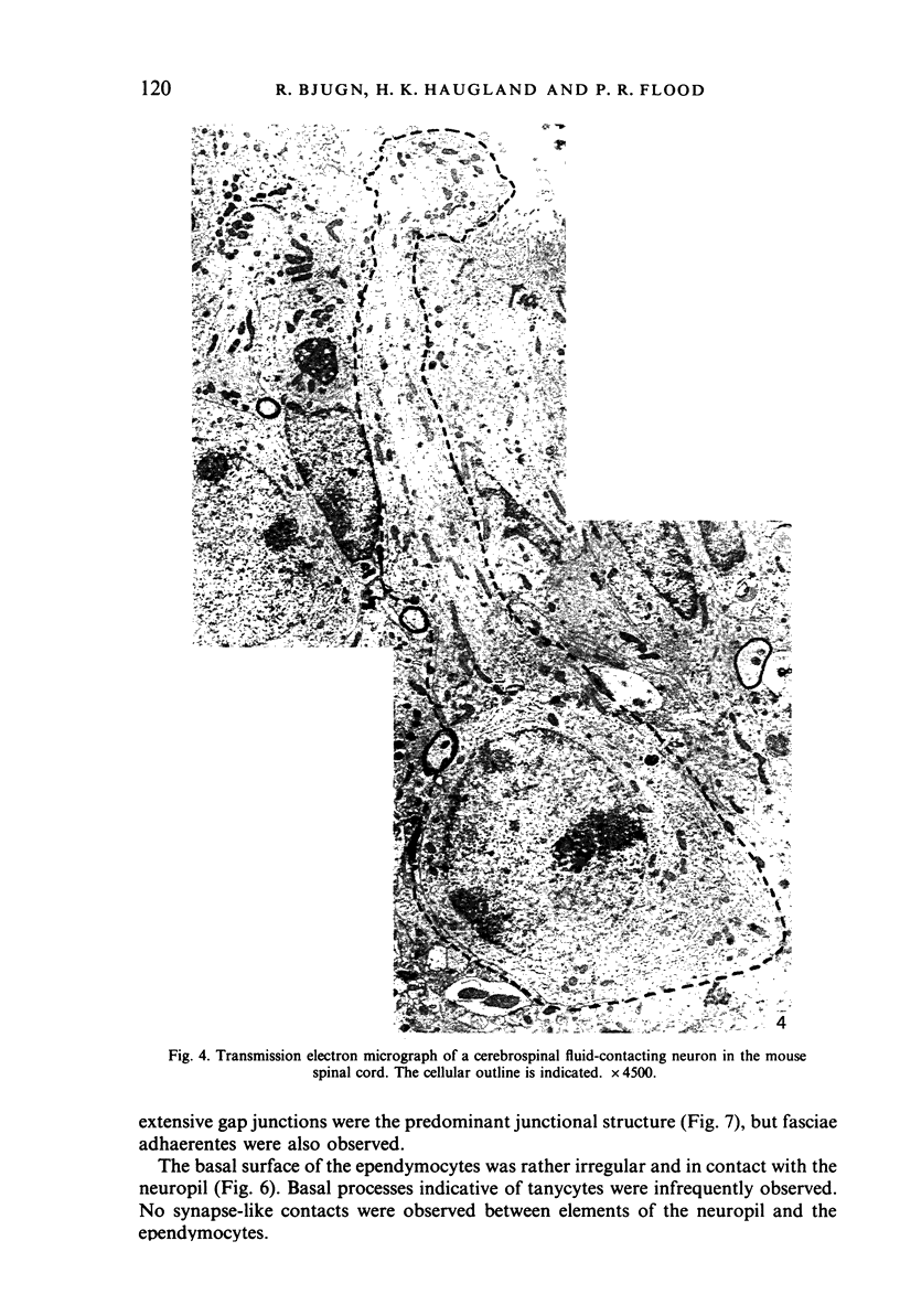
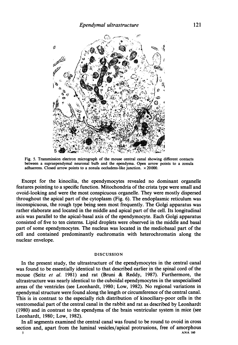
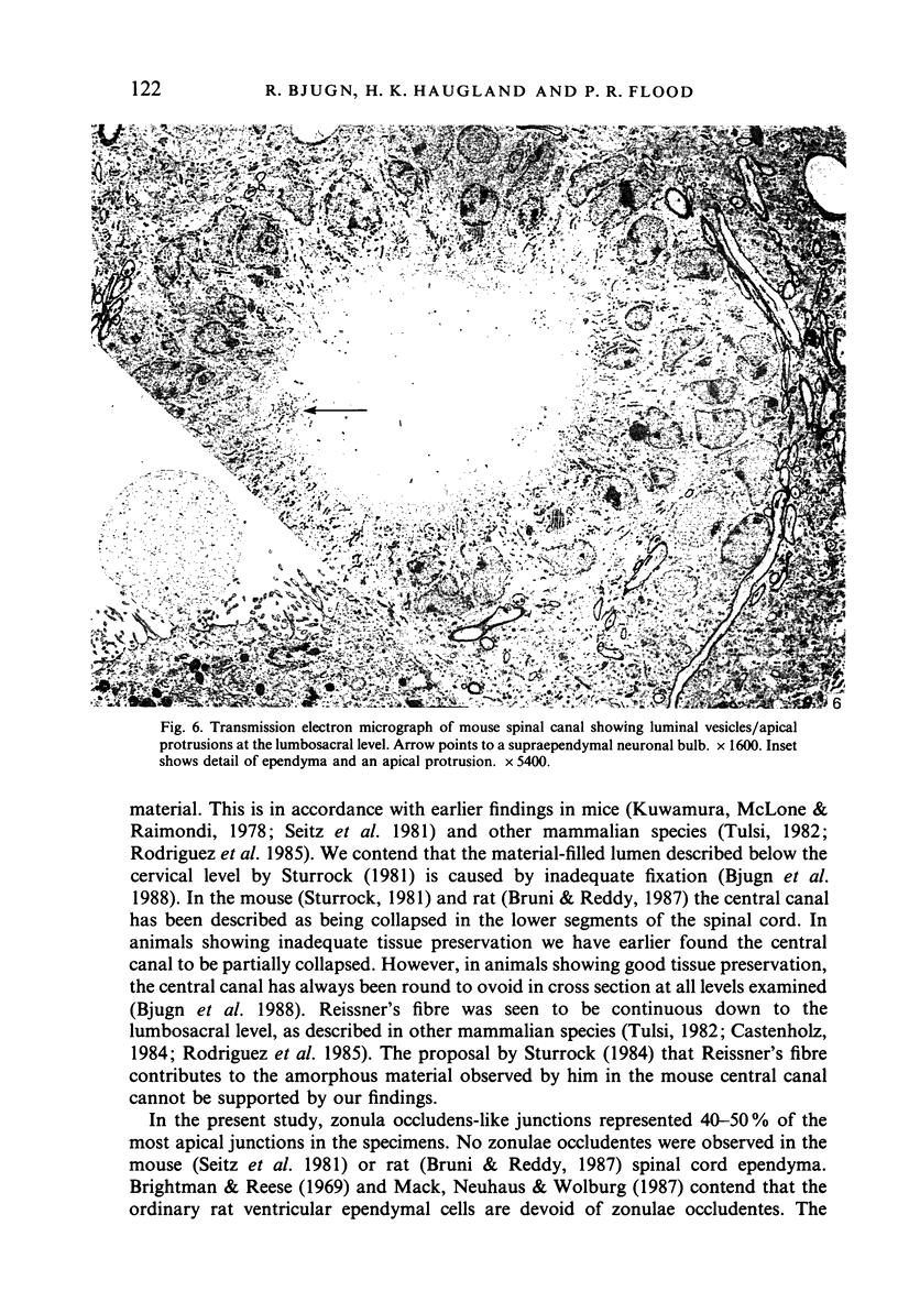
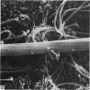
Images in this article
Selected References
These references are in PubMed. This may not be the complete list of references from this article.
- Agnew W. F., Alvarez R. B., Yuen T. G., Crews A. K. Protein synthesis and transport by the rat choroid plexus and ependyma: an autoradiographic study. Cell Tissue Res. 1980;208(2):261–281. doi: 10.1007/BF00234876. [DOI] [PubMed] [Google Scholar]
- Bjugn R., Haugland H. K., Flood P. R. Preservation of the ependyma of the mouse spinal cord by various vascular perfusion procedures. J Neurosci Methods. 1988 Feb;23(1):49–55. doi: 10.1016/0165-0270(88)90021-0. [DOI] [PubMed] [Google Scholar]
- Booz K. H. Secretory phenomena at the ependyma of the IIIrd ventricle of the embryonic rat. Anat Embryol (Berl) 1975 Aug 9;147(2):143–159. doi: 10.1007/BF00306729. [DOI] [PubMed] [Google Scholar]
- Brightman M. W., Reese T. S. Junctions between intimately apposed cell membranes in the vertebrate brain. J Cell Biol. 1969 Mar;40(3):648–677. doi: 10.1083/jcb.40.3.648. [DOI] [PMC free article] [PubMed] [Google Scholar]
- Castenholz A. Scanning electron-microscopic study of Reissner's fiber in the caudal spinal cord of the cat and rabbit. Cell Tissue Res. 1984;237(1):181–183. doi: 10.1007/BF00229214. [DOI] [PubMed] [Google Scholar]
- Gonzalez-Santander R. Electron-microscopic study of the secretion of the ependymal cells in the domestic cat (ependymin-beta cells). Acta Anat (Basel) 1979;103(3):266–277. doi: 10.1159/000145024. [DOI] [PubMed] [Google Scholar]
- Hetzel W. Ependyma and ependymal protrusions of the lateral ventricles of the rabbit brain. Cell Tissue Res. 1978 Sep 26;192(3):475–488. doi: 10.1007/BF00212327. [DOI] [PubMed] [Google Scholar]
- Kuwamura K., McLone D. G., Raimondi A. J. The central (spinal) canal in congenital murine hydrocephalus: morphological and physiological aspects. Childs Brain. 1978;4(4):216–234. doi: 10.1159/000119779. [DOI] [PubMed] [Google Scholar]
- Low F. N. The central nervous system in scanning electron microscopy. Scan Electron Microsc. 1982;(Pt 2):869–890. [PubMed] [Google Scholar]
- Mack A., Neuhaus J., Wolburg H. Relationship between orthogonal arrays of particles and tight junctions as demonstrated in cells of the ventricular wall of the rat brain. Cell Tissue Res. 1987 Jun;248(3):619–625. doi: 10.1007/BF00216492. [DOI] [PubMed] [Google Scholar]
- Rodríguez S., Hein S., Yulis R., Delannoy L., Siegmund I., Rodríguez E. Reissner's fiber and the wall of the central canal in the lumbo-sacral region of the bovine spinal cord. Comparative immunocytochemical and ultrastructural study. Cell Tissue Res. 1985;240(3):649–662. doi: 10.1007/BF00216353. [DOI] [PubMed] [Google Scholar]
- Schechter J., Weiner R. Ultrastructural changes in the ependymal lining of the median eminence following the intraventricular administration of catecholamine. Anat Rec. 1972 Apr;172(4):643–650. doi: 10.1002/ar.1091720404. [DOI] [PubMed] [Google Scholar]
- Seitz R., Löhler J., Schwendemann G. Ependyma and meninges of the spinal cord of the mouse. A light-and electron-microscopic study. Cell Tissue Res. 1981;220(1):61–72. doi: 10.1007/BF00209966. [DOI] [PubMed] [Google Scholar]
- Stumpf W. E., Hellreich M. A., Aumüller G., Lamb J. C., 4th, Sar M. The collicular recess organ: evidence for structural and secretory specialization of the ventricular lining in the collicular recess. Cell Tissue Res. 1977 Oct 21;184(1):29–44. doi: 10.1007/BF00220525. [DOI] [PubMed] [Google Scholar]
- Sturrock R. R. A scanning electron microscopic study of Reissner's fibre in elderly mice. Anat Anz. 1984;156(1):21–30. [PubMed] [Google Scholar]
- Sturrock R. R. An electron microscopic study of the development of the ependyma of the central canal of the mouse spinal cord. J Anat. 1981 Jan;132(Pt 1):119–136. [PMC free article] [PubMed] [Google Scholar]
- Tulsi R. S. Reissner's fiber in the sacral cord and filum terminale of the possum Trichosurus vulpecula: a light, scanning, and electron microscopic study. J Comp Neurol. 1982 Oct 10;211(1):11–20. doi: 10.1002/cne.902110103. [DOI] [PubMed] [Google Scholar]


