Abstract
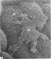
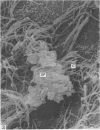
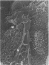
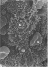
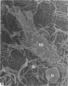
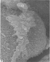
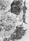
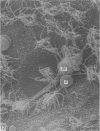
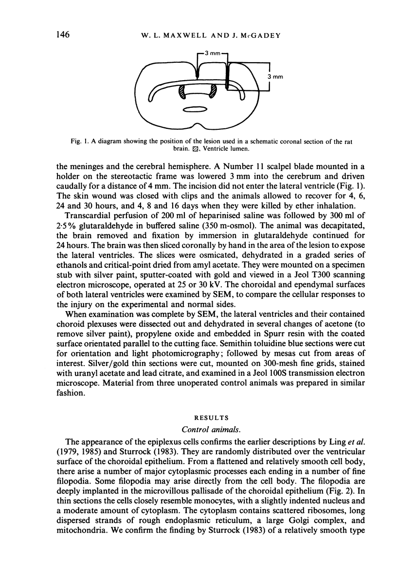
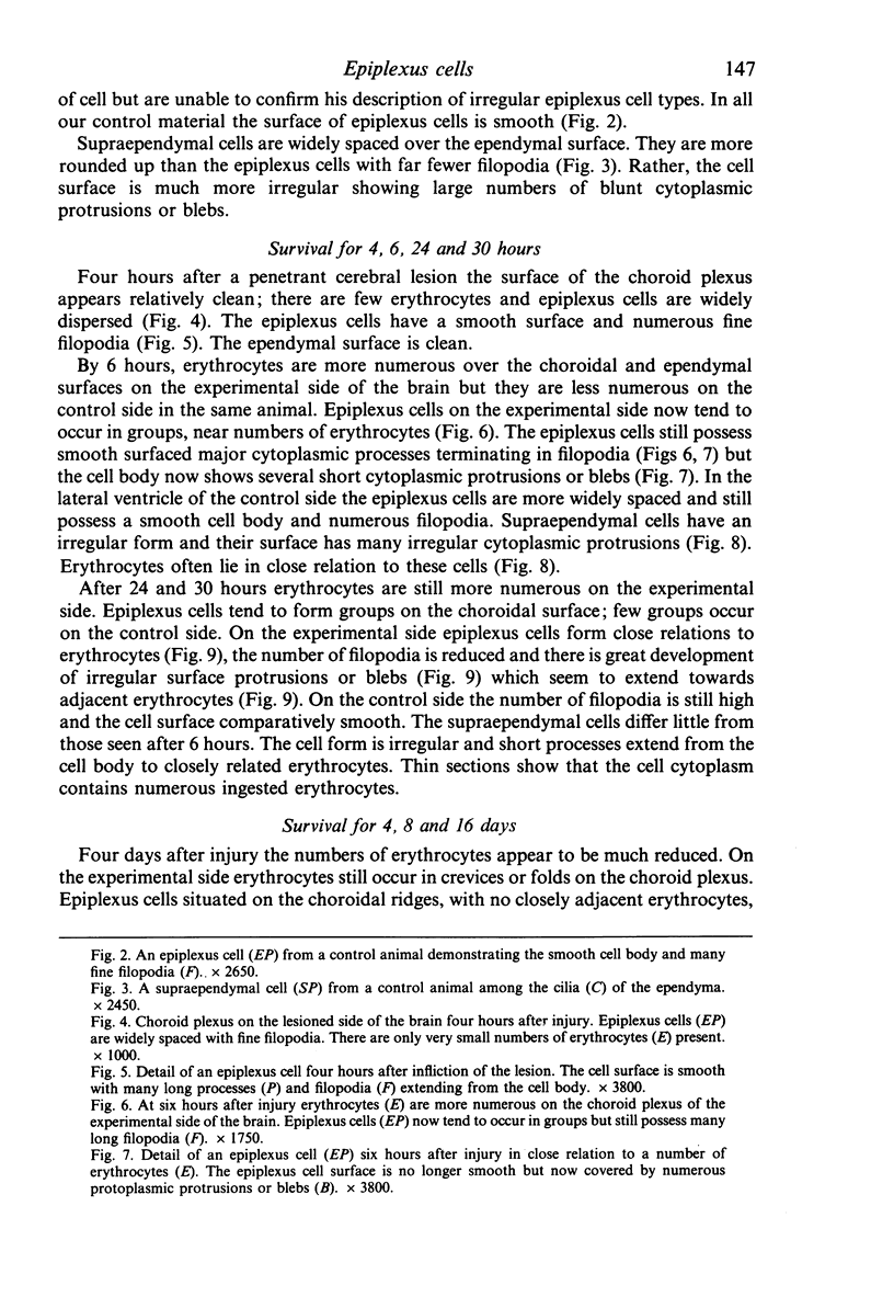
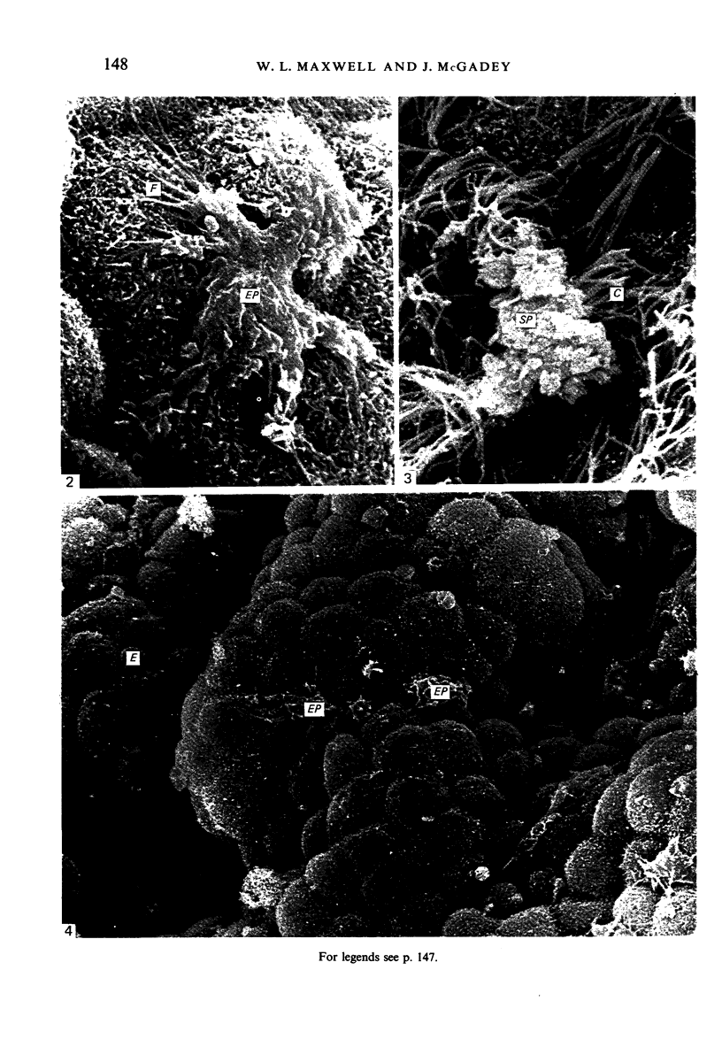
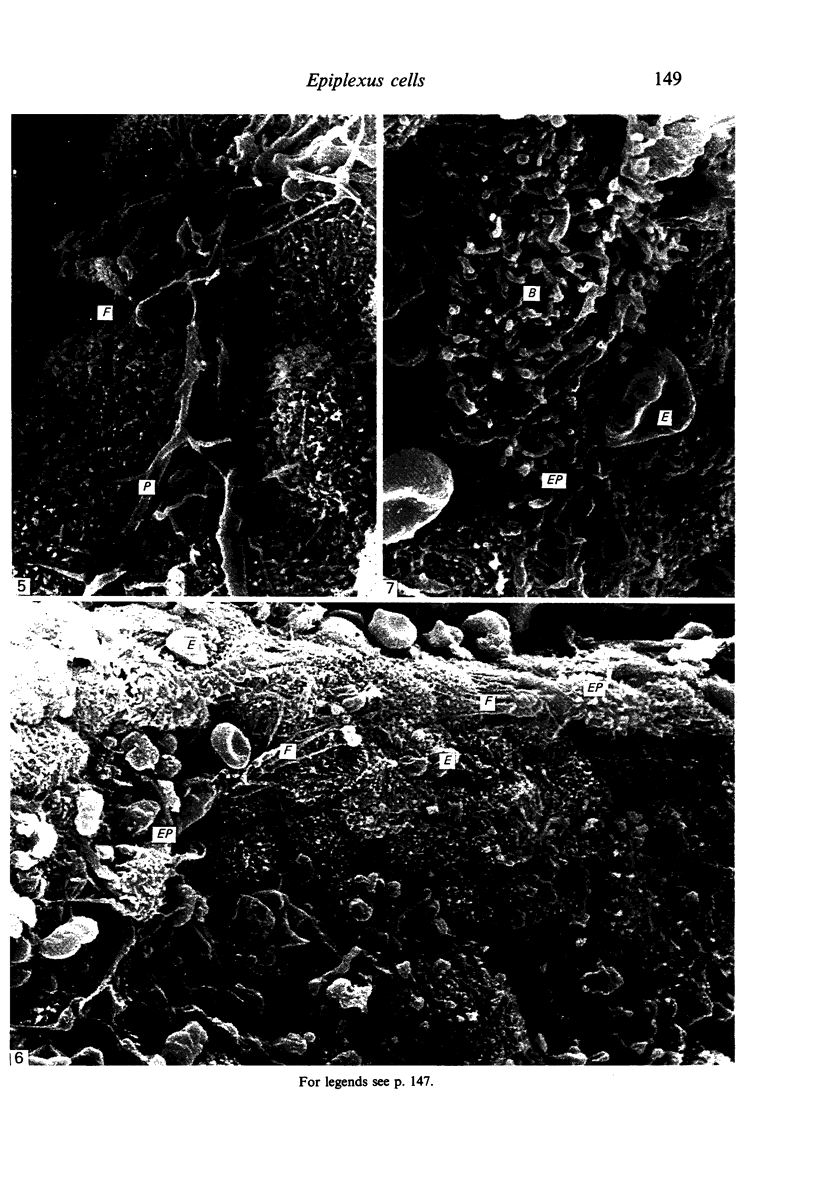
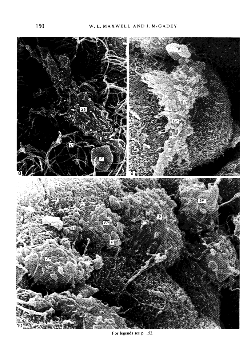
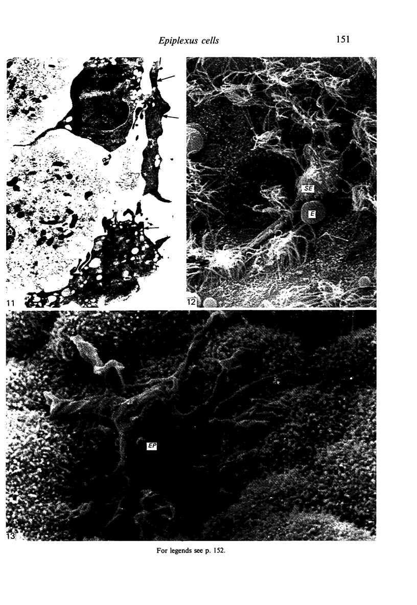
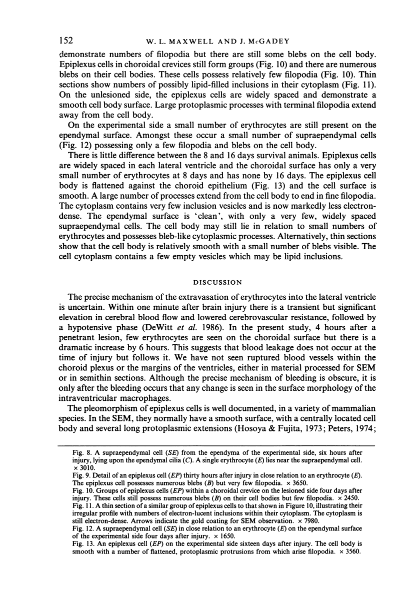
The response of epiplexus and supraependymal cells to extravasated blood after a penetrant cerebral lesion was investigated. Epiplexus cells respond more actively than supraependymal cells. The epiplexus cells tend to aggregate near areas of extravasation of erythrocytes, this being most marked 6 hours after injury. Epiplexus cells lose their smooth surface appearance, retract their filopodia and adopt a more spherical form, with short microvilli or blebs. Numerous inclusion vesicles develop; some contain disrupted erythrocytes 6-12 hours after injury and these are still present 24-30 hours after injury. By 8-16 days after injury epiplexus cells resume a smooth surface appearance and the number of inclusion vesicles is much reduced. This suggests reversion to a quiescent state, from an earlier active state.
Full text
PDF
Images in this article
Selected References
These references are in PubMed. This may not be the complete list of references from this article.
- Allen D. J. Scanning electron microscopy of epiplexus macrophages (Kolmer cells) in the dog. J Comp Neurol. 1975 May 15;161(2):197–213. doi: 10.1002/cne.901610205. [DOI] [PubMed] [Google Scholar]
- Bleier R., Albrecht R. Supraependymal macrophages of third ventricle of hamster: morphological, functional and histochemical characterization in situ and in culture. J Comp Neurol. 1980 Aug 1;192(3):489–504. doi: 10.1002/cne.901920308. [DOI] [PubMed] [Google Scholar]
- Carpenter S. J., McCarthy L. E., Borison H. L. Electron microscopic study of the epiplexus (Kolmer) cells of the cat choroid plexus. Z Zellforsch Mikrosk Anat. 1970;110(4):471–486. doi: 10.1007/BF00330099. [DOI] [PubMed] [Google Scholar]
- Coates P. W. Scanning electron microscopy of a second type of supraependymal cell in the monkey third ventricle. Anat Rec. 1975 Jul;182(3):275–287. doi: 10.1002/ar.1091820302. [DOI] [PubMed] [Google Scholar]
- Coates P. W. Supraependymal cells in recesses of the monkey third ventricle. Am J Anat. 1973 Apr;136(4):533–539. doi: 10.1002/aja.1001360410. [DOI] [PubMed] [Google Scholar]
- Coates P. W. Supraependymal cells: light and transmission electron microscopy extends scanning electron microscopic demonstration. Brain Res. 1973 Jul 27;57(2):502–507. doi: 10.1016/0006-8993(73)90157-1. [DOI] [PubMed] [Google Scholar]
- Coates P. W. The third ventricle of monkeys; Scanning electron microscopy of surface features in mature males and females. Cell Tissue Res. 1977 Feb 15;177(3):307–316. doi: 10.1007/BF00220306. [DOI] [PubMed] [Google Scholar]
- DeWitt D. S., Jenkins L. W., Wei E. P., Lutz H., Becker D. P., Kontos H. A. Effects of fluid-percussion brain injury on regional cerebral blood flow and pial arteriolar diameter. J Neurosurg. 1986 May;64(5):787–794. doi: 10.3171/jns.1986.64.5.0787. [DOI] [PubMed] [Google Scholar]
- Hosoya Y., Fujita T. Scanning electron microscope observation of intraventricular macrophages (Kolmer cells) in the rat brain. Arch Histol Jpn. 1973 Jan;35(2):133–140. doi: 10.1679/aohc1950.35.133. [DOI] [PubMed] [Google Scholar]
- Jennes L., Sikora K., Simonsberger P., Adam H. Ventrikuläre Topographie des diencephalen Ependyms bei Rattus rattus (L.)--eine rasterelektronenmikroskopische Untersuchung. J Hirnforsch. 1977;18(6):501–520. [PubMed] [Google Scholar]
- Ling E. A. Scanning electron microscopic study of epiplexus cells in the lateral ventricles of the monkey (Macaca fascicularis). J Anat. 1983 Dec;137(Pt 4):645–652. [PMC free article] [PubMed] [Google Scholar]
- Ling E. A., Tseng C. Y., Wong W. C. An electron microscopical study of epiplexus and supraependymal cells in the prenatal rat brain following a maternal injection of 6-aminonicotinamide. J Anat. 1985 Jan;140(Pt 1):119–129. [PMC free article] [PubMed] [Google Scholar]
- Ling E. A. Ultrastruct and origin of epiplexus cells in the telencephalic choroid plexus of postnatal rats studied by intravenous injection of carbon particles. J Anat. 1979 Oct;129(Pt 3):479–492. [PMC free article] [PubMed] [Google Scholar]
- Ling E. A. Ultrastructure and mode of formation of epiplexus cells in the choroid plexus in the lateral ventricles of the monkey (Macaca fascicularis). J Anat. 1981 Dec;133(Pt 4):555–569. [PMC free article] [PubMed] [Google Scholar]
- Malloy J. J., Low F. N. Scanning electron microscopy of the subarachnoid space in the dog. IV. Subarachnoid macrophages. J Comp Neurol. 1976 Jun 1;167(3):257–283. doi: 10.1002/cne.901670302. [DOI] [PubMed] [Google Scholar]
- Merchant R. E., Low F. N. Identification of challenged subarachnoid free cells. Am J Anat. 1977 Jan;148(1):143–148. doi: 10.1002/aja.1001480112. [DOI] [PubMed] [Google Scholar]
- Merchant R. E., Low F. N. Scanning electron microscopy of the subarachnoid space in the dog. V. Macrophages challenged by bacillus Calmette-Guerin. J Comp Neurol. 1977 Apr 1;172(3):381–407. doi: 10.1002/cne.901720302. [DOI] [PubMed] [Google Scholar]
- Merchant R. E. Scanning electron microscopy of epiplexus macrophages responding to challenge by bacillus Calmette-Guerin. Acta Neuropathol. 1979 Aug;47(3):183–188. doi: 10.1007/BF00690545. [DOI] [PubMed] [Google Scholar]
- Nielsen S. L., Gauger G. E. Experimental hydrocephalus: surface alterations of the lateral ventricle. Scanning electron microscopic studies. Lab Invest. 1974 May;30(5):618–625. [PubMed] [Google Scholar]
- Peters A., Swan R. C. The choroid plexus of the mature and aging rat: the choroidal epithelium. Anat Rec. 1979 Jul;194(3):325–353. doi: 10.1002/ar.1091940303. [DOI] [PubMed] [Google Scholar]
- Peters A. The surface fine structure of the choroid plexus and ependymal lining of the rat lateral ventricle. J Neurocytol. 1974 Mar;3(1):99–108. doi: 10.1007/BF01111935. [DOI] [PubMed] [Google Scholar]
- Sturrock R. R. A developmental study of epiplexus cells and supraependymal cells and their possible relationship to microglia. Neuropathol Appl Neurobiol. 1978 Sep-Oct;4(5):307–322. doi: 10.1111/j.1365-2990.1978.tb01345.x. [DOI] [PubMed] [Google Scholar]
- Sturrock R. R. A light microscopic and scanning electron microscopic study of intraventricular macrophages in the brains of aged mice. J Anat. 1983 Jun;136(Pt 4):761–771. [PMC free article] [PubMed] [Google Scholar]
- Sturrock R. R. A semithin light microscopic, transmission electron microscopic and scanning electron microscopic study of macrophages in the lateral ventricle of mice from embryonic to adult life. J Anat. 1979 Aug;129(Pt 1):31–44. [PMC free article] [PubMed] [Google Scholar]
- Tseng C. Y., Ling E. A., Wong W. C. A scanning and transmission electron microscopic study of amoeboid microglial cells in the prenatal rat brain following a maternal injection of 6-aminonicotinamide. J Anat. 1984 Jun;138(Pt 4):733–743. [PMC free article] [PubMed] [Google Scholar]


