Abstract
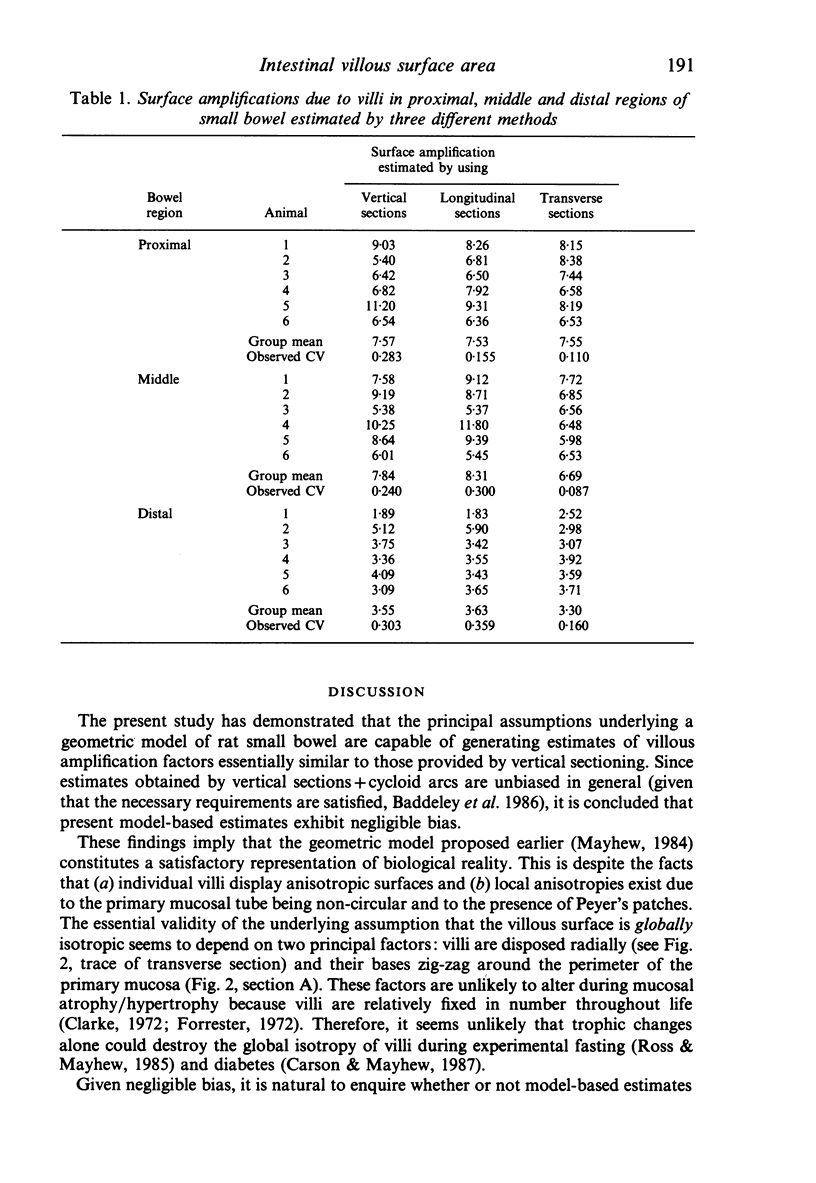
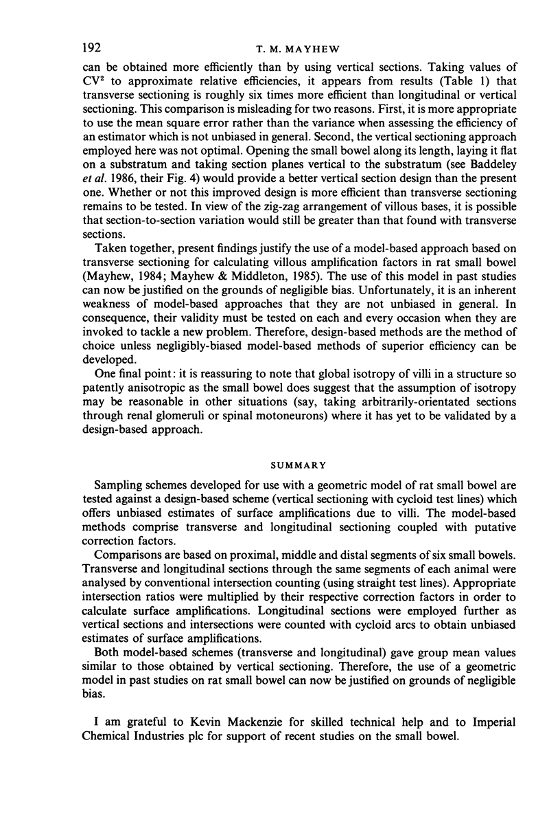
Sampling schemes developed for use with a geometric model of rat small bowel are tested against a design-based scheme (vertical sectioning with cycloid test lines) which offers unbiased estimates of surface amplifications due to villi. The model-based methods comprise transverse and longitudinal sectioning coupled with putative correction factors. Comparisons are based on proximal, middle and distal segments of six small bowels. Transverse and longitudinal sections through the same segments of each animal were analysed by conventional intersection counting (using straight test lines). Appropriate intersection ratios were multiplied by their respective correction factors in order to calculate surface amplifications. Longitudinal sections were employed further as vertical sections and intersections were counted with cycloid arcs to obtain unbiased estimates of surface amplifications. Both model-based schemes (transverse and longitudinal) gave group mean values similar to those obtained by vertical sectioning. Therefore, the use of a geometric model in past studies on rat small bowel can now be justified on grounds of negligible bias.
Full text
PDF
Selected References
These references are in PubMed. This may not be the complete list of references from this article.
- Baddeley A. J., Gundersen H. J., Cruz-Orive L. M. Estimation of surface area from vertical sections. J Microsc. 1986 Jun;142(Pt 3):259–276. doi: 10.1111/j.1365-2818.1986.tb04282.x. [DOI] [PubMed] [Google Scholar]
- Clarke R. M. The effect of growth and of fasting on the number of villi and crypts in the small intestine of the albino rat. J Anat. 1972 May;112(Pt 1):27–33. [PMC free article] [PubMed] [Google Scholar]
- Cruz-Orive L. M., Hunziker E. B. Stereology for anisotropic cells: application to growth cartilage. J Microsc. 1986 Jul;143(Pt 1):47–80. doi: 10.1111/j.1365-2818.1986.tb02765.x. [DOI] [PubMed] [Google Scholar]
- Mattfeldt T., Möbius H. J., Mall G. Orthogonal triplet probes: an efficient method for unbiased estimation of length and surface of objects with unknown orientation in space. J Microsc. 1985 Sep;139(Pt 3):279–289. doi: 10.1111/j.1365-2818.1985.tb02644.x. [DOI] [PubMed] [Google Scholar]
- Mayhew T. M. Geometric model of the rat intestinal mucosa for stereological evaluation of villus amplification factors. J Microsc. 1984 Sep;135(Pt 3):337–346. doi: 10.1111/j.1365-2818.1984.tb02538.x. [DOI] [PubMed] [Google Scholar]
- Mayhew T. M., Middleton C. Crypts, villi and microvilli in the small intestine of the rat. A stereological study of their variability within and between animals. J Anat. 1985 Aug;141:1–17. [PMC free article] [PubMed] [Google Scholar]
- Ross G. A., Mayhew T. M. Effects of fasting on mucosal dimensions in the duodenum, jejunum and ileum of the rat. J Anat. 1985 Oct;142:191–200. [PMC free article] [PubMed] [Google Scholar]


