Abstract
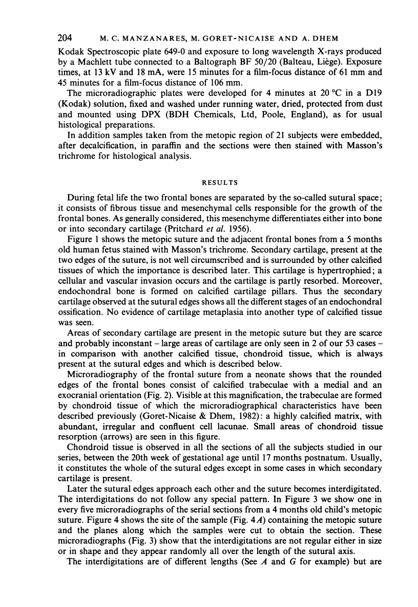
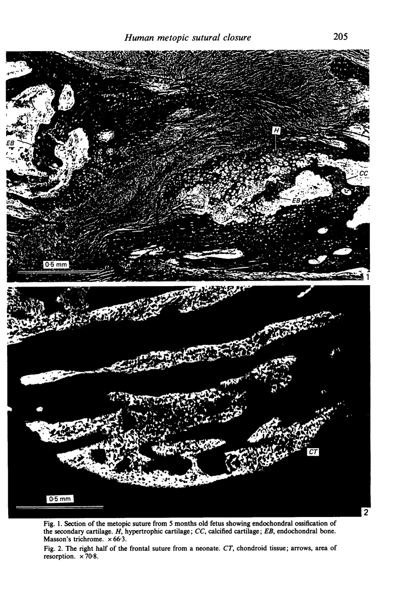
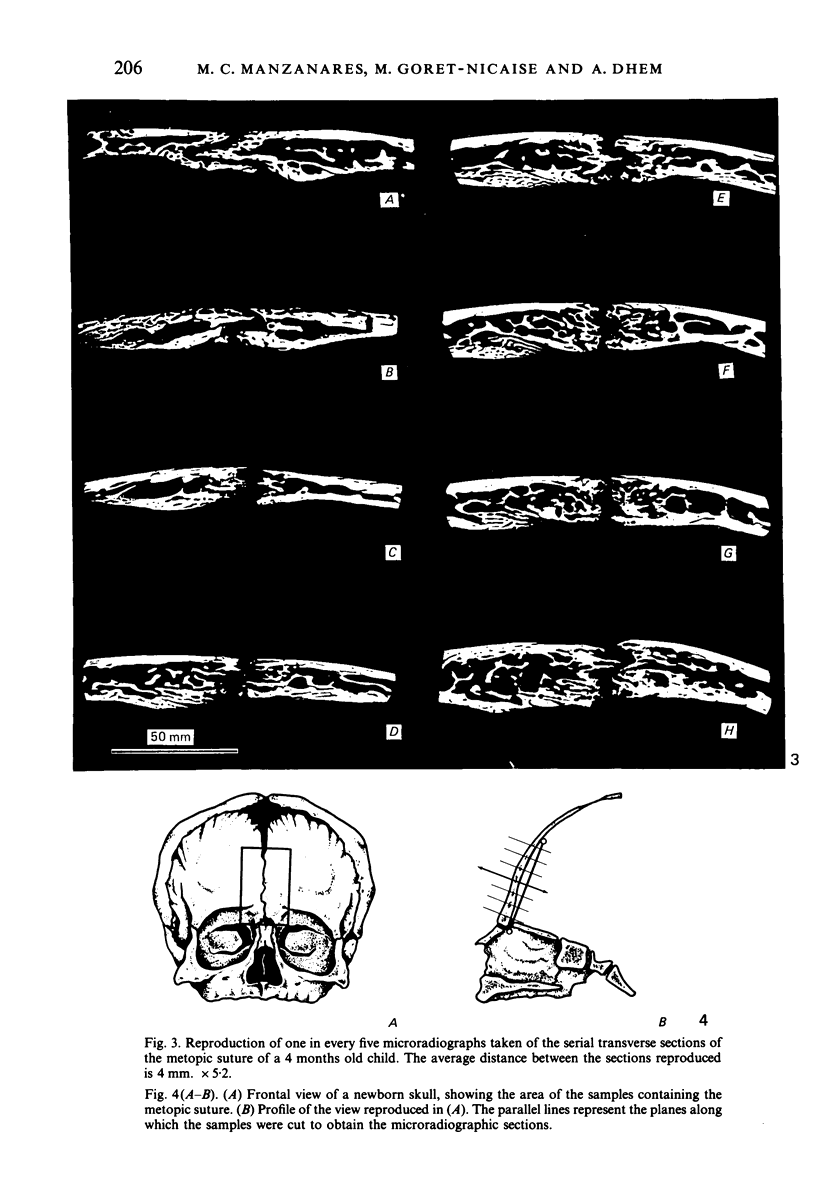
The present study reveals the presence in the sutural area of secondary cartilage, assuring the passive growth of the bones and undergoing an endochondral ossification, but without playing a direct role in the synostosis. The chondroid tissue is responsible for the growth of each frontal bone towards the other and constitutes the first bridge of union between the two bones. It is the most important finding in this study, which provides a description of the closure of the metopic suture and of the maintenance of an open sutural space by a process of active resorption. This new knowledge will help to understand better the whole process of suture closure and its pathology.
Full text
PDF


Images in this article
Selected References
These references are in PubMed. This may not be the complete list of references from this article.
- Babler W. J., Persing J. A., Nagorsky M. J., Jane J. A. Restricted growth at the frontonasal suture: alterations in craniofacial growth in rabbits. Am J Anat. 1987 Jan;178(1):90–98. doi: 10.1002/aja.1001780112. [DOI] [PubMed] [Google Scholar]
- Dhem A., Dambrain R., Thauvoy C., Stricker M. Contribution to the histological and microradiographic study of the craniostenosis. Acta Neurochir (Wien) 1983;69(3-4):259–272. doi: 10.1007/BF01401813. [DOI] [PubMed] [Google Scholar]
- Engström C., Linde A., Thilander B. Craniofacial morphology and growth in the rat. Cephalometric analysis of the effects of a low calcium and vitamin D-deficient diet. J Anat. 1982 Mar;134(Pt 2):299–314. [PMC free article] [PubMed] [Google Scholar]
- Goret-Nicaise M., Dhem A. Comparison of the calcium content of different tissues present in the human mandible. Acta Anat (Basel) 1985;124(3-4):167–172. doi: 10.1159/000146113. [DOI] [PubMed] [Google Scholar]
- Goret-Nicaise M., Dhem A. Electron microscopic study of chondroid tissue in the cat mandible. Calcif Tissue Int. 1987 Apr;40(4):219–223. doi: 10.1007/BF02556625. [DOI] [PubMed] [Google Scholar]
- Goret-Nicaise M., Dhem A. Presence of chondroid tissue in the symphyseal region of the growing human mandible. Acta Anat (Basel) 1982;113(3):189–195. doi: 10.1159/000145554. [DOI] [PubMed] [Google Scholar]
- Goret-Nicaise M. Identification of collagen type I and type II in chondroid tissue. Calcif Tissue Int. 1984 Dec;36(6):682–689. doi: 10.1007/BF02405390. [DOI] [PubMed] [Google Scholar]
- Hall B. K. Immobilization and cartilage transformation into bone in the embryonic chick. Anat Rec. 1972 Aug;173(4):391–403. doi: 10.1002/ar.1091730402. [DOI] [PubMed] [Google Scholar]
- Hall B. K. The formation of adventitious cartilage by membrane bones under the influence of mechanical stimulation applied in vitro. Life Sci. 1967 Mar 15;6(6):663–667. doi: 10.1016/0024-3205(67)90104-x. [DOI] [PubMed] [Google Scholar]
- Herring S. W. A biometric study of suture fusion and skull growth in peccaries. Anat Embryol (Berl) 1974;146(2):167–180. doi: 10.1007/BF00315593. [DOI] [PubMed] [Google Scholar]
- Herring S. W. Sutures--a tool in functional cranial analysis. Acta Anat (Basel) 1972;83(2):222–247. doi: 10.1159/000143860. [DOI] [PubMed] [Google Scholar]
- Hinton D. R., Becker L. E., Muakkassa K. F., Hoffman H. J. Lambdoid synostosis. Part 1. The lambdoid suture: normal development and pathology of "synostosis". J Neurosurg. 1984 Aug;61(2):333–339. doi: 10.3171/jns.1984.61.2.0333. [DOI] [PubMed] [Google Scholar]
- Jane J. A., Edgerton M. T., Futrell J. W., Park T. S. Immediate correction of sagittal synostosis. J Neurosurg. 1978 Nov;49(5):705–710. doi: 10.3171/jns.1978.49.5.0705. [DOI] [PubMed] [Google Scholar]
- KNESE K. H., BIERMANN H. Die Knochenbildung an Sehnen- und Bandansătzen im Bereich ursprünglich chondraier Apophysen. Z Zellforsch Mikrosk Anat. 1958;49(2):142–187. [PubMed] [Google Scholar]
- Koskinen L. Adaptive sutures. Changes after unilateral masticatory muscle resection in rats. A microscopic study. Proc Finn Dent Soc. 1977;73 (Suppl 10-11):3–80. [PubMed] [Google Scholar]
- Koskinen L., Isotupa K., Koski K. A note on craniofacial sutural growth. Am J Phys Anthropol. 1976 Nov;45(3 Pt 1):511–516. doi: 10.1002/ajpa.1330450312. [DOI] [PubMed] [Google Scholar]
- Latham R. A., Burston W. R. The postnatal pattern of growth at the sutures of the human skull. An histological survey. Dent Pract Dent Rec. 1966 Oct;17(2):61–67. [PubMed] [Google Scholar]
- MILES A. E. W. Chondrosarcoma of the maxilla. Br Dent J. 1950 May 19;88(10):257–269. [PubMed] [Google Scholar]
- MOHAMMED C. I. Growth pattern of the rat maxilla from 16 days insemination age to 30 days after birth. Am J Anat. 1957 Jan;100(1):115–165. doi: 10.1002/aja.1001000106. [DOI] [PubMed] [Google Scholar]
- MOSS M. L. Experimental alteration of sutural area morphology. Anat Rec. 1957 Mar;127(3):569–589. doi: 10.1002/ar.1091270307. [DOI] [PubMed] [Google Scholar]
- MOSS M. L. Fusion of the frontal suture in the rat. Am J Anat. 1958 Jan;102(1):141–165. doi: 10.1002/aja.1001020107. [DOI] [PubMed] [Google Scholar]
- MOSS M. L. Inhibition and stimulation of sutural fusion in the rat calvaria. Anat Rec. 1960 Apr;136:457–467. doi: 10.1002/ar.1091360405. [DOI] [PubMed] [Google Scholar]
- Markens I. S., Oudhof H. A. Morphological changes in the coronal suture after replantation. Acta Anat (Basel) 1980;107(3):289–296. doi: 10.1159/000145253. [DOI] [PubMed] [Google Scholar]
- PRITCHARD J. J., SCOTT J. H., GIRGIS F. G. The structure and development of cranial and facial sutures. J Anat. 1956 Jan;90(1):73–86. [PMC free article] [PubMed] [Google Scholar]
- Persing J. A., Babler W. J., Jane J. A., Duckworth P. F. Experimental unilateral coronal synostosis in rabbits. Plast Reconstr Surg. 1986 Mar;77(3):369–377. doi: 10.1097/00006534-198603000-00003. [DOI] [PubMed] [Google Scholar]
- Persson M., Magnusson B. C., Thilander B. Sutural closure in rabbit and man: a morphological and histochemical study. J Anat. 1978 Feb;125(Pt 2):313–321. [PMC free article] [PubMed] [Google Scholar]
- Sarnat B. G. Something of the nature of gross sutural growth. Ann Plast Surg. 1986 Oct;17(4):339–349. doi: 10.1097/00000637-198610000-00013. [DOI] [PubMed] [Google Scholar]
- Schmahl W., Meyer I., Kriegel H., Tempel K. H. Cartilaginous metaplasia and overgrowth of neurocranium skull after X-irradiation in utero. Virchows Arch A Pathol Anat Histol. 1979;384(2):173–184. doi: 10.1007/BF00427254. [DOI] [PubMed] [Google Scholar]
- Thilander B., Ingervall B. The human spheno-occipital synchondrosis. II. A histological and microradiographic study of its growth. Acta Odontol Scand. 1973;31(5):323–334. doi: 10.3109/00016357309002520. [DOI] [PubMed] [Google Scholar]
- Thorogood P. In vitro studies on skeletogenic potential of membrane bone periosteal cells. J Embryol Exp Morphol. 1979 Dec;54:185–207. [PubMed] [Google Scholar]
- Trevisan R. A., Scapino R. P. Secondary cartilages in growth and development of the symphysis menti in the hamster. Acta Anat (Basel) 1976;94(1):40–58. doi: 10.1159/000144543. [DOI] [PubMed] [Google Scholar]
- Watzek G., Grundschober F., Plenk H., Jr, Eschberger J. Experimental investigations into the clinical significance of bone growth at viscerocranial sutures. J Maxillofac Surg. 1982 May;10(2):61–79. doi: 10.1016/s0301-0503(82)80016-7. [DOI] [PubMed] [Google Scholar]
- YOUNG R. W. The influence of cranial contents on postnatal growth of the skull in the rat. Am J Anat. 1959 Nov;105:383–415. doi: 10.1002/aja.1001050304. [DOI] [PubMed] [Google Scholar]


