Abstract
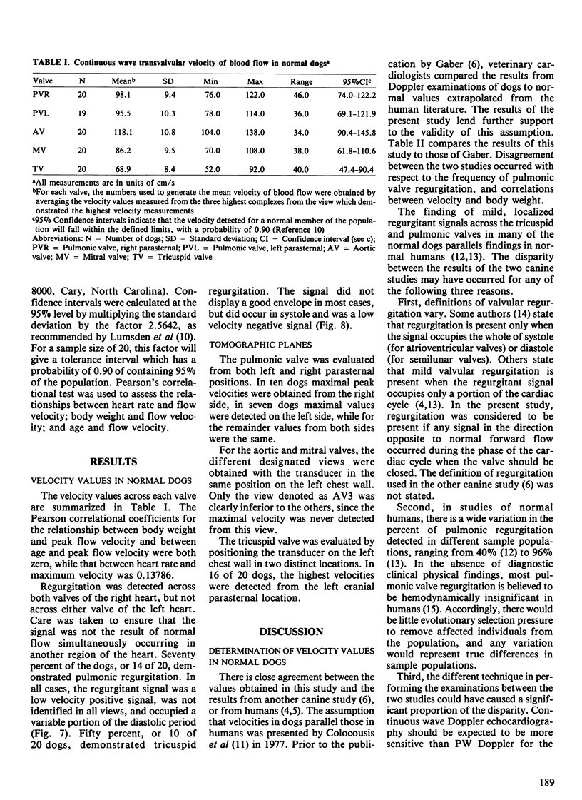
Doppler echocardiography is a relatively new procedure used to assess certain cardiovascular disorders in the dog. The objectives of this study were to determine the range of values for the maximal peak velocity of blood flow across each of the four cardiac valves in a sample population of normal adult dogs, using duplex continuous wave Doppler echocardiography, and to determine the optimal tomographic planes to use for an adequate continuous wave Doppler evaluation of the canine heart. Twenty normal dogs were examined to obtain values for peak transvalvular velocity for each of the four cardiac valves. The mean values +/- 1 SD, in cm/s were: 98.1 +/- 9.4 for the pulmonary valve imaged from the left side of the chest, 95.5 +/- 10.3 for the pulmonary valve imaged from the right side of the chest (n = 19), 118.1 +/- 10.8 for the aortic valve, 86.2 +/- 9.5 for the mitral valve and 68.9 +/- 8.4 for the tricuspid valve. Regurgitation was detected across the pulmonic valve in 14 of the 20 dogs, and across the tricuspid valve in ten dogs. The analysis of the tomographic images confirmed that for a complete assessment of a given intracardiac valve, the valve must be examined from all possible directions to obtain maximum values for peak velocity.
Full text
PDF

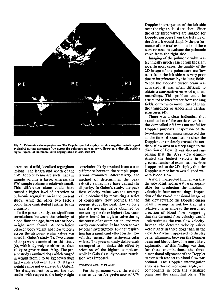
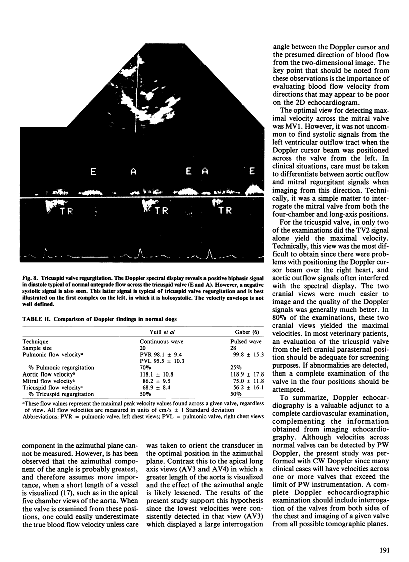
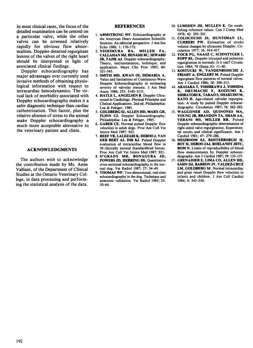
Images in this article
Selected References
These references are in PubMed. This may not be the complete list of references from this article.
- Akasaka T., Yoshikawa J., Yoshida K., Okumachi F., Koizumi K., Shiratori K., Takao S., Shakudo M., Kato H. Age-related valvular regurgitation: a study by pulsed Doppler echocardiography. Circulation. 1987 Aug;76(2):262–265. doi: 10.1161/01.cir.76.2.262. [DOI] [PubMed] [Google Scholar]
- Armstrong W. F. Echocardiography at the American Heart Association Scientific Sessions: an editorial perspective. J Am Soc Echocardiogr. 1988 Mar-Apr;1(2):170–172. doi: 10.1016/s0894-7317(88)80104-4. [DOI] [PubMed] [Google Scholar]
- Colocousis J. S., Huntsman L. L., Curreri P. W. Estimation of stroke volume changes by ultrasonic doppler. Circulation. 1977 Dec;56(6):914–917. doi: 10.1161/01.cir.56.6.914. [DOI] [PubMed] [Google Scholar]
- Grenadier E., Oliveira Lima C., Allen H. D., Sahn D. J., Vargas Barron J., Valdes-Cruz L. M., Goldberg S. J. Normal intracardiac and great vessel Doppler flow velocities in infants and children. J Am Coll Cardiol. 1984 Aug;4(2):343–350. doi: 10.1016/s0735-1097(84)80224-7. [DOI] [PubMed] [Google Scholar]
- Kostucki W., Vandenbossche J. L., Friart A., Englert M. Pulsed Doppler regurgitant flow patterns of normal valves. Am J Cardiol. 1986 Aug 1;58(3):309–313. doi: 10.1016/0002-9149(86)90068-8. [DOI] [PubMed] [Google Scholar]
- Lumsden J. H., Mullen K. On establishing reference values. Can J Comp Med. 1978 Jul;42(3):293–301. [PMC free article] [PubMed] [Google Scholar]
- Meijboom E. J., Rijsterborgh H., Bot H., De Boo J. A., Roelandt J. R., Bom N. Limits of reproducibility of blood flow measurements by Doppler echocardiography. Am J Cardiol. 1987 Jan 1;59(1):133–137. doi: 10.1016/s0002-9149(87)80085-1. [DOI] [PubMed] [Google Scholar]
- Nishimura R. A., Miller F. A., Jr, Callahan M. J., Benassi R. C., Seward J. B., Tajik A. J. Doppler echocardiography: theory, instrumentation, technique, and application. Mayo Clin Proc. 1985 May;60(5):321–343. doi: 10.1016/s0025-6196(12)60540-0. [DOI] [PubMed] [Google Scholar]
- Smith M. D., Kwan O. L., DeMaria A. N. Value and limitations of continuous-wave Doppler echocardiography in estimating severity of valvular stenosis. JAMA. 1986 Jun 13;255(22):3145–3151. [PubMed] [Google Scholar]
- Waggoner A. D., Quinones M. A., Young J. B., Brandon T. A., Shah A. A., Verani M. S., Miller R. R. Pulsed Doppler echocardiographic detection of right-sided valve regurgitation. Experimental results and clinical significance. Am J Cardiol. 1981 Feb;47(2):279–286. doi: 10.1016/0002-9149(81)90398-2. [DOI] [PubMed] [Google Scholar]


