Full text
PDF
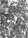
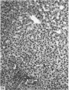
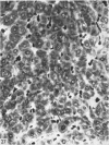
Images in this article
Selected References
These references are in PubMed. This may not be the complete list of references from this article.
- Bischoff M. B., Richter W. R., Stein R. J. Ultrastructural changes in pig hepatocytes during the transitional period from late foetal to early neonatal life. J Cell Sci. 1969 Mar;4(2):381–395. doi: 10.1242/jcs.4.2.381. [DOI] [PubMed] [Google Scholar]
- ELIAS H., BENGELSDORF H. The structure of the liver of vertebrates. Acta Anat (Basel) 1952;14(4):297–337. doi: 10.1159/000140715. [DOI] [PubMed] [Google Scholar]
- EMERY J. L. The distribution of haemopoietic foci in the infantile human liver. J Anat. 1956 Apr;90(2):293–297. [PMC free article] [PubMed] [Google Scholar]
- Flaks B. Observations on the fine structure of the normal porcine liver. J Anat. 1971 Apr;108(Pt 3):563–577. [PMC free article] [PubMed] [Google Scholar]
- Greengard O., Federman M., Knox W. E. Cytomorphometry of developing rat liver and its application to enzymic differentiation. J Cell Biol. 1972 Feb;52(2):261–272. doi: 10.1083/jcb.52.2.261. [DOI] [PMC free article] [PubMed] [Google Scholar]
- LeBouton A. V., Marchand R. Changes in the distribution of thymidine-3H labeled cells in the growing liver acinus of neonatal rats. Dev Biol. 1970 Dec;23(4):524–533. doi: 10.1016/0012-1606(70)90138-7. [DOI] [PubMed] [Google Scholar]
- Leeson C. R., Cutts J. H. The postnatal development of the rabbit liver. Biol Neonate. 1972;20(5):404–413. doi: 10.1159/000240482. [DOI] [PubMed] [Google Scholar]
- Leeson C. R., Leeson T. S. Staining methods for sections of epon-embedded tissues for light microscopy. Can J Zool. 1970 Jan;48(1):189–191. doi: 10.1139/z70-026. [DOI] [PubMed] [Google Scholar]
- McKELLAR M. The postnatal growth and mitotic activity of the liver of the albino rat. Am J Anat. 1949 Sep;85(2):263-307, incl 6 pl. doi: 10.1002/aja.1000850205. [DOI] [PubMed] [Google Scholar]
- Nichols J. E., Simmons K. R. A quantitative histologic analysis of selected tissues in growing mice. Dev Biol. 1970 Sep;23(1):113–127. doi: 10.1016/s0012-1606(70)80009-4. [DOI] [PubMed] [Google Scholar]
- SORENSON G. D. An electron microscopic study of hematopoiesis in the liver of the fetal rabbit. Am J Anat. 1960 Jan;106:27–40. doi: 10.1002/aja.1001060103. [DOI] [PubMed] [Google Scholar]
- Severn C. B. A morphological study of the development of the human liver. I. Development of the hepatic diverticulum. Am J Anat. 1971 Jun;131(2):133–158. doi: 10.1002/aja.1001310202. [DOI] [PubMed] [Google Scholar]
- TEIR H., RAVANTI K. Mitotic activity and growth factors in the liver of the white rat. Exp Cell Res. 1953 Dec;5(2):500–507. doi: 10.1016/0014-4827(53)90236-5. [DOI] [PubMed] [Google Scholar]
- Wheatley D. N. Binucleation in mammalian liver. Studies on the control of cytokinesis in vivo. Exp Cell Res. 1972 Oct;74(2):455–465. doi: 10.1016/0014-4827(72)90401-6. [DOI] [PubMed] [Google Scholar]
- White E. G. Some observations on the liver of the pig: the hepatic lobule and liver cell during post-natal growth. J Anat. 1939 Apr;73(Pt 3):365–386.1. [PMC free article] [PubMed] [Google Scholar]


