

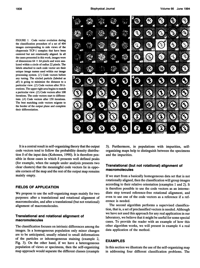
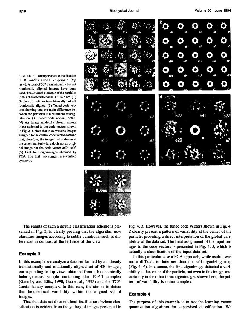
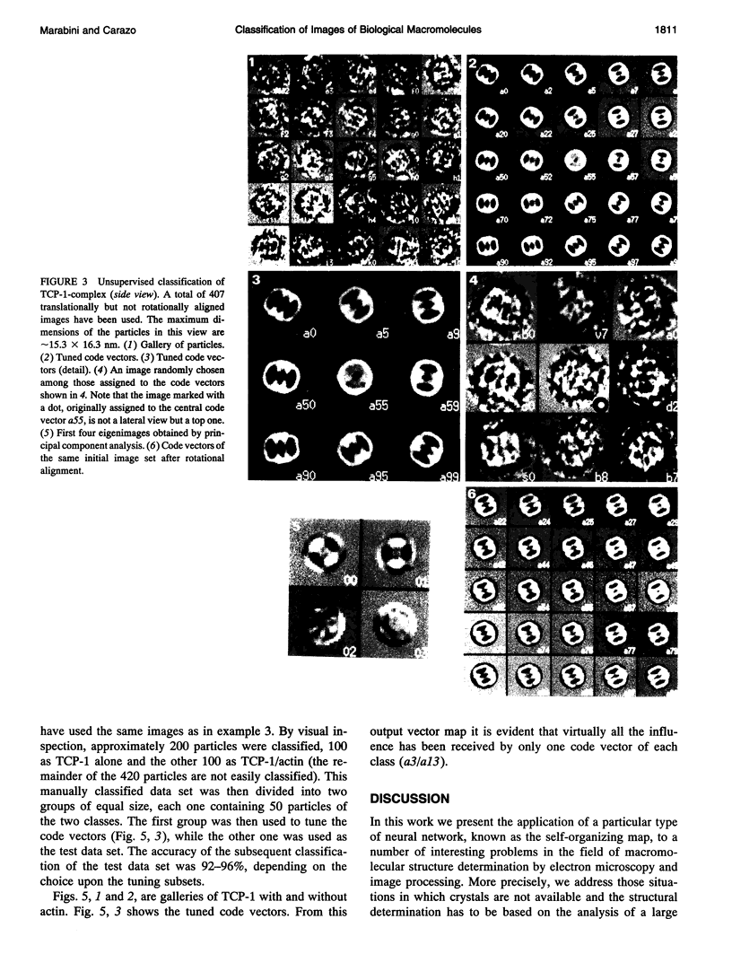
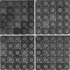
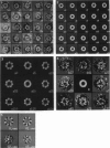
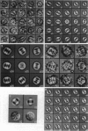
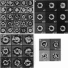
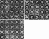
Abstract
The goal of this work was to analyze an image data set and to detect the structural variability within this set. Two algorithms for pattern recognition based on neural networks are presented, one that performs an unsupervised classification (the self-organizing map) and the other a supervised classification (the learning vector quantization). The approach has a direct impact in current strategies for structural determination from electron microscopic images of biological macromolecules. In this work we performed a classification of both aligned but heterogeneous image data sets as well as basically homogeneous but otherwise rotationally misaligned image populations, in the latter case completely avoiding the typical reference dependency of correlation-based alignment methods. A number of examples on chaperonins are presented. The approach is computationally fast and robust with respect to noise. Programs are available through ftp.
Full text
PDF
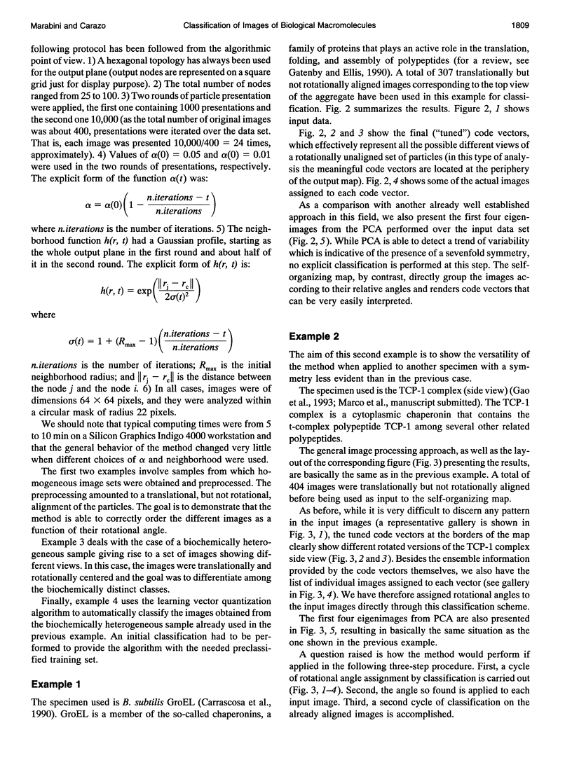

Images in this article
Selected References
These references are in PubMed. This may not be the complete list of references from this article.
- Boekema E. J., Berden J. A., van Heel M. G. Structure of mitochondrial F1-ATPase studied by electron microscopy and image processing. Biochim Biophys Acta. 1986 Oct 8;851(3):353–360. doi: 10.1016/0005-2728(86)90071-x. [DOI] [PubMed] [Google Scholar]
- Carazo J. M., Rivera F. F., Zapata E. L., Radermacher M., Frank J. Fuzzy sets-based classification of electron microscopy images of biological macromolecules with an application to ribosomal particles. J Microsc. 1990 Feb;157(Pt 2):187–203. doi: 10.1111/j.1365-2818.1990.tb02958.x. [DOI] [PubMed] [Google Scholar]
- Carazo J. M., Wagenknecht T., Frank J. Variations of the three-dimensional structure of the Escherichia coli ribosome in the range of overlap views. An application of the methods of multicone and local single-cone three-dimensional reconstruction. Biophys J. 1989 Mar;55(3):465–477. doi: 10.1016/S0006-3495(89)82840-1. [DOI] [PMC free article] [PubMed] [Google Scholar]
- Carrascosa J. L., Abella G., Marco S., Carazo J. M. Three-dimensional reconstruction of the sevenfolded form of Bacillus subtilis Gro EL Chaperonin. J Struct Biol. 1990 Jul-Sep;104(1-3):2–8. doi: 10.1016/1047-8477(90)90051-d. [DOI] [PubMed] [Google Scholar]
- Cottrell M., Fort J. C. A stochastic model of retinotopy: a self organizing process. Biol Cybern. 1986;53(6):405–411. doi: 10.1007/BF00318206. [DOI] [PubMed] [Google Scholar]
- Dube P., Tavares P., Lurz R., van Heel M. The portal protein of bacteriophage SPP1: a DNA pump with 13-fold symmetry. EMBO J. 1993 Apr;12(4):1303–1309. doi: 10.1002/j.1460-2075.1993.tb05775.x. [DOI] [PMC free article] [PubMed] [Google Scholar]
- Frank J., Bretaudiere J. P., Carazo J. M., Verschoor A., Wagenknecht T. Classification of images of biomolecular assemblies: a study of ribosomes and ribosomal subunits of Escherichia coli. J Microsc. 1988 May;150(Pt 2):99–115. doi: 10.1111/j.1365-2818.1988.tb04602.x. [DOI] [PubMed] [Google Scholar]
- Frank J. Classification of macromolecular assemblies studied as 'single particles'. Q Rev Biophys. 1990 Aug;23(3):281–329. doi: 10.1017/s0033583500005564. [DOI] [PubMed] [Google Scholar]
- Frank J., Penczek P., Grassucci R., Srivastava S. Three-dimensional reconstruction of the 70S Escherichia coli ribosome in ice: the distribution of ribosomal RNA. J Cell Biol. 1991 Nov;115(3):597–605. doi: 10.1083/jcb.115.3.597. [DOI] [PMC free article] [PubMed] [Google Scholar]
- Frank J., Radermacher M., Wagenknecht T., Verschoor A. Studying ribosome structure by electron microscopy and computer-image processing. Methods Enzymol. 1988;164:3–35. doi: 10.1016/s0076-6879(88)64032-8. [DOI] [PubMed] [Google Scholar]
- Gao Y., Vainberg I. E., Chow R. L., Cowan N. J. Two cofactors and cytoplasmic chaperonin are required for the folding of alpha- and beta-tubulin. Mol Cell Biol. 1993 Apr;13(4):2478–2485. doi: 10.1128/mcb.13.4.2478. [DOI] [PMC free article] [PubMed] [Google Scholar]
- Gatenby A. A., Ellis R. J. Chaperone function: the assembly of ribulose bisphosphate carboxylase-oxygenase. Annu Rev Cell Biol. 1990;6:125–149. doi: 10.1146/annurev.cb.06.110190.001013. [DOI] [PubMed] [Google Scholar]
- Radermacher M., Frank J. Use of nonlinear mapping in multivariate image analysis of molecule projections. Ultramicroscopy. 1985;17(2):117–126. doi: 10.1016/0304-3991(85)90004-x. [DOI] [PubMed] [Google Scholar]
- Schatz M., van Heel M. Invariant classification of molecular views in electron micrographs. Ultramicroscopy. 1990 Mar-Apr;32(3):255–264. doi: 10.1016/0304-3991(90)90003-5. [DOI] [PubMed] [Google Scholar]
- Stewart P. L., Burnett R. M., Cyrklaff M., Fuller S. D. Image reconstruction reveals the complex molecular organization of adenovirus. Cell. 1991 Oct 4;67(1):145–154. doi: 10.1016/0092-8674(91)90578-m. [DOI] [PubMed] [Google Scholar]
- Wagenknecht T., Carazo J. M., Radermacher M., Frank J. Three-dimensional reconstruction of the ribosome from Escherichia coli. Biophys J. 1989 Mar;55(3):455–464. doi: 10.1016/S0006-3495(89)82839-5. [DOI] [PMC free article] [PubMed] [Google Scholar]