Abstract
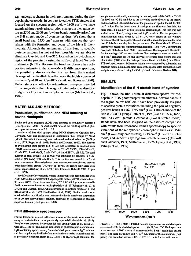
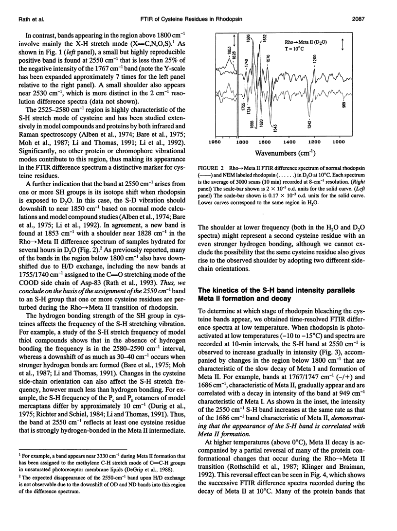
FTIR difference spectroscopy has been used to study the role of cysteine residues in the photoactivation of rhodopsin. A positive band near 2550 cm-1 with a low frequency shoulder is detected during rhodopsin photobleaching, which is assigned on the basis of its frequency and isotope shift to the S-H stretching mode of one or more cysteine residues. Time-resolved studies at low temperature show that the intensity of this band correlates with the formation and decay kinetics of the Meta II intermediate. Modification of rhodopsin with the reagent NEM, which selectively reacts with the SH groups of Cys-140 and Cys-316 on the cytoplasmic surface of rhodopsin, has no effect on the appearance of this band. Four other cysteine residues are also unlikely to contribute to this band because they are either thio-palmitylated (Cys-322 and Cys-323) or form a disulfide bond (Cys-110 and Cys-187). On this basis, it is likely that at least one of the four remaining cysteine residues in rhodopsin is structurally active during rhodopsin photoactivation. The possibility is also considered that this band arises from a transient cleavage of the disulfide bond between cysteine residues 110 and 187.
Full text
PDF

Selected References
These references are in PubMed. This may not be the complete list of references from this article.
- Alben J. O., Bare G. H., Bromberg P. A. Sulphydryl groups as a new molecular probe at the alpha1 beta1 interface in haemoglobin using Fourier transform infrared spectroscopy. Nature. 1974 Dec 20;252(5485):736–738. doi: 10.1038/252736a0. [DOI] [PubMed] [Google Scholar]
- Baenziger J. E., Miller K. W., Rothschild K. J. Incorporation of the nicotinic acetylcholine receptor into planar multilamellar films: characterization by fluorescence and Fourier transform infrared difference spectroscopy. Biophys J. 1992 Apr;61(4):983–992. doi: 10.1016/S0006-3495(92)81905-7. [DOI] [PMC free article] [PubMed] [Google Scholar]
- Bagley K. A., Balogh-Nair V., Croteau A. A., Dollinger G., Ebrey T. G., Eisenstein L., Hong M. K., Nakanishi K., Vittitow J. Fourier-transform infrared difference spectroscopy of rhodopsin and its photoproducts at low temperature. Biochemistry. 1985 Oct 22;24(22):6055–6071. doi: 10.1021/bi00343a006. [DOI] [PubMed] [Google Scholar]
- Bagley K., Dollinger G., Eisenstein L., Singh A. K., Zimányi L. Fourier transform infrared difference spectroscopy of bacteriorhodopsin and its photoproducts. Proc Natl Acad Sci U S A. 1982 Aug;79(16):4972–4976. doi: 10.1073/pnas.79.16.4972. [DOI] [PMC free article] [PubMed] [Google Scholar]
- Bare G. H., Alben J. O., Bromberg P. A. Sulfhydryl groups in hemoglobin. A new molecular probe at the alpha1 beta 1 interface studied by Fourier transform infrared spectroscopy. Biochemistry. 1975 Apr 22;14(8):1578–1583. doi: 10.1021/bi00679a005. [DOI] [PubMed] [Google Scholar]
- Birge R. R. Nature of the primary photochemical events in rhodopsin and bacteriorhodopsin. Biochim Biophys Acta. 1990 Apr 26;1016(3):293–327. doi: 10.1016/0005-2728(90)90163-x. [DOI] [PubMed] [Google Scholar]
- Chen Y. S., Hubbell W. L. Reactions of the sulfhydryl groups of membrane-bound bovine rhodopsin. Membr Biochem. 1978;1(1-2):107–130. doi: 10.3109/09687687809064162. [DOI] [PubMed] [Google Scholar]
- Clark N. A., Rothschild K. J., Luippold D. A., Simon B. A. Surface-induced lamellar orientation of multilayer membrane arrays. Theoretical analysis and a new method with application to purple membrane fragments. Biophys J. 1980 Jul;31(1):65–96. doi: 10.1016/S0006-3495(80)85041-7. [DOI] [PMC free article] [PubMed] [Google Scholar]
- Cohen G. B., Oprian D. D., Robinson P. R. Mechanism of activation and inactivation of opsin: role of Glu113 and Lys296. Biochemistry. 1992 Dec 22;31(50):12592–12601. doi: 10.1021/bi00165a008. [DOI] [PubMed] [Google Scholar]
- De Grip W. J., Bonting S. L., Daemen F. J. Biochemical aspects of the visual process. XXVIII. Classification of sulfhydryl groups in phodopsin and other photoreceptor membrane proteins. Biochim Biophys Acta. 1975 Jul 8;396(1):104–115. doi: 10.1016/0005-2728(75)90193-0. [DOI] [PubMed] [Google Scholar]
- De Grip W. J., Daemen F. J., Bonting S. L. Isolation and purification of bovine rhodopsin. Methods Enzymol. 1980;67:301–320. doi: 10.1016/s0076-6879(80)67038-4. [DOI] [PubMed] [Google Scholar]
- De Grip W. J., Daemen F. J. Sulfhydryl chemistry of rhodopsin. Methods Enzymol. 1982;81:223–236. doi: 10.1016/s0076-6879(82)81035-5. [DOI] [PubMed] [Google Scholar]
- DeGrip W. J., Gray D., Gillespie J., Bovee P. H., Van den Berg E. M., Lugtenburg J., Rothschild K. J. Photoexcitation of rhodopsin: conformation changes in the chromophore, protein and associated lipids as determined by FTIR difference spectroscopy. Photochem Photobiol. 1988 Oct;48(4):497–504. doi: 10.1111/j.1751-1097.1988.tb02852.x. [DOI] [PubMed] [Google Scholar]
- Eyring G., Curry B., Broek A., Lugtenburg J., Mathies R. Assignment and interpretation of hydrogen out-of-plane vibrations in the resonance Raman spectra of rhodopsin and bathorhodopsin. Biochemistry. 1982 Jan 19;21(2):384–393. doi: 10.1021/bi00531a028. [DOI] [PubMed] [Google Scholar]
- Fahmy K., Jäger F., Beck M., Zvyaga T. A., Sakmar T. P., Siebert F. Protonation states of membrane-embedded carboxylic acid groups in rhodopsin and metarhodopsin II: a Fourier-transform infrared spectroscopy study of site-directed mutants. Proc Natl Acad Sci U S A. 1993 Nov 1;90(21):10206–10210. doi: 10.1073/pnas.90.21.10206. [DOI] [PMC free article] [PubMed] [Google Scholar]
- Farahbakhsh Z. T., Altenbach C., Hubbell W. L. Spin labeled cysteines as sensors for protein-lipid interaction and conformation in rhodopsin. Photochem Photobiol. 1992 Dec;56(6):1019–1033. doi: 10.1111/j.1751-1097.1992.tb09725.x. [DOI] [PubMed] [Google Scholar]
- Farahbakhsh Z. T., Hideg K., Hubbell W. L. Photoactivated conformational changes in rhodopsin: a time-resolved spin label study. Science. 1993 Nov 26;262(5138):1416–1419. doi: 10.1126/science.8248781. [DOI] [PubMed] [Google Scholar]
- Ganter U. M., Schmid E. D., Perez-Sala D., Rando R. R., Siebert F. Removal of the 9-methyl group of retinal inhibits signal transduction in the visual process. A Fourier transform infrared and biochemical investigation. Biochemistry. 1989 Jul 11;28(14):5954–5962. doi: 10.1021/bi00440a036. [DOI] [PubMed] [Google Scholar]
- Green B. H., Monger T. G., Alfano R. R., Aton B., Callender R. H. Cis-trans isomerisation in rhodopsin occurs in picoseconds. Nature. 1977 Sep 8;269(5624):179–180. doi: 10.1038/269179a0. [DOI] [PubMed] [Google Scholar]
- Hargrave P. A., McDowell J. H. Rhodopsin and phototransduction. Int Rev Cytol. 1992;137B:49–97. doi: 10.1016/s0074-7696(08)62600-5. [DOI] [PubMed] [Google Scholar]
- Hargrave P. A., McDowell J. H. Rhodopsin and phototransduction: a model system for G protein-linked receptors. FASEB J. 1992 Mar;6(6):2323–2331. doi: 10.1096/fasebj.6.6.1544542. [DOI] [PubMed] [Google Scholar]
- Karnik S. S., Khorana H. G. Assembly of functional rhodopsin requires a disulfide bond between cysteine residues 110 and 187. J Biol Chem. 1990 Oct 15;265(29):17520–17524. [PubMed] [Google Scholar]
- Khorana H. G. Rhodopsin, photoreceptor of the rod cell. An emerging pattern for structure and function. J Biol Chem. 1992 Jan 5;267(1):1–4. [PubMed] [Google Scholar]
- Kibelbek J., Mitchell D. C., Beach J. M., Litman B. J. Functional equivalence of metarhodopsin II and the Gt-activating form of photolyzed bovine rhodopsin. Biochemistry. 1991 Jul 9;30(27):6761–6768. doi: 10.1021/bi00241a019. [DOI] [PubMed] [Google Scholar]
- Klinger A. L., Braiman M. S. Structural comparison of metarhodopsin II, metarhodopsin III, and opsin based on kinetic analysis of Fourier transform infrared difference spectra. Biophys J. 1992 Nov;63(5):1244–1255. doi: 10.1016/S0006-3495(92)81700-9. [DOI] [PMC free article] [PubMed] [Google Scholar]
- Liggett S. B., Bouvier M., O'Dowd B. F., Caron M. G., Lefkowitz R. J., DeBlasi A. Substitution of an extracellular cysteine in the beta 2-adrenergic receptor enhances agonist-promoted phosphorylation and receptor desensitization. Biochem Biophys Res Commun. 1989 Nov 30;165(1):257–263. doi: 10.1016/0006-291x(89)91063-2. [DOI] [PubMed] [Google Scholar]
- MATTHEWS R. G., HUBBARD R., BROWN P. K., WALD G. TAUTOMERIC FORMS OF METARHODOPSIN. J Gen Physiol. 1963 Nov;47:215–240. doi: 10.1085/jgp.47.2.215. [DOI] [PMC free article] [PubMed] [Google Scholar]
- Mathies R., Oseroff A. R., Stryer L. Rapid-flow resonance Raman spectroscopy of photolabile molecules: rhodopsin and isorhodopsin. Proc Natl Acad Sci U S A. 1976 Jan;73(1):1–5. doi: 10.1073/pnas.73.1.1. [DOI] [PMC free article] [PubMed] [Google Scholar]
- McDowell J. H., Mas M. T., Griffith K. D., Hargrave P. A. The reactivity of the sulfhydryl groups of rhodopsin in rod outer segment membranes. Vision Res. 1979;19(10):1143–1145. doi: 10.1016/0042-6989(79)90010-5. [DOI] [PubMed] [Google Scholar]
- Moh P. P., Fiamingo F. G., Alben J. O. Conformational sensitivity of beta-93 cysteine SH to ligation of hemoglobin observed by FT-IR spectroscopy. Biochemistry. 1987 Sep 22;26(19):6243–6249. doi: 10.1021/bi00393a044. [DOI] [PubMed] [Google Scholar]
- Nakayama T. A., Khorana H. G. Mapping of the amino acids in membrane-embedded helices that interact with the retinal chromophore in bovine rhodopsin. J Biol Chem. 1991 Mar 5;266(7):4269–4275. [PubMed] [Google Scholar]
- Nathans J. Determinants of visual pigment absorbance: identification of the retinylidene Schiff's base counterion in bovine rhodopsin. Biochemistry. 1990 Oct 16;29(41):9746–9752. doi: 10.1021/bi00493a034. [DOI] [PubMed] [Google Scholar]
- Oprian D. D. The ligand-binding domain of rhodopsin and other G protein-linked receptors. J Bioenerg Biomembr. 1992 Apr;24(2):211–217. doi: 10.1007/BF00762679. [DOI] [PubMed] [Google Scholar]
- Oseroff A. R., Callender R. H. Resonance Raman spectroscopy of rhodopsin in retinal disk membranes. Biochemistry. 1974 Sep 24;13(20):4243–4248. doi: 10.1021/bi00717a027. [DOI] [PubMed] [Google Scholar]
- Ostroy S. E. Rhodopsin and the visual process. Biochim Biophys Acta. 1977 Jun 21;463(1):91–125. doi: 10.1016/0304-4173(77)90004-0. [DOI] [PubMed] [Google Scholar]
- Ovchinnikov YuA, Abdulaev N. G., Bogachuk A. S. Two adjacent cysteine residues in the C-terminal cytoplasmic fragment of bovine rhodopsin are palmitylated. FEBS Lett. 1988 Mar 28;230(1-2):1–5. doi: 10.1016/0014-5793(88)80628-8. [DOI] [PubMed] [Google Scholar]
- Palings I., Pardoen J. A., van den Berg E., Winkel C., Lugtenburg J., Mathies R. A. Assignment of fingerprint vibrations in the resonance Raman spectra of rhodopsin, isorhodopsin, and bathorhodopsin: implications for chromophore structure and environment. Biochemistry. 1987 May 5;26(9):2544–2556. doi: 10.1021/bi00383a021. [DOI] [PubMed] [Google Scholar]
- Rath P., DeCaluwé L. L., Bovee-Geurts P. H., DeGrip W. J., Rothschild K. J. Fourier transform infrared difference spectroscopy of rhodopsin mutants: light activation of rhodopsin causes hydrogen-bonding change in residue aspartic acid-83 during meta II formation. Biochemistry. 1993 Oct 5;32(39):10277–10282. doi: 10.1021/bi00090a001. [DOI] [PubMed] [Google Scholar]
- Regan C. M., de Grip W. J., Daemen F. J., Bonting S. L. Biochemical aspects of the visual process. XXXIX. Sulfhydryl group reactivity as a probe of transient protein conformational changes during rhodopsin photolysis. Biochim Biophys Acta. 1978 Nov 20;537(1):145–152. doi: 10.1016/0005-2795(78)90609-8. [DOI] [PubMed] [Google Scholar]
- Rothschild K. J., Braiman M. S., Mogi T., Stern L. J., Khorana H. G. Conserved amino acids in F-helix of bacteriorhodopsin form part of a retinal binding pocket. FEBS Lett. 1989 Jul 3;250(2):448–452. doi: 10.1016/0014-5793(89)80774-4. [DOI] [PubMed] [Google Scholar]
- Rothschild K. J., Cantore W. A., Marrero H. Fourier transform infrared difference spectra of intermediates in rhodopsin bleaching. Science. 1983 Mar 18;219(4590):1333–1335. doi: 10.1126/science.6828860. [DOI] [PubMed] [Google Scholar]
- Rothschild K. J. FTIR difference spectroscopy of bacteriorhodopsin: toward a molecular model. J Bioenerg Biomembr. 1992 Apr;24(2):147–167. doi: 10.1007/BF00762674. [DOI] [PubMed] [Google Scholar]
- Rothschild K. J., Gillespie J., DeGrip W. J. Evidence for rhodopsin refolding during the decay of Meta II. Biophys J. 1987 Feb;51(2):345–350. doi: 10.1016/S0006-3495(87)83341-6. [DOI] [PMC free article] [PubMed] [Google Scholar]
- Rothschild K. J., Zagaeski M., Cantore W. A. Conformational changes of bacteriorhodopsin detected by Fourier transform infrared difference spectroscopy. Biochem Biophys Res Commun. 1981 Nov 30;103(2):483–489. doi: 10.1016/0006-291x(81)90478-2. [DOI] [PubMed] [Google Scholar]
- Sakmar T. P., Franke R. R., Khorana H. G. Glutamic acid-113 serves as the retinylidene Schiff base counterion in bovine rhodopsin. Proc Natl Acad Sci U S A. 1989 Nov;86(21):8309–8313. doi: 10.1073/pnas.86.21.8309. [DOI] [PMC free article] [PubMed] [Google Scholar]
- Schoenlein R. W., Peteanu L. A., Mathies R. A., Shank C. V. The first step in vision: femtosecond isomerization of rhodopsin. Science. 1991 Oct 18;254(5030):412–415. doi: 10.1126/science.1925597. [DOI] [PubMed] [Google Scholar]
- Siebert F., Mäntele W., Gerwert K. Fourier-transform infrared spectroscopy applied to rhodopsin. The problem of the protonation state of the retinylidene Schiff base re-investigated. Eur J Biochem. 1983 Oct 17;136(1):119–127. doi: 10.1111/j.1432-1033.1983.tb07714.x. [DOI] [PubMed] [Google Scholar]
- Siebert F., Mäntele W. Investigation of the primary photochemistry of bacteriorhodopsin by low-temperature Fourier-transform infrared spectroscopy. Eur J Biochem. 1983 Feb 15;130(3):565–573. doi: 10.1111/j.1432-1033.1983.tb07187.x. [DOI] [PubMed] [Google Scholar]
- Skulachev V. P. Interrelations of bioenergetic and sensory functions of the retinal proteins. Q Rev Biophys. 1993 May;26(2):177–199. doi: 10.1017/s0033583500004066. [DOI] [PubMed] [Google Scholar]
- Suzuki T., Callender R. H. Primary photochemistry and photoisomerization of retinal at 77 degrees K in cattle and squid rhodopsins. Biophys J. 1981 May;34(2):261–270. doi: 10.1016/S0006-3495(81)84848-5. [DOI] [PMC free article] [PubMed] [Google Scholar]
- Vuong T. M., Chabre M., Stryer L. Millisecond activation of transducin in the cyclic nucleotide cascade of vision. Nature. 1984 Oct 18;311(5987):659–661. doi: 10.1038/311659a0. [DOI] [PubMed] [Google Scholar]
- Wald G. Molecular basis of visual excitation. Science. 1968 Oct 11;162(3850):230–239. doi: 10.1126/science.162.3850.230. [DOI] [PubMed] [Google Scholar]
- YOSHIZAWA T., WALD G. Pre-lumirhodopsin and the bleaching of visual pigments. Nature. 1963 Mar 30;197:1279–1286. doi: 10.1038/1971279a0. [DOI] [PubMed] [Google Scholar]
- Yan M., Manor D., Weng G., Chao H., Rothberg L., Jedju T. M., Alfano R. R., Callender R. H. Ultrafast spectroscopy of the visual pigment rhodopsin. Proc Natl Acad Sci U S A. 1991 Nov 1;88(21):9809–9812. doi: 10.1073/pnas.88.21.9809. [DOI] [PMC free article] [PubMed] [Google Scholar]
- Zhukovsky E. A., Oprian D. D. Effect of carboxylic acid side chains on the absorption maximum of visual pigments. Science. 1989 Nov 17;246(4932):928–930. doi: 10.1126/science.2573154. [DOI] [PubMed] [Google Scholar]
- de Grip W. J., Gillespie J., Rothschild K. J. Carboxyl group involvement in the meta I and meta II stages in rhodopsin bleaching. A Fourier transform infrared spectroscopic study. Biochim Biophys Acta. 1985 Aug 28;809(1):97–106. doi: 10.1016/0005-2728(85)90172-0. [DOI] [PubMed] [Google Scholar]
- de Grip W. J., van de Laar G. L., Daemen F. J., Bonting S. L. Biochemical aspects of the visual process. 23. Sulfhydryl groups and rhodopsin photolysis. Biochim Biophys Acta. 1973 Nov 22;325(2):315–322. doi: 10.1016/0005-2728(73)90107-2. [DOI] [PubMed] [Google Scholar]
- van Breugel P. J., Bovee-Geurts P. H., Bonting S. L., Daemen F. J. Biochemical aspects of the visual process. XL. Spectral and chemical analysis of metarhodopsin III in photoreceptor membrane suspensions. Biochim Biophys Acta. 1979 Oct 19;557(1):188–198. doi: 10.1016/0005-2736(79)90101-9. [DOI] [PubMed] [Google Scholar]


