Abstract
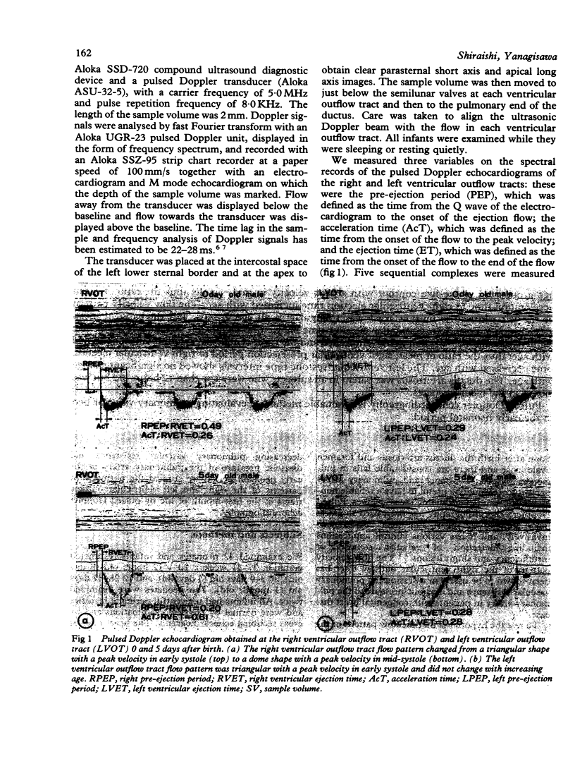
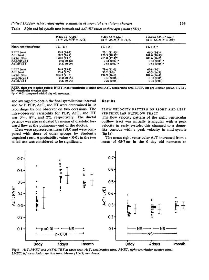
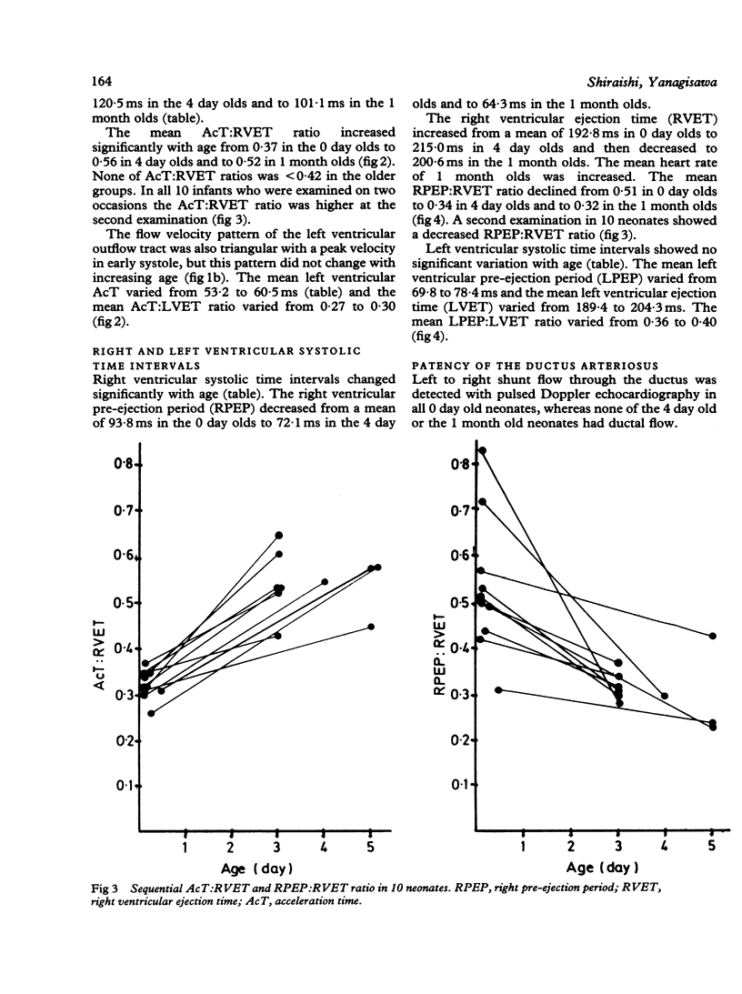
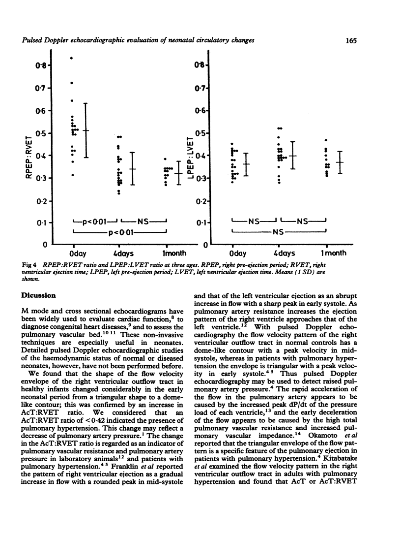
Pulsed Doppler echocardiograms were obtained from 42 normal fullterm neonates at less than 12 hours (20 subjects), 4 days (20 subjects), and 33 days (12 subjects). The acceleration time of the flow velocity and ventricular systolic time intervals were measured on recordings obtained at the right and left ventricular outflow tract, and the patency of the ductus arteriosus was evaluated by the flow at the pulmonary end of the ductus. The flow velocity pattern of the right ventricular outflow tract changed from a triangular shape with a peak velocity in early systole in the younger age groups to a dome-like contour with a peak velocity in mid-systole; thus the ratio of mean acceleration time to right ventricular ejection time increased with age. The flow velocity pattern of the left ventricular outflow tract was triangular in all age groups, and the ratio of mean acceleration time to left ventricular ejection time showed no significant change with age. The right ventricular pre-ejection period shortened and the right ventricular ejection time lengthened with age; thus the ratio of mean right ventricular pre-ejection period to right ventricular ejection time decreased with age. The left ventricular systolic time intervals showed no significant change with age. The ductus arteriosus was patent in all subjects who were less than 12 hours old but was closed in the older neonates. Pulsed Doppler echocardiography is a valuable method of evaluating pulmonary vascular bed in the early neonatal period.
Full text
PDF
Images in this article
Selected References
These references are in PubMed. This may not be the complete list of references from this article.
- FRANKLIN D. L., VAN CITTERS R. L., RUSHMER R. F. Balance between right and left ventricular output. Circ Res. 1962 Jan;10:17–26. doi: 10.1161/01.res.10.1.17. [DOI] [PubMed] [Google Scholar]
- GLEASON W. L., BRAUNWALD E. Studies on the first derivative of the ventricular pressure pulse in man. J Clin Invest. 1962 Jan;41:80–91. doi: 10.1172/JCI104469. [DOI] [PMC free article] [PubMed] [Google Scholar]
- Gentile R., Stevenson G., Dooley T., Franklin D., Kawabori I., Pearlman A. Pulsed Doppler echocardiographic determination of time of ductal closure in normal newborn infants. J Pediatr. 1981 Mar;98(3):443–448. doi: 10.1016/s0022-3476(81)80719-6. [DOI] [PubMed] [Google Scholar]
- Gutgesell H. P., Pinsky W. W., Duff D. F., Adams J., McNamara D. G. Left and right ventricular systolic time intervals in the newborn. Usefulness and limitation in distinguishing respiratory disease from transposition of the great arteries. Br Heart J. 1979 Jul;42(1):27–34. doi: 10.1136/hrt.42.1.27. [DOI] [PMC free article] [PubMed] [Google Scholar]
- Hirschfeld S., Meyer R., Schwartz D. C., Kofhagen J., Kaplan S. The echocardiographic assessment of pulmonary artery pressure and pulmonary vascular resistance. Circulation. 1975 Oct;52(4):642–650. doi: 10.1161/01.cir.52.4.642. [DOI] [PubMed] [Google Scholar]
- Huhta J. C., Cohen M., Gutgesell H. P. Patency of the ductus arteriosus in normal neonates: two-dimensional echocardiography versus Doppler assessment. J Am Coll Cardiol. 1984 Sep;4(3):561–564. doi: 10.1016/s0735-1097(84)80102-3. [DOI] [PubMed] [Google Scholar]
- Kitabatake A., Inoue M., Asao M., Masuyama T., Tanouchi J., Morita T., Mishima M., Uematsu M., Shimazu T., Hori M. Noninvasive evaluation of pulmonary hypertension by a pulsed Doppler technique. Circulation. 1983 Aug;68(2):302–309. doi: 10.1161/01.cir.68.2.302. [DOI] [PubMed] [Google Scholar]
- Leighton R. F., Weissler A. M., Weinstein P. B., Wooley C. F. Right and left ventricular systolic time intervals. Effects of heart rate, respiration and atrial pacing. Am J Cardiol. 1971 Jan;27(1):66–72. doi: 10.1016/0002-9149(71)90084-1. [DOI] [PubMed] [Google Scholar]
- Macartney F. J. Cross sectional echocardiographic diagnosis of congenital heart disease in infants. Br Heart J. 1983 Dec;50(6):501–505. doi: 10.1136/hrt.50.6.501. [DOI] [PMC free article] [PubMed] [Google Scholar]
- Matsuo H., Morita H. [Current status of pulsed Doppler method]. J Cardiogr Suppl. 1984;(2):27–39. [PubMed] [Google Scholar]
- Okamoto M., Miyatake K., Kinoshita N., Sakakibara H., Nimura Y. Analysis of blood flow in pulmonary hypertension with the pulsed Doppler flowmeter combined with cross sectional echocardiography. Br Heart J. 1984 Apr;51(4):407–415. doi: 10.1136/hrt.51.4.407. [DOI] [PMC free article] [PubMed] [Google Scholar]
- Pearlman A. S., Lighty G. W., Jr Clinical applications of two-dimensional/Doppler echocardiography. Cardiovasc Clin. 1983;13(3):201–222. [PubMed] [Google Scholar]
- Reuben S. R. Compliance of the human pulmonary arterial system in disease. Circ Res. 1971 Jul;29(1):40–50. doi: 10.1161/01.res.29.1.40. [DOI] [PubMed] [Google Scholar]
- Riggs T., Hirschfeld S., Bormuth C., Fanaroff A., Liebman J. Neonatal circulatory changes: an echocardiographic study. Pediatrics. 1977 Mar;59(3):338–344. [PubMed] [Google Scholar]
- Riggs T., Hirschfeld S., Fanaroff A., Liebman J., Fletcher B., Meyer R. Persistence of fetal circulation syndrome: an echocardiographic study. J Pediatr. 1977 Oct;91(4):626–631. doi: 10.1016/s0022-3476(77)80521-0. [DOI] [PubMed] [Google Scholar]


