Abstract
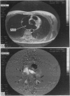
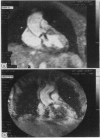
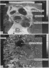
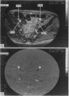
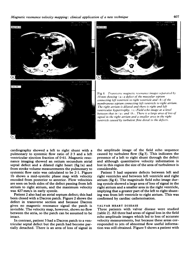
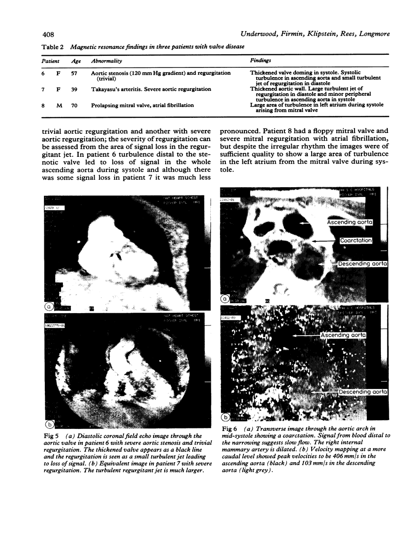
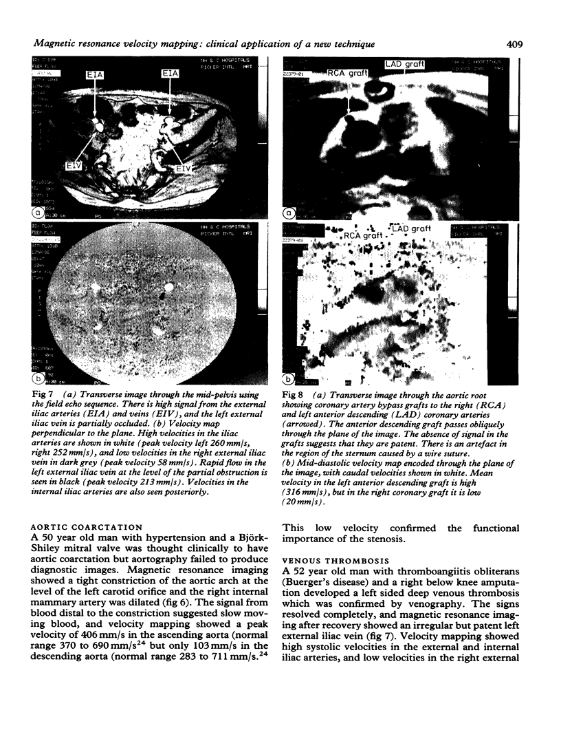
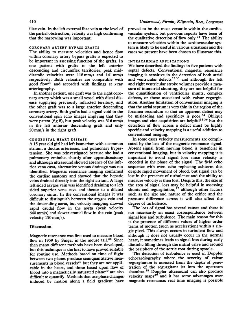
Magnetic resonance velocity mapping is a new technique which provides a display of velocity within the cardiovascular system at any point of the cardiac cycle. A short field echo sequence with even echo rephasing is used to obtain a signal from rapidly moving blood and a cine display is provided by rapid repetition of the sequence. The amplitude image shows the anatomy, with blood giving a high signal and areas of turbulent flow no signal. The phase image is a map of velocities at each point in the image plane. Thirteen cases are described in which the technique either provided a diagnosis or helped in functional assessment. Flow through atrial and ventricular septal defects was seen, although turbulent flow distal to the ventricular shunts led to some loss of quantitative information. In three patients with valve disease jets of abnormal flow were seen because of signal loss and it is suggested that the size of the area of turbulence may be used to quantify the severity of regurgitation. Velocities were measured in four coronary artery bypass grafts in two patients, and low velocity was seen in a graft with distal disease that supplied the infarcted territory. Velocity was reduced distal to an aortic coarctation and it was increased at the site of narrowing caused by thrombosis in a deep vein. The speed and direction of flow in the central vessels in a patient with complex congenital heart disease helped to establish the anatomy. The technique provides useful information in a wide range of disorders of the cardiovascular system, and in some cases may avoid the need for invasive investigation.
Full text
PDF
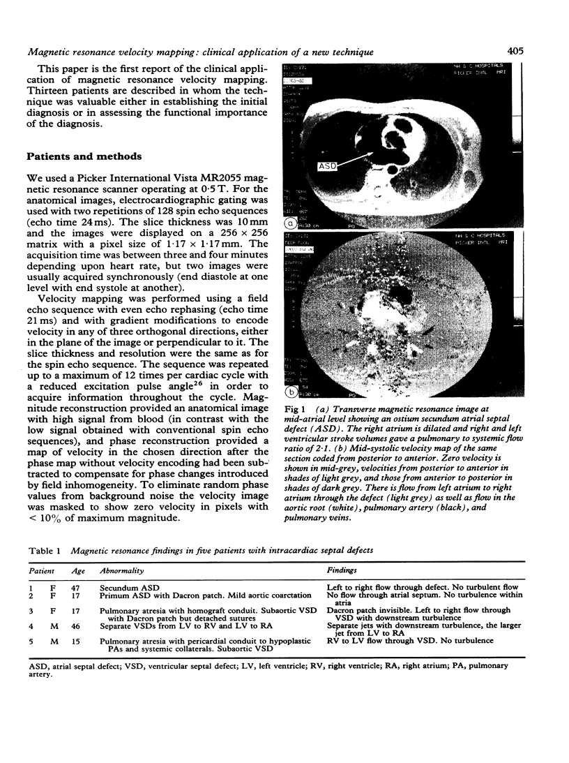
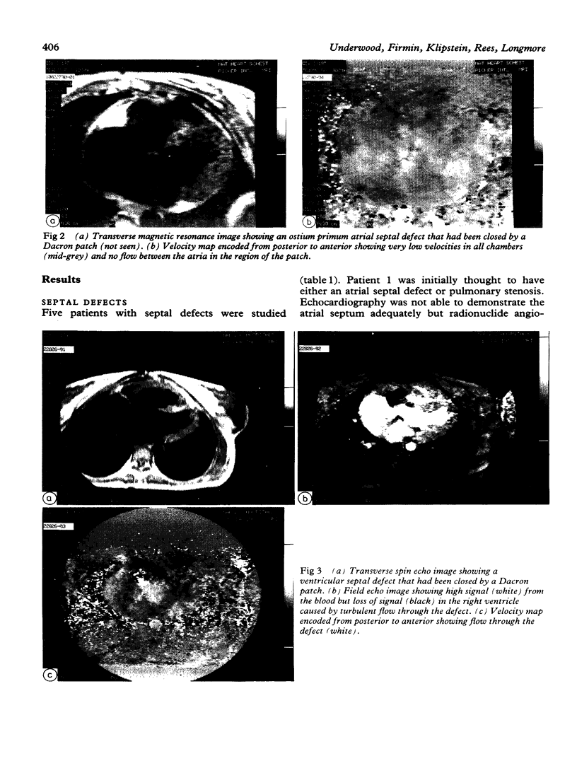
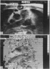
Images in this article
Selected References
These references are in PubMed. This may not be the complete list of references from this article.
- Amparo E. G., Higgins C. B., Shafton E. P. Demonstration of coarctation of the aorta by magnetic resonance imaging. AJR Am J Roentgenol. 1984 Dec;143(6):1192–1194. doi: 10.2214/ajr.143.6.1192. [DOI] [PubMed] [Google Scholar]
- Bryant D. J., Payne J. A., Firmin D. N., Longmore D. B. Measurement of flow with NMR imaging using a gradient pulse and phase difference technique. J Comput Assist Tomogr. 1984 Aug;8(4):588–593. doi: 10.1097/00004728-198408000-00002. [DOI] [PubMed] [Google Scholar]
- Didier D., Higgins C. B., Fisher M. R., Osaki L., Silverman N. H., Cheitlin M. D. Congenital heart disease: gated MR imaging in 72 patients. Radiology. 1986 Jan;158(1):227–235. doi: 10.1148/radiology.158.1.3940387. [DOI] [PubMed] [Google Scholar]
- Dinsmore R. E., Wismer G. L., Guyer D., Thompson R., Liu P., Stratemeier E., Miller S., Okada R., Brady T. Magnetic resonance imaging of the interatrial septum and atrial septal defects. AJR Am J Roentgenol. 1985 Oct;145(4):697–703. doi: 10.2214/ajr.145.4.697. [DOI] [PubMed] [Google Scholar]
- Feiglin D. H., George C. R., MacIntyre W. J., O'Donnell J. K., Go R. T., Pavlicek W., Meaney T. F. Gated cardiac magnetic resonance structural imaging: optimization by electronic axial rotation. Radiology. 1985 Jan;154(1):129–132. doi: 10.1148/radiology.154.1.3155478. [DOI] [PubMed] [Google Scholar]
- Feinberg D. A., Crooks L., Hoenninger J., 3rd, Arakawa M., Watts J. Pulsatile blood velocity in human arteries displayed by magnetic resonance imaging. Radiology. 1984 Oct;153(1):177–180. doi: 10.1148/radiology.153.1.6473779. [DOI] [PubMed] [Google Scholar]
- Fisher M. R., Hricak H., Higgins C. B. Magnetic resonance imaging of developmental venous anomalies. AJR Am J Roentgenol. 1985 Oct;145(4):705–709. doi: 10.2214/ajr.145.4.705. [DOI] [PubMed] [Google Scholar]
- Fletcher B. D., Jacobstein M. D., Nelson A. D., Riemenschneider T. A., Alfidi R. J. Gated magnetic resonance imaging of congenital cardiac malformations. Radiology. 1984 Jan;150(1):137–140. doi: 10.1148/radiology.150.1.6689753. [DOI] [PubMed] [Google Scholar]
- Glazer H. S., Gutierrez F. R., Levitt R. G., Lee J. K., Murphy W. A. The thoracic aorta studied by MR imaging. Radiology. 1985 Oct;157(1):149–155. doi: 10.1148/radiology.157.1.2863853. [DOI] [PubMed] [Google Scholar]
- Goldman M. E. Real-time two-dimensional Doppler flow imaging: a word of caution. J Am Coll Cardiol. 1986 Jan;7(1):89–90. doi: 10.1016/s0735-1097(86)80264-9. [DOI] [PubMed] [Google Scholar]
- Hatle L., Angelsen B. A., Tromsdal A. Non-invasive assessment of aortic stenosis by Doppler ultrasound. Br Heart J. 1980 Mar;43(3):284–292. doi: 10.1136/hrt.43.3.284. [DOI] [PMC free article] [PubMed] [Google Scholar]
- Hatle L., Brubakk A., Tromsdal A., Angelsen B. Noninvasive assessment of pressure drop in mitral stenosis by Doppler ultrasound. Br Heart J. 1978 Feb;40(2):131–140. doi: 10.1136/hrt.40.2.131. [DOI] [PMC free article] [PubMed] [Google Scholar]
- Herfkens R. J., Higgins C. B., Hricak H., Lipton M. J., Crooks L. E., Sheldon P. E., Kaufman L. Nuclear magnetic resonance imaging of atherosclerotic disease. Radiology. 1983 Jul;148(1):161–166. doi: 10.1148/radiology.148.1.6856827. [DOI] [PubMed] [Google Scholar]
- Higgins C. B., Byrd B. F., 2nd, McNamara M. T., Lanzer P., Lipton M. J., Botvinick E., Schiller N. B., Crooks L. E., Kaufman L. Magnetic resonance imaging of the heart: a review of the experience in 172 subjects. Radiology. 1985 Jun;155(3):671–679. doi: 10.1148/radiology.155.3.3159039. [DOI] [PubMed] [Google Scholar]
- Higgins C. B., Lanzer P., Stark D., Botvinick E., Schiller N. B., Crooks L., Kaufman L., Lipton M. J. Imaging by nuclear magnetic resonance in patients with chronic ischemic heart disease. Circulation. 1984 Mar;69(3):523–531. doi: 10.1161/01.cir.69.3.523. [DOI] [PubMed] [Google Scholar]
- Jacobstein M. D., Fletcher B. D., Goldstein S., Riemenschneider T. A. Evaluation of atrioventricular septal defect by magnetic resonance imaging. Am J Cardiol. 1985 Apr 15;55(9):1158–1161. doi: 10.1016/0002-9149(85)90655-1. [DOI] [PubMed] [Google Scholar]
- Klipstein R. H., Firmin D. N., Underwood S. R., Nayler G. L., Rees R. S., Longmore D. B. Colour display of quantitative blood flow and cardiac anatomy in a single magnetic resonance cine loop. Br J Radiol. 1987 Feb;60(710):105–111. doi: 10.1259/0007-1285-60-710-105. [DOI] [PubMed] [Google Scholar]
- Lieberman J. M., Alfidi R. J., Nelson A. D., Botti R. E., Moir T. W., Haaga J. R., Kopiwoda S., Miraldi F. D., Cohen A. M., Butler H. E. Gated magnetic resonance imaging of the normal and diseased heart. Radiology. 1984 Aug;152(2):465–470. doi: 10.1148/radiology.152.2.6739817. [DOI] [PubMed] [Google Scholar]
- Longmore D. B., Klipstein R. H., Underwood S. R., Firmin D. N., Hounsfield G. N., Watanabe M., Bland C., Fox K., Poole-Wilson P. A., Rees R. S. Dimensional accuracy of magnetic resonance in studies of the heart. Lancet. 1985 Jun 15;1(8442):1360–1362. doi: 10.1016/s0140-6736(85)91786-6. [DOI] [PubMed] [Google Scholar]
- Lowell D. G., Turner D. A., Smith S. M., Bucheleres G. H., Santucci B. A., Gresick R. J., Jr, Monson D. O. The detection of atrial and ventricular septal defects with electrocardiographically synchronized magnetic resonance imaging. Circulation. 1986 Jan;73(1):89–94. doi: 10.1161/01.cir.73.1.89. [DOI] [PubMed] [Google Scholar]
- McLennan F. M., Haites N. E., Mackenzie J. D., Daniel M. K., Rawles J. M. Reproducibility of linear cardiac output measurement by Doppler ultrasound alone. Br Heart J. 1986 Jan;55(1):25–31. doi: 10.1136/hrt.55.1.25. [DOI] [PMC free article] [PubMed] [Google Scholar]
- McMurdo K. K., de Geer G., Webb W. R., Gamsu G. Normal and occluded mediastinal veins: MR imaging. Radiology. 1986 Apr;159(1):33–38. doi: 10.1148/radiology.159.1.3952327. [DOI] [PubMed] [Google Scholar]
- McNamara M. T., Higgins C. B., Schechtmann N., Botvinick E., Lipton M. J., Chatterjee K., Amparo E. G. Detection and characterization of acute myocardial infarction in man with use of gated magnetic resonance. Circulation. 1985 Apr;71(4):717–724. doi: 10.1161/01.cir.71.4.717. [DOI] [PubMed] [Google Scholar]
- Miyatake K., Izumi S., Okamoto M., Kinoshita N., Asonuma H., Nakagawa H., Yamamoto K., Takamiya M., Sakakibara H., Nimura Y. Semiquantitative grading of severity of mitral regurgitation by real-time two-dimensional Doppler flow imaging technique. J Am Coll Cardiol. 1986 Jan;7(1):82–88. doi: 10.1016/s0735-1097(86)80263-7. [DOI] [PubMed] [Google Scholar]
- Moran P. R. A flow velocity zeugmatographic interlace for NMR imaging in humans. Magn Reson Imaging. 1982;1(4):197–203. doi: 10.1016/0730-725x(82)90170-9. [DOI] [PubMed] [Google Scholar]
- Moran P. R., Moran R. A., Karstaedt N. Verification and evaluation of internal flow and motion. True magnetic resonance imaging by the phase gradient modulation method. Radiology. 1985 Feb;154(2):433–441. doi: 10.1148/radiology.154.2.3966130. [DOI] [PubMed] [Google Scholar]
- Nayler G. L., Firmin D. N., Longmore D. B. Blood flow imaging by cine magnetic resonance. J Comput Assist Tomogr. 1986 Sep-Oct;10(5):715–722. doi: 10.1097/00004728-198609000-00001. [DOI] [PubMed] [Google Scholar]
- Redpath T. W., Norris D. G., Jones R. A., Hutchison J. M. A new method of NMR flow imaging. Phys Med Biol. 1984 Jul;29(7):891–895. doi: 10.1088/0031-9155/29/7/013. [DOI] [PubMed] [Google Scholar]
- Singer J. R. Blood Flow Rates by Nuclear Magnetic Resonance Measurements. Science. 1959 Dec 11;130(3389):1652–1653. doi: 10.1126/science.130.3389.1652. [DOI] [PubMed] [Google Scholar]
- Singer J. R., Crooks L. E. Nuclear magnetic resonance blood flow measurements in the human brain. Science. 1983 Aug 12;221(4611):654–656. doi: 10.1126/science.6867733. [DOI] [PubMed] [Google Scholar]
- Soulen R. L., Stark D. D., Higgins C. B. Magnetic resonance imaging of constrictive pericardial disease. Am J Cardiol. 1985 Feb 1;55(4):480–484. doi: 10.1016/0002-9149(85)90398-4. [DOI] [PubMed] [Google Scholar]
- Stark D. D., Higgins C. B., Lanzer P., Lipton M. J., Schiller N., Crooks L. E., Botvinick E. B., Kaufman L. Magnetic resonance imaging of the pericardium: normal and pathologic findings. Radiology. 1984 Feb;150(2):469–474. doi: 10.1148/radiology.150.2.6691103. [DOI] [PubMed] [Google Scholar]
- Steiner R. E., Bydder G. M., Selwyn A., Deanfield J., Longmore D. B., Klipsten R. H., Firmin D. Nuclear magnetic resonance imaging of the heart. Current status and future prospects. Br Heart J. 1983 Sep;50(3):202–208. doi: 10.1136/hrt.50.3.202. [DOI] [PMC free article] [PubMed] [Google Scholar]
- Underwood S. R., Klipstein R. H., Firmin D. N., Fox K. M., Poole-Wilson P. A., Rees R. S., Longmore D. B. Magnetic resonance assessment of aortic and mitral regurgitation. Br Heart J. 1986 Nov;56(5):455–462. doi: 10.1136/hrt.56.5.455. [DOI] [PMC free article] [PubMed] [Google Scholar]
- Underwood S. R., Rees R. S., Savage P. E., Klipstein R. H., Firmin D. N., Fox K. M., Poole-Wilson P. A., Longmore D. B. Assessment of regional left ventricular function by magnetic resonance. Br Heart J. 1986 Oct;56(4):334–340. doi: 10.1136/hrt.56.4.334. [DOI] [PMC free article] [PubMed] [Google Scholar]
- Wedeen V. J., Rosen B. R., Chesler D., Brady T. J. MR velocity imaging by phase display. J Comput Assist Tomogr. 1985 May-Jun;9(3):530–536. doi: 10.1097/00004728-198505000-00023. [DOI] [PubMed] [Google Scholar]
- Wesbey G. E., Higgins C. B., Amparo E. G., Hale J. D., Kaufman L., Pogany A. C. Peripheral vascular disease: correlation of MR imaging and angiography. Radiology. 1985 Sep;156(3):733–739. doi: 10.1148/radiology.156.3.4023235. [DOI] [PubMed] [Google Scholar]
- van Dijk P. Direct cardiac NMR imaging of heart wall and blood flow velocity. J Comput Assist Tomogr. 1984 Jun;8(3):429–436. doi: 10.1097/00004728-198406000-00012. [DOI] [PubMed] [Google Scholar]


